Loading
PDBj
メニューPDBj@FacebookPDBj@TwitterPDBj@YouTubewwPDB FoundationwwPDB
RCSB PDBPDBeBMRBAdv. SearchSearch help
PDB: 445 件

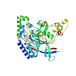
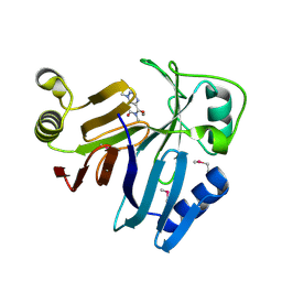
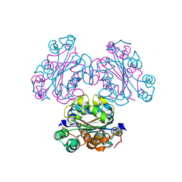
6IMP
DownloadVisualize
BU of 6imp by Molmil
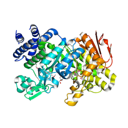
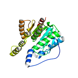
Crystal structure of alpha-beta hydrolase (ABH) from Vibrio vulnificus
分子名称: RTX toxin RtxA
著者Kim, M.H, Hwang, J.
登録日2018-10-23
公開日2019-08-21
最終更新日2024-03-27
実験手法X-RAY DIFFRACTION (2.62 Å)
主引用文献Makes caterpillars floppy-like effector-containing MARTX toxins require host ADP-ribosylation factor (ARF) proteins for systemic pathogenicity.
Proc.Natl.Acad.Sci.USA, 116, 2019
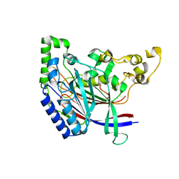
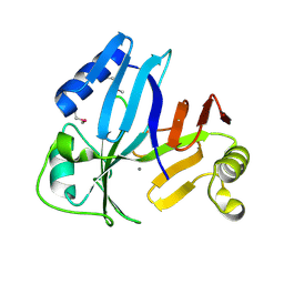
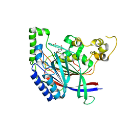
5K5U
DownloadVisualize
BU of 5k5u by Molmil
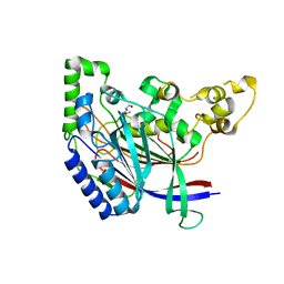
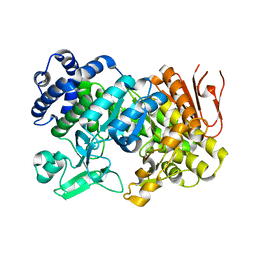
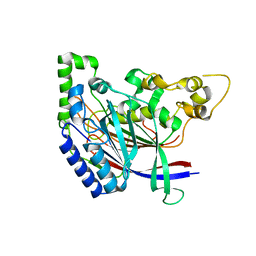
Crystal structure of N-terminal amidase
分子名称: Nta1p
著者Kim, M.K, Oh, S.-J, Lee, B.-G, Song, H.K.
登録日2016-05-24
公開日2017-01-11
最終更新日2024-03-20
実験手法X-RAY DIFFRACTION (2.7 Å)
主引用文献Structural basis for dual specificity of yeast N-terminal amidase in the N-end rule pathway.
Proc. Natl. Acad. Sci. U.S.A., 113, 2016
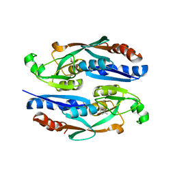
5K63
DownloadVisualize
BU of 5k63 by Molmil
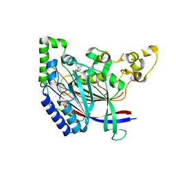
Crystal structure of N-terminal amidase C187S
分子名称: ASPARAGINE, GLYCINE, Nta1p
著者Kim, M.K, Oh, S.-J, Lee, B.-G, Song, H.K.
登録日2016-05-24
公開日2017-01-11
最終更新日2024-03-20
実験手法X-RAY DIFFRACTION (2.5 Å)
主引用文献Structural basis for dual specificity of yeast N-terminal amidase in the N-end rule pathway.
Proc. Natl. Acad. Sci. U.S.A., 113, 2016
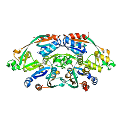
5K66
DownloadVisualize
BU of 5k66 by Molmil
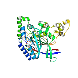
Crystal structure of N-terminal amidase with Asn-Glu peptide
分子名称: ASPARAGINE, GLUTAMIC ACID, Nta1p
著者Kim, M.K, Oh, S.-J, Lee, B.-G, Song, H.K.
登録日2016-05-24
公開日2017-01-11
最終更新日2024-03-20
実験手法X-RAY DIFFRACTION (2.002 Å)
主引用文献Structural basis for dual specificity of yeast N-terminal amidase in the N-end rule pathway.
Proc. Natl. Acad. Sci. U.S.A., 113, 2016
5K60
DownloadVisualize
BU of 5k60 by Molmil
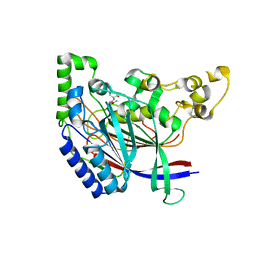
Crystal structure of N-terminal amidase with Gln-Val peptide
分子名称: GLUTAMINE, Nta1p, VALINE
著者Kim, M.K, Oh, S.-J, Lee, B.-G, Song, H.K.
登録日2016-05-24
公開日2017-01-11
最終更新日2024-03-20
実験手法X-RAY DIFFRACTION (1.9 Å)
主引用文献Structural basis for dual specificity of yeast N-terminal amidase in the N-end rule pathway.
Proc. Natl. Acad. Sci. U.S.A., 113, 2016
5K62
DownloadVisualize
BU of 5k62 by Molmil
Crystal structure of N-terminal amidase C187S
分子名称: ASPARAGINE, Nta1p, VALINE
著者Kim, M.K, Oh, S.-J, Lee, B.-G, Song, H.K.
登録日2016-05-24
公開日2017-01-11
最終更新日2024-03-20
実験手法X-RAY DIFFRACTION (1.899 Å)
主引用文献Structural basis for dual specificity of yeast N-terminal amidase in the N-end rule pathway.
Proc. Natl. Acad. Sci. U.S.A., 113, 2016
5K61
DownloadVisualize
BU of 5k61 by Molmil
Crystal structure of N-terminal amidase with Gln-Gly peptide
分子名称: GLUTAMINE, Nta1p
著者Kim, M.K, Oh, S.-J, Lee, B.-G, Song, H.K.
登録日2016-05-24
公開日2017-04-19
最終更新日2024-03-20
実験手法X-RAY DIFFRACTION (2.001 Å)
主引用文献Structural basis for dual specificity of yeast N-terminal amidase in the N-end rule pathway.
Proc. Natl. Acad. Sci. U.S.A., 113, 2016
5K5V
DownloadVisualize
BU of 5k5v by Molmil
Crystal structure of N-terminal amidase C187S
分子名称: Nta1p
著者Kim, M.K, Oh, S.-J, Lee, B.-G, Song, H.K.
登録日2016-05-24
公開日2017-04-05
最終更新日2024-03-20
実験手法X-RAY DIFFRACTION (1.947 Å)
主引用文献Structural basis for dual specificity of yeast N-terminal amidase in the N-end rule pathway.
Proc. Natl. Acad. Sci. U.S.A., 113, 2016
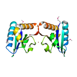
4TN5
DownloadVisualize
BU of 4tn5 by Molmil
Crystal Structure of Predicted Fructose Specific IIB from Escherichia Coli
分子名称: Fructose-like phosphotransferase enzyme IIB component 3, NICKEL (II) ION
著者Kim, M.S, Shin, D.H.
登録日2014-06-03
公開日2015-06-10
最終更新日2024-10-16
実験手法X-RAY DIFFRACTION (2.285 Å)
主引用文献High-accuracy protein modeling and its application to molecular replacement of crystallographic phasing
To Be Published
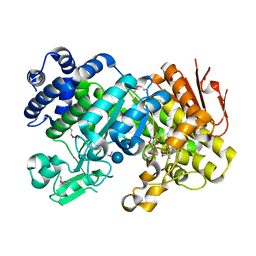
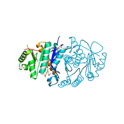
3CZG
DownloadVisualize
BU of 3czg by Molmil
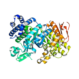
Crystal Structure Analysis of Sucrose hydrolase (SUH)-glucose complex
分子名称: Sucrose hydrolase, alpha-D-glucopyranose
著者Kim, M.I, Rhee, S.
登録日2008-04-29
公開日2008-07-15
最終更新日2024-10-16
実験手法X-RAY DIFFRACTION (1.8 Å)
主引用文献Crystal structures and mutagenesis of sucrose hydrolase from Xanthomonas axonopodis pv. glycines: insight into the exclusively hydrolytic amylosucrase fold.
J.Mol.Biol., 380, 2008
3CZE
DownloadVisualize
BU of 3cze by Molmil
Crystal Structure Analysis of Sucrose hydrolase (SUH)- Tris complex
分子名称: 2-AMINO-2-HYDROXYMETHYL-PROPANE-1,3-DIOL, Sucrose hydrolase
著者Kim, M.I, Rhee, S.
登録日2008-04-29
公開日2008-07-15
最終更新日2024-10-16
実験手法X-RAY DIFFRACTION (1.9 Å)
主引用文献Crystal structures and mutagenesis of sucrose hydrolase from Xanthomonas axonopodis pv. glycines: insight into the exclusively hydrolytic amylosucrase fold.
J.Mol.Biol., 380, 2008
3CZK
DownloadVisualize
BU of 3czk by Molmil
Crystal Structure Analysis of Sucrose hydrolase(SUH) E322Q-sucrose complex
分子名称: Sucrose hydrolase, beta-D-fructofuranose-(2-1)-alpha-D-glucopyranose
著者Kim, M.I, Rhee, S.
登録日2008-04-29
公開日2008-07-15
最終更新日2023-11-01
実験手法X-RAY DIFFRACTION (2.2 Å)
主引用文献Crystal structures and mutagenesis of sucrose hydrolase from Xanthomonas axonopodis pv. glycines: insight into the exclusively hydrolytic amylosucrase fold.
J.Mol.Biol., 380, 2008
7DR4
DownloadVisualize
BU of 7dr4 by Molmil
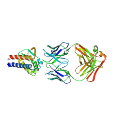
Complex of anti-human IL-2 antibody and human IL-2
分子名称: Interleukin-2, anti-human IL-2 antibody, mouse Ig G, ...
著者Kim, M.S, Kim, J.E.
登録日2020-12-25
公開日2021-04-14
最終更新日2023-11-29
実験手法X-RAY DIFFRACTION (2.49 Å)
主引用文献Crystal structure of human interleukin-2 in complex with TCB2, a new antibody-drug candidate with antitumor activity.
Oncoimmunology, 10, 2021
3CZL
DownloadVisualize
BU of 3czl by Molmil
Crystal Structure Analysis of Sucrose hydrolase(SUH) E322Q-glucose complex
分子名称: alpha-D-glucopyranose, sucrose hydrolase
著者Kim, M.I, Rhee, S.
登録日2008-04-29
公開日2008-07-15
最終更新日2023-11-01
実験手法X-RAY DIFFRACTION (2 Å)
主引用文献Crystal structures and mutagenesis of sucrose hydrolase from Xanthomonas axonopodis pv. glycines: insight into the exclusively hydrolytic amylosucrase fold.
J.Mol.Biol., 380, 2008
3OUM
DownloadVisualize
BU of 3oum by Molmil
Crystal Structure of toxoflavin-degrading enzyme in complex with toxoflavin
分子名称: 1,6-dimethylpyrimido[5,4-e][1,2,4]triazine-5,7(1H,6H)-dione, MANGANESE (II) ION, toxoflavin-degrading enzyme
著者Kim, M.I, Rhee, S.
登録日2010-09-15
公開日2011-08-10
最終更新日2024-04-03
実験手法X-RAY DIFFRACTION (2 Å)
主引用文献Structural and functional analysis of phytotoxin toxoflavin-degrading enzyme
Plos One, 6, 2011
3OUL
DownloadVisualize
BU of 3oul by Molmil
Crystal Structure of toxoflavin-degrading enzyme in a substrate-free form
分子名称: MANGANESE (II) ION, Toxoflavin-degrading enzyme
著者Kim, M.I, Rhee, S.
登録日2010-09-15
公開日2011-08-10
最終更新日2024-04-03
実験手法X-RAY DIFFRACTION (1.6 Å)
主引用文献Structural and functional analysis of phytotoxin toxoflavin-degrading enzyme
Plos One, 6, 2011
5IE9
DownloadVisualize
BU of 5ie9 by Molmil
Crystal structure of the Bacillus-conserved MazG protein, a nucleotide pyrophosphohydrolase
分子名称: MANGANESE (II) ION, Nucleotide pyrophosphohydrolase
著者Kim, M, Hong, M.
登録日2016-02-25
公開日2016-03-30
最終更新日2023-11-08
実験手法X-RAY DIFFRACTION (2.8 Å)
主引用文献Crystal structure of the Bacillus-conserved MazG protein, a nucleotide pyrophosphohydrolase.
Biochem.Biophys.Res.Commun., 472, 2016
5B6A
DownloadVisualize
BU of 5b6a by Molmil
Structure of Pyridoxal Kinasefrom Pseudomonas Aeruginosa
分子名称: 2-AMINO-2-HYDROXYMETHYL-PROPANE-1,3-DIOL, MAGNESIUM ION, PHOSPHATE ION, ...
著者Kim, M.I, Hong, M.
登録日2016-05-25
公開日2016-08-10
最終更新日2023-11-08
実験手法X-RAY DIFFRACTION (2 Å)
主引用文献Crystal structure and catalytic mechanism of pyridoxal kinase from Pseudomonas aeruginosa
Biochem.Biophys.Res.Commun., 478, 2016
8U2F
DownloadVisualize
BU of 8u2f by Molmil
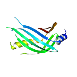
Vibrio parahaemolyticus ToxS periplasmic domain
分子名称: Transmembrane regulatory protein ToxS
著者Kim, M, Kull, F.J, Midgett, C.R.
登録日2023-09-06
公開日2024-09-11
実験手法X-RAY DIFFRACTION (2 Å)
主引用文献Vibrio parahaemolyticus ToxS periplasmic domain
To Be Published
4ENO
DownloadVisualize
BU of 4eno by Molmil
Crystal structure of oxidized human nm23-H1
分子名称: Nucleoside diphosphate kinase A, PHOSPHATE ION
著者Kim, M.-S, Shin, D.-H.
登録日2012-04-13
公開日2013-03-27
最終更新日2024-10-16
実験手法X-RAY DIFFRACTION (2.8 Å)
主引用文献Structure of Nm23-H1 under oxidative conditions.
Acta Crystallogr.,Sect.D, 69, 2013
5B62
DownloadVisualize
BU of 5b62 by Molmil
Crystal structure of N-terminal amidase with Asn-Glu-Ala peptide
分子名称: ASN-GLU-ALA, Nta1p
著者Kim, M.K, Oh, S.-J, Lee, B.-G, Song, H.K.
登録日2016-05-24
公開日2017-01-11
最終更新日2024-03-20
実験手法X-RAY DIFFRACTION (3.042 Å)
主引用文献Structural basis for dual specificity of yeast N-terminal amidase in the N-end rule pathway.
Proc. Natl. Acad. Sci. U.S.A., 113, 2016
8H5A
DownloadVisualize
BU of 8h5a by Molmil
Crystal structure of YhaJ effector binding domain (ligand-bound)
分子名称: 2-methylbenzene-1,4-diol, HTH-type transcriptional regulator YhaJ, SODIUM ION
著者Kim, M, Ryu, S.E.
登録日2022-10-12
公開日2023-10-25
最終更新日2024-03-06
実験手法X-RAY DIFFRACTION (2.803 Å)
主引用文献Structural basis of transcription factor YhaJ for DNT detection.
Iscience, 26, 2023
8H58
DownloadVisualize
BU of 8h58 by Molmil
Crystal structure of YhaJ effector binding domain
分子名称: HTH-type transcriptional regulator YhaJ, SODIUM ION
著者Kim, M, Ryu, S.E.
登録日2022-10-12
公開日2023-10-25
最終更新日2024-03-06
実験手法X-RAY DIFFRACTION (2.639 Å)
主引用文献Structural basis of transcription factor YhaJ for DNT detection.
Iscience, 26, 2023
2BQQ
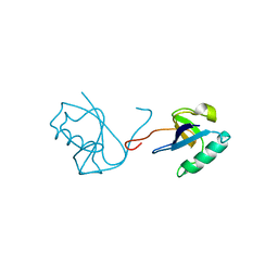
DownloadVisualize
BU of 2bqq by Molmil
X-ray Structure of the N-terminal Domain of Human Doublecortin
分子名称: NEURONAL MIGRATION PROTEIN DOUBLECORTIN
著者Kim, M.H, Cooper, D.R, Derewenda, U, Derewenda, Z.S.
登録日2005-04-27
公開日2006-07-19
最終更新日2023-12-13
実験手法X-RAY DIFFRACTION (2.2 Å)
主引用文献The Dc-Module of Doublecortin: Dynamics, Domain Boundaries, and Functional Implications.
Proteins, 64, 2006
3ODN
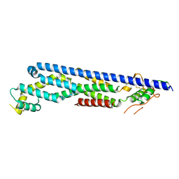
DownloadVisualize
BU of 3odn by Molmil
The crystal structure of Drosophila Dally-Like Protein core domain
分子名称: Dally-like protein
著者Kim, M.-S, Leahy, D.J.
登録日2010-08-11
公開日2011-07-27
最終更新日2024-10-30
実験手法X-RAY DIFFRACTION (2.4 Å)
主引用文献Structure of the protein core of the glypican Dally-like and localization of a region important for hedgehog signaling.
Proc.Natl.Acad.Sci.USA, 108, 2011

226707

件を2024-10-30に公開中

PDB statisticsPDBj update infoContact PDBjnumon