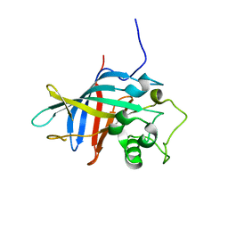
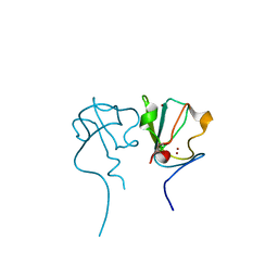
5YP8
 
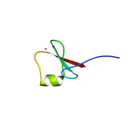 | | p62/SQSTM1 ZZ domain with Arg-peptide | | 分子名称: | 78 kDa glucose-regulated protein,Sequestosome-1, ZINC ION | | 著者 | Kwon, D.H, Kim, L, Song, H.K. | | 登録日 | 2017-11-01 | | 公開日 | 2018-08-29 | | 最終更新日 | 2024-03-27 | | 実験手法 | X-RAY DIFFRACTION (1.448 Å) | | 主引用文献 | Insights into degradation mechanism of N-end rule substrates by p62/SQSTM1 autophagy adapter.
Nat Commun, 9, 2018
|
|
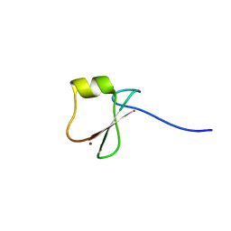
5YPG
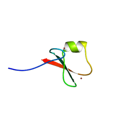 | | p62/SQSTM1 ZZ domain with Leu-peptide | | 分子名称: | 78 kDa glucose-regulated protein,Sequestosome-1, ZINC ION | | 著者 | Kwon, D.H, Kim, L, Song, H.K. | | 登録日 | 2017-11-01 | | 公開日 | 2018-08-29 | | 最終更新日 | 2024-03-27 | | 実験手法 | X-RAY DIFFRACTION (2.199 Å) | | 主引用文献 | Insights into degradation mechanism of N-end rule substrates by p62/SQSTM1 autophagy adapter.
Nat Commun, 9, 2018
|
|
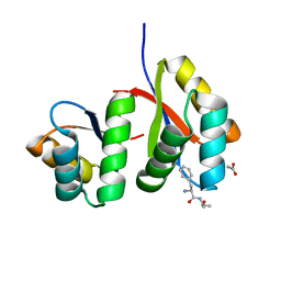
5YPA
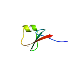 | | p62/SQSTM1 ZZ domain with Lys-peptide | | 分子名称: | 78 kDa glucose-regulated protein,Sequestosome-1, ZINC ION | | 著者 | Kwon, D.H, Kim, L, Song, H.K. | | 登録日 | 2017-11-01 | | 公開日 | 2018-08-29 | | 最終更新日 | 2024-03-27 | | 実験手法 | X-RAY DIFFRACTION (2.502 Å) | | 主引用文献 | Insights into degradation mechanism of N-end rule substrates by p62/SQSTM1 autophagy adapter.
Nat Commun, 9, 2018
|
|
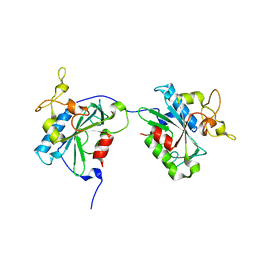
5YP7
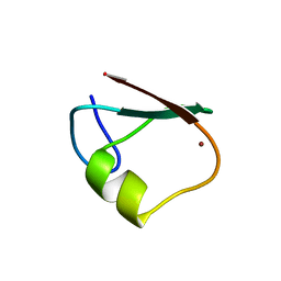 | | p62/SQSTM1 ZZ domain | | 分子名称: | Sequestosome-1, ZINC ION | | 著者 | Kwon, D.H, Kim, L, Song, H.K. | | 登録日 | 2017-11-01 | | 公開日 | 2018-08-29 | | 最終更新日 | 2024-03-27 | | 実験手法 | X-RAY DIFFRACTION (1.424 Å) | | 主引用文献 | Insights into degradation mechanism of N-end rule substrates by p62/SQSTM1 autophagy adapter.
Nat Commun, 9, 2018
|
|
7VGW
 | | Yeast gid10 with Pro-peptide | | 分子名称: | BJ4_G0041530.mRNA.1.CDS.1 | | 著者 | Shin, J.S, Park, S.H, Kim, L, Heo, J, Song, H.K. | | 登録日 | 2021-09-19 | | 公開日 | 2022-07-27 | | 最終更新日 | 2023-11-29 | | 実験手法 | X-RAY DIFFRACTION (2.8 Å) | | 主引用文献 | Crystal structure of yeast Gid10 in complex with Pro/N-degron.
Biochem.Biophys.Res.Commun., 582, 2021
|
|
6KHZ
 | | p62/SQSTM1 ZZ domain with Gly-peptide | | 分子名称: | Sequestosome-1, ZINC ION | | 著者 | Kwon, D.H, Kim, L, Song, H.K. | | 登録日 | 2019-07-16 | | 公開日 | 2020-01-22 | | 最終更新日 | 2023-11-22 | | 実験手法 | X-RAY DIFFRACTION (2.8 Å) | | 主引用文献 | Use of the LC3B-fusion technique for biochemical and structural studies of proteins involved in the N-degron pathway.
J.Biol.Chem., 295, 2020
|
|
7D34
 | | AtClpS1-peptide complex | | 分子名称: | ACETIC ACID, ALANINE, ATP-dependent Clp protease adapter protein CLPS1, ... | | 著者 | Heo, J, Kim, L, Kwon, D.H, Song, H.K. | | 登録日 | 2020-09-18 | | 公開日 | 2021-04-28 | | 最終更新日 | 2023-11-29 | | 実験手法 | X-RAY DIFFRACTION (2.007 Å) | | 主引用文献 | Structural basis for the N-degron specificity of ClpS1 from Arabidopsis thaliana.
Protein Sci., 30, 2021
|
|
6KGJ
 | | M1Q-hNTAQ1 C28S | | 分子名称: | Protein N-terminal glutamine amidohydrolase | | 著者 | Park, M.R, Kim, L, Kwon, D.H, Song, H.K. | | 登録日 | 2019-07-11 | | 公開日 | 2020-01-22 | | 最終更新日 | 2023-11-22 | | 実験手法 | X-RAY DIFFRACTION (1.8 Å) | | 主引用文献 | Use of the LC3B-fusion technique for biochemical and structural studies of proteins involved in the N-degron pathway.
J.Biol.Chem., 295, 2020
|
|
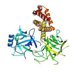
6KGI
 | | RLGS-yUbr1 Ubr box | | 分子名称: | E3 ubiquitin-protein ligase UBR1, ZINC ION | | 著者 | Heo, J, Kwon, D.H, Kim, L, Song, H.K. | | 登録日 | 2019-07-11 | | 公開日 | 2020-01-22 | | 最終更新日 | 2023-11-22 | | 実験手法 | X-RAY DIFFRACTION (1.04 Å) | | 主引用文献 | Use of the LC3B-fusion technique for biochemical and structural studies of proteins involved in the N-degron pathway.
J.Biol.Chem., 295, 2020
|
|
8EW4
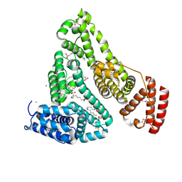 | | Human Serum Albumin with Cobalt (II) and Myristic Acid - crystal 1 | | 分子名称: | COBALT (II) ION, MYRISTIC ACID, Serum albumin | | 著者 | Gucwa, M, Cooper, D.R, Unciano, J, Lea, K, Kim, L, Lenkiewicz, J, Starban, I, Stewart, A.J, Minor, W, Center for Structural Genomics of Infectious Diseases (CSGID), Center for Structural Biology of Infectious Diseases (CSBID) | | 登録日 | 2022-10-21 | | 公開日 | 2022-11-09 | | 最終更新日 | 2024-06-26 | | 実験手法 | X-RAY DIFFRACTION (2.4 Å) | | 主引用文献 | Structural and biochemical characterisation of Co2+-binding sites on serum albumins and their interplay with fatty acids
Chem Sci, 14, 2023
|
|
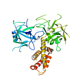
1TD5
 | | Crystal Structure of the Ligand Binding Domain of E. coli IclR. | | 分子名称: | Acetate operon repressor | | 著者 | Walker, J.R, Evdokimova, L, Zhang, R.-G, Bochkarev, A, Joachimiak, A, Arrowsmith, C, Edwards, A, Savchenko, A, Midwest Center for Structural Genomics (MCSG) | | 登録日 | 2004-05-21 | | 公開日 | 2004-07-13 | | 最終更新日 | 2011-07-13 | | 実験手法 | X-RAY DIFFRACTION (2.3 Å) | | 主引用文献 | Structural Analyses of the Ligand Binding Sites of the IclR family of transcriptional regulators
To be Published
|
|
2XOA
 | |
4I0Y
 | |


