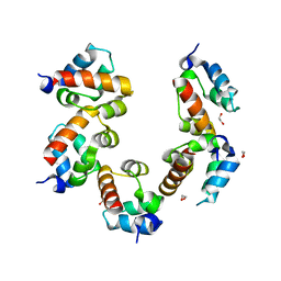
Loading
PDBj
MenuPDBj@FacebookPDBj@TwitterPDBj@YouTubewwPDB FoundationwwPDB
RCSB PDBPDBeBMRBAdv. SearchSearch help
PDB: 291 results

7L99
DownloadVisualize
BU of 7l99 by Molmil
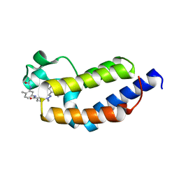
Crystal structure of BRDT bromodomain 2 in complex with CDD-1302
Descriptor: Bromodomain testis-specific protein, N-[3-(acetylamino)-4-methylphenyl]-3-(4-amino-2-methylphenyl)-1-methyl-1H-indazole-5-carboxamide, O-(O-(2-AMINOPROPYL)-O'-(2-METHOXYETHYL)POLYPROPYLENE GLYCOL 500)
Authors:Sharma, R, Yu, Z, Ku, A.F, Anglin, J.L, Ucisik, M.N, Faver, J.C, Sankaran, B, Kim, C, Matzuk, M.M.
Deposit date:2021-01-03
Release date:2021-06-30
Last modified:2023-10-18
Method:X-RAY DIFFRACTION (1.9 Å)
Cite:Discovery and characterization of bromodomain 2-specific inhibitors of BRDT.
Proc.Natl.Acad.Sci.USA, 118, 2021
3DQH
DownloadVisualize
BU of 3dqh by Molmil
Structure of the Yellow Fluorescent Protein Citrine Frozen at 1000 Atmospheres Number 5: Structure 9 in a Series of 26 High Pressure Structures
Descriptor: Green fluorescent protein
Authors:Barstow, B, Kim, C.U.
Deposit date:2008-07-09
Release date:2008-09-23
Last modified:2023-11-15
Method:X-RAY DIFFRACTION (1.45 Å)
Cite:Alteration of citrine structure by hydrostatic pressure explains the accompanying spectral shift.
Proc.Natl.Acad.Sci.Usa, 105, 2008
3DQ9
DownloadVisualize
BU of 3dq9 by Molmil
Structure of the Yellow Fluorescent Protein Citrine Frozen at 1500 Atmospheres Number 1: Structure 15 in a Series of 26 High Pressure Structures
Descriptor: Green fluorescent protein
Authors:Barstow, B, Kim, C.U.
Deposit date:2008-07-09
Release date:2008-09-23
Last modified:2023-11-15
Method:X-RAY DIFFRACTION (1.4 Å)
Cite:Alteration of citrine structure by hydrostatic pressure explains the accompanying spectral shift.
Proc.Natl.Acad.Sci.Usa, 105, 2008
3DQM
DownloadVisualize
BU of 3dqm by Molmil
Structure of the Yellow Fluorescent Protein Citrine Frozen at 750 Atmospheres: Structure 4 in a Series of 26 High Pressure Structures
Descriptor: Green fluorescent protein
Authors:Barstow, B, Kim, C.U.
Deposit date:2008-07-09
Release date:2008-09-23
Last modified:2023-11-15
Method:X-RAY DIFFRACTION (1.44 Å)
Cite:Alteration of citrine structure by hydrostatic pressure explains the accompanying spectral shift.
Proc.Natl.Acad.Sci.Usa, 105, 2008
3DQ2
DownloadVisualize
BU of 3dq2 by Molmil
Structure of the Yellow Fluorescent Protein Citrine Frozen at 4000 Atmospheres Number 1: Structure 23 in a Series of 26 High Pressure Structures
Descriptor: Green fluorescent protein
Authors:Barstow, B, Kim, C.U.
Deposit date:2008-07-09
Release date:2008-09-23
Last modified:2023-11-15
Method:X-RAY DIFFRACTION (1.6 Å)
Cite:Alteration of citrine structure by hydrostatic pressure explains the accompanying spectral shift.
Proc.Natl.Acad.Sci.Usa, 105, 2008
3DQC
DownloadVisualize
BU of 3dqc by Molmil
Structure of the Yellow Fluorescent Protein Citrine Frozen at 1250 Atmospheres Number 3: Structure 13 in a Series of 26 High Pressure Structures
Descriptor: Green fluorescent protein
Authors:Barstow, B, Kim, C.U.
Deposit date:2008-07-09
Release date:2008-09-23
Last modified:2023-11-15
Method:X-RAY DIFFRACTION (1.49 Å)
Cite:Alteration of citrine structure by hydrostatic pressure explains the accompanying spectral shift.
Proc.Natl.Acad.Sci.Usa, 105, 2008
3DQ4
DownloadVisualize
BU of 3dq4 by Molmil
Structure of the Yellow Fluorescent Protein Citrine Frozen at 2000 Atmospheres Number 2: Structure 20 in a Series of 26 High Pressure Structures
Descriptor: Green fluorescent protein
Authors:Barstow, B, Kim, C.U.
Deposit date:2008-07-09
Release date:2008-09-23
Last modified:2023-11-15
Method:X-RAY DIFFRACTION (1.47 Å)
Cite:Alteration of citrine structure by hydrostatic pressure explains the accompanying spectral shift.
Proc.Natl.Acad.Sci.Usa, 105, 2008
3DQF
DownloadVisualize
BU of 3dqf by Molmil
Structure of the Yellow Fluorescent Protein Citrine Frozen at 1000 Atmospheres Number 6: Structure 10 in a Series of 26 High Pressure Structures
Descriptor: Green fluorescent protein
Authors:Barstow, B, Kim, C.U.
Deposit date:2008-07-09
Release date:2008-09-23
Last modified:2023-11-15
Method:X-RAY DIFFRACTION (1.46 Å)
Cite:Alteration of citrine structure by hydrostatic pressure explains the accompanying spectral shift.
Proc.Natl.Acad.Sci.Usa, 105, 2008
3DPX
DownloadVisualize
BU of 3dpx by Molmil
Structure of the Yellow Fluorescent Protein Citrine Frozen at 5000 Atmospheres: Structure 26 in a Series of 26 High Pressure Structures
Descriptor: Green fluorescent protein
Authors:Barstow, B, Kim, C.U.
Deposit date:2008-07-09
Release date:2008-09-23
Last modified:2023-11-15
Method:X-RAY DIFFRACTION (1.5 Å)
Cite:Alteration of citrine structure by hydrostatic pressure explains the accompanying spectral shift.
Proc.Natl.Acad.Sci.Usa, 105, 2008
3DQ5
DownloadVisualize
BU of 3dq5 by Molmil
Structure of the Yellow Fluorescent Protein Citrine Frozen at 1960 Atmospheres: Structure 19 in a Series of 26 High Pressure Structures
Descriptor: Green fluorescent protein
Authors:Barstow, B, Kim, C.U.
Deposit date:2008-07-09
Release date:2008-09-23
Last modified:2023-11-15
Method:X-RAY DIFFRACTION (1.5 Å)
Cite:Alteration of citrine structure by hydrostatic pressure explains the accompanying spectral shift.
Proc.Natl.Acad.Sci.Usa, 105, 2008
3DQE
DownloadVisualize
BU of 3dqe by Molmil
Structure of the Yellow Fluorescent Protein Citrine Frozen at 1250 Atmospheres Number 1: Structure 11 in a Series of 26 High Pressure Structures
Descriptor: Green fluorescent protein
Authors:Barstow, B, Kim, C.U.
Deposit date:2008-07-09
Release date:2008-09-23
Last modified:2023-11-15
Method:X-RAY DIFFRACTION (1.43 Å)
Cite:Alteration of citrine structure by hydrostatic pressure explains the accompanying spectral shift.
Proc.Natl.Acad.Sci.Usa, 105, 2008
3DQI
DownloadVisualize
BU of 3dqi by Molmil
Structure of the Yellow Fluorescent Protein Citrine Frozen at 1000 Atmospheres Number 4: Structure 8 in a Series of 26 High Pressure Structures
Descriptor: Green fluorescent protein
Authors:Barstow, B, Kim, C.U.
Deposit date:2008-07-09
Release date:2008-09-23
Last modified:2023-11-15
Method:X-RAY DIFFRACTION (1.42 Å)
Cite:Alteration of citrine structure by hydrostatic pressure explains the accompanying spectral shift.
Proc.Natl.Acad.Sci.Usa, 105, 2008
3DQU
DownloadVisualize
BU of 3dqu by Molmil
Structure of the Yellow Fluorescent Protein Citrine Frozen at 2000 Atmospheres Number 1: Structure 20 in a Series of 26 High Pressure Structures
Descriptor: Green fluorescent protein
Authors:Barstow, B, Kim, C.U.
Deposit date:2008-07-09
Release date:2008-09-23
Last modified:2023-11-15
Method:X-RAY DIFFRACTION (1.42 Å)
Cite:Alteration of citrine structure by hydrostatic pressure explains the accompanying spectral shift.
Proc.Natl.Acad.Sci.Usa, 105, 2008
3DQJ
DownloadVisualize
BU of 3dqj by Molmil
Structure of the Yellow Fluorescent Protein Citrine Frozen at 1000 Atmospheres Number 3: Structure 7 in a Series of 26 High Pressure Structures
Descriptor: Green fluorescent protein
Authors:Barstow, B, Kim, C.U.
Deposit date:2008-07-09
Release date:2008-09-23
Last modified:2023-11-15
Method:X-RAY DIFFRACTION (1.51 Å)
Cite:Alteration of citrine structure by hydrostatic pressure explains the accompanying spectral shift.
Proc.Natl.Acad.Sci.Usa, 105, 2008
3DQ7
DownloadVisualize
BU of 3dq7 by Molmil
Structure of the Yellow Fluorescent Protein Citrine Frozen at 1920 Atmospheres Number 1: Structure 17 in a Series of 26 High Pressure Structures
Descriptor: Green fluorescent protein
Authors:Barstow, B, Kim, C.U.
Deposit date:2008-07-09
Release date:2008-09-23
Last modified:2023-11-15
Method:X-RAY DIFFRACTION (1.23 Å)
Cite:Alteration of citrine structure by hydrostatic pressure explains the accompanying spectral shift.
Proc.Natl.Acad.Sci.Usa, 105, 2008
3DQK
DownloadVisualize
BU of 3dqk by Molmil
Structure of the Yellow Fluorescent Protein Citrine Frozen at 1000 Atmospheres Number 2: Structure 6 in a Series of 26 High Pressure Structures
Descriptor: Green fluorescent protein
Authors:Barstow, B, Kim, C.U.
Deposit date:2008-07-09
Release date:2008-09-23
Last modified:2023-11-15
Method:X-RAY DIFFRACTION (1.4 Å)
Cite:Alteration of citrine structure by hydrostatic pressure explains the accompanying spectral shift.
Proc.Natl.Acad.Sci.Usa, 105, 2008
3DPZ
DownloadVisualize
BU of 3dpz by Molmil
Structure of the Yellow Fluorescent Protein Citrine Frozen at 4000 Atmospheres Number 3: Structure 25 in a Series of 26 High Pressure Structures
Descriptor: Green fluorescent protein
Authors:Barstow, B, Kim, C.U.
Deposit date:2008-07-09
Release date:2008-09-23
Last modified:2023-11-15
Method:X-RAY DIFFRACTION (1.7 Å)
Cite:Alteration of citrine structure by hydrostatic pressure explains the accompanying spectral shift.
Proc.Natl.Acad.Sci.Usa, 105, 2008
3DQ8
DownloadVisualize
BU of 3dq8 by Molmil
Structure of the Yellow Fluorescent Protein Citrine Frozen at 1500 Atmospheres Number 2: Structure 16 in a Series of 26 High Pressure Structures
Descriptor: Green fluorescent protein
Authors:Barstow, B, Kim, C.U.
Deposit date:2008-07-09
Release date:2008-09-23
Last modified:2023-11-15
Method:X-RAY DIFFRACTION (1.51 Å)
Cite:Alteration of citrine structure by hydrostatic pressure explains the accompanying spectral shift.
Proc.Natl.Acad.Sci.Usa, 105, 2008
2QO3
DownloadVisualize
BU of 2qo3 by Molmil
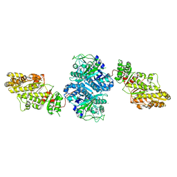
Crystal Structure of [KS3][AT3] didomain from module 3 of 6-deoxyerthronolide B synthase
Descriptor: (2S, 3R)-3-HYDROXY-4-OXO-7,10-TRANS,TRANS-DODECADIENAMIDE, ACETATE ION, ...
Authors:Khosla, C, Cane, E.D, Tang, Y, Chen, Y.A, Kim, C.Y.
Deposit date:2007-07-19
Release date:2007-09-04
Last modified:2011-07-13
Method:X-RAY DIFFRACTION (2.59 Å)
Cite:Structural and mechanistic analysis of protein interactions in module 3 of the 6-deoxyerythronolide B synthase
Chem.Biol., 14, 2007
3IDB
DownloadVisualize
BU of 3idb by Molmil
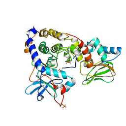
Crystal structure of (108-268)RIIb:C holoenzyme of cAMP-dependent protein kinase
Descriptor: MANGANESE (II) ION, PHOSPHOAMINOPHOSPHONIC ACID-ADENYLATE ESTER, cAMP-dependent protein kinase catalytic subunit alpha, ...
Authors:Brown, S.H.J, Wu, J, Kim, C, Alberto, K, Taylor, S.S.
Deposit date:2009-07-20
Release date:2009-09-29
Last modified:2023-09-06
Method:X-RAY DIFFRACTION (1.62 Å)
Cite:Novel isoform-specific interfaces revealed by PKA RIIbeta holoenzyme structures.
J.Mol.Biol., 393, 2009
3IDC
DownloadVisualize
BU of 3idc by Molmil
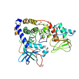
Crystal structure of (102-265)RIIb:C holoenzyme of cAMP-dependent protein kinase
Descriptor: MANGANESE (II) ION, PHOSPHOAMINOPHOSPHONIC ACID-ADENYLATE ESTER, cAMP-dependent protein kinase catalytic subunit alpha, ...
Authors:Brown, S.H.J, Wu, J, Kim, C, Alberto, K, Taylor, S.S.
Deposit date:2009-07-20
Release date:2009-09-29
Last modified:2023-09-06
Method:X-RAY DIFFRACTION (2.7 Å)
Cite:Novel isoform-specific interfaces revealed by PKA RIIbeta holoenzyme structures.
J.Mol.Biol., 393, 2009
4PZN
DownloadVisualize
BU of 4pzn by Molmil
Crystal structure of PHC3 SAM L971E
Descriptor: 1,2-ETHANEDIOL, Polyhomeotic-like protein 3
Authors:Nanyes, D.R, Junco, S.E, Taylor, A.B, Robinson, A.K, Patterson, N.L, Shivarajpur, A, Halloran, J, Hale, S.M, Kaur, Y, Hart, P.J, Kim, C.A.
Deposit date:2014-03-31
Release date:2014-07-30
Last modified:2023-09-20
Method:X-RAY DIFFRACTION (2.3 Å)
Cite:Multiple polymer architectures of human polyhomeotic homolog 3 sterile alpha motif.
Proteins, 82, 2014
3IXS
DownloadVisualize
BU of 3ixs by Molmil
Ring1B C-terminal domain/RYBP C-terminal domain Complex
Descriptor: 1,2-ETHANEDIOL, 2-[N-CYCLOHEXYLAMINO]ETHANE SULFONIC ACID, E3 ubiquitin-protein ligase RING2, ...
Authors:Wang, R, Taylor, A.B, Kim, C.A.
Deposit date:2009-09-04
Release date:2010-08-25
Last modified:2024-02-21
Method:X-RAY DIFFRACTION (1.7 Å)
Cite:Polycomb Group Targeting through Different Binding Partners of RING1B C-Terminal Domain.
Structure, 18, 2010
4PZO
DownloadVisualize
BU of 4pzo by Molmil
Crystal structure of PHC3 SAM L967R
Descriptor: Polyhomeotic-like protein 3
Authors:Nanyes, D.R, Junco, S.E, Taylor, A.B, Robinson, A.K, Patterson, N.L, Shivarajpur, A, Halloran, J, Hale, S.M, Kaur, Y, Hart, P.J, Kim, C.A.
Deposit date:2014-03-31
Release date:2014-07-30
Last modified:2023-09-20
Method:X-RAY DIFFRACTION (2.25 Å)
Cite:Multiple polymer architectures of human polyhomeotic homolog 3 sterile alpha motif.
Proteins, 82, 2014
6BDL
DownloadVisualize
BU of 6bdl by Molmil
Crystal structure of cGMP-dependent protein kinase Ialpha (PKG Ialpha) catalytic domain in apo state
Descriptor: IMIDAZOLE, cGMP-dependent protein kinase 1
Authors:Qin, L, Sankaran, B, Kim, C.
Deposit date:2017-10-23
Release date:2018-10-24
Last modified:2023-10-04
Method:X-RAY DIFFRACTION (1.96 Å)
Cite:Crystal structure of cGMP-dependent protein kinase Ialpha (PKG Ialpha) catalytic domain
to be published

222624

数据于2024-07-17公开中

PDB statisticsPDBj update infoContact PDBjnumon