Loading
PDBj
メニューPDBj@FacebookPDBj@TwitterPDBj@YouTubewwPDB FoundationwwPDB
RCSB PDBPDBeBMRBAdv. SearchSearch help
PDB: 214 件

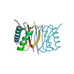
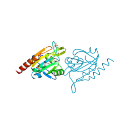
4XFI
DownloadVisualize
BU of 4xfi by Molmil
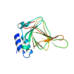
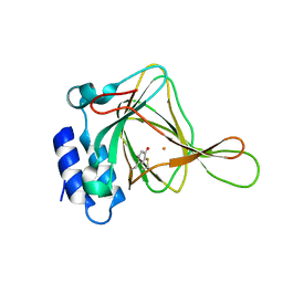
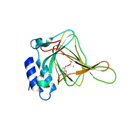
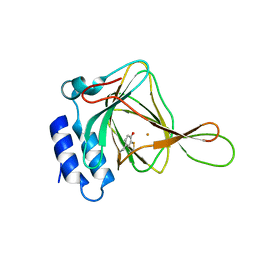
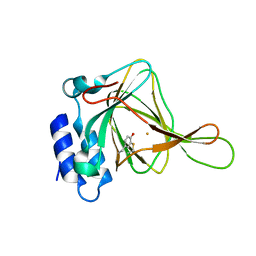
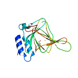
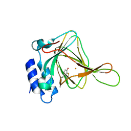
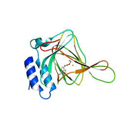
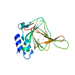
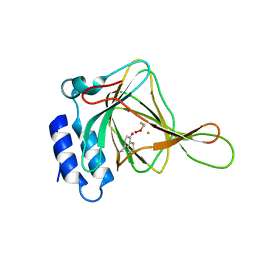
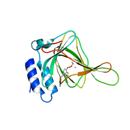
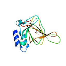
cysteine dioxygenase variant - Y157F at pH 6.2 with homocysteine
分子名称: 2-AMINO-4-MERCAPTO-BUTYRIC ACID, Cysteine dioxygenase type 1, FE (III) ION
著者Driggers, C.M, Karplus, P.A.
登録日2014-12-26
公開日2016-02-24
最終更新日2024-04-03
実験手法X-RAY DIFFRACTION (1.4 Å)
主引用文献Structure-Based Insights into the Role of the Cys-Tyr Crosslink and Inhibitor Recognition by Mammalian Cysteine Dioxygenase.
J. Mol. Biol., 428, 2016
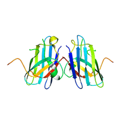
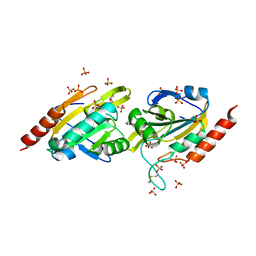
4XS1
DownloadVisualize
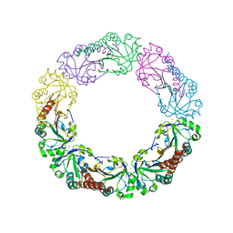
BU of 4xs1 by Molmil
Salmonella typhimurium AhpC T43V mutant
分子名称: Alkyl hydroperoxide reductase subunit C, CHLORIDE ION, POTASSIUM ION, ...
著者Perkins, A, Nelson, K, Parsonage, D, Poole, L, Karplus, P.A.
登録日2015-01-21
公開日2016-01-27
最終更新日2023-09-27
実験手法X-RAY DIFFRACTION (2.1 Å)
主引用文献Experimentally Dissecting the Origins of Peroxiredoxin Catalysis.
Antioxid.Redox Signal., 28, 2018
2P2T
DownloadVisualize
BU of 2p2t by Molmil
Crystal structure of dynein light chain LC8 bound to residues 123-138 of intermediate chain IC74
分子名称: ACETATE ION, Dynein intermediate chain peptide, Dynein light chain 1, ...
著者Benison, G, Karplus, P.A, Barbar, E.
登録日2007-03-07
公開日2008-01-15
最終更新日2024-02-21
実験手法X-RAY DIFFRACTION (3 Å)
主引用文献Structure and dynamics of LC8 complexes with KXTQT-motif peptides: swallow and dynein intermediate chain compete for a common site.
J.Mol.Biol., 371, 2007
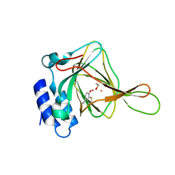
2R27
DownloadVisualize
BU of 2r27 by Molmil
Constitutively zinc-deficient mutant of human superoxide dismutase (SOD), C6A, H80S, H83S, C111S
分子名称: COPPER (II) ION, Superoxide dismutase [Cu-Zn]
著者Roberts, B.R, Getzoff, E.D, Karplus, P.A, Beckman, J.S, Tainer, J.A.
登録日2007-08-24
公開日2007-12-11
最終更新日2023-08-30
実験手法X-RAY DIFFRACTION (2 Å)
主引用文献Structural characterization of zinc-deficient human superoxide dismutase and implications for ALS.
J.Mol.Biol., 373, 2007
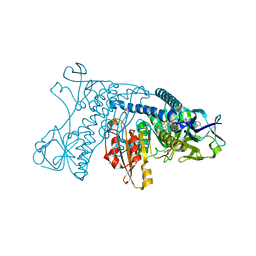
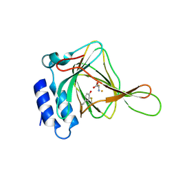
3GRS
DownloadVisualize
BU of 3grs by Molmil
REFINED STRUCTURE OF GLUTATHIONE REDUCTASE AT 1.54 ANGSTROMS RESOLUTION
分子名称: FLAVIN-ADENINE DINUCLEOTIDE, GLUTATHIONE REDUCTASE, PHOSPHATE ION
著者Schulz, G.E, Karplus, P.A.
登録日1988-02-05
公開日1988-04-16
最終更新日2024-06-05
実験手法X-RAY DIFFRACTION (1.54 Å)
主引用文献Refined structure of glutathione reductase at 1.54 A resolution.
J.Mol.Biol., 195, 1987
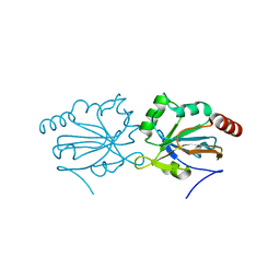
4G2E
DownloadVisualize
BU of 4g2e by Molmil
Crystal structure of a dimeric PrxQ from Sulfolobus tokodaii
分子名称: Peroxiredoxin
著者Perkins, A, Gretes, M.C, Karplus, P.A.
登録日2012-07-11
公開日2012-10-24
最終更新日2023-09-13
実験手法X-RAY DIFFRACTION (1.4 Å)
主引用文献Mapping the Active Site Helix-to-Strand Conversion of CxxxxC Peroxiredoxin Q Enzymes.
Biochemistry, 51, 2012
4GQF
DownloadVisualize
BU of 4gqf by Molmil
Aeropyrum pernix Peroxiredoxin Q Enzyme in the Locally Unfolded Conformation
分子名称: GLYCEROL, SULFATE ION, Thiol peroxidase
著者Perkins, A, Karplus, P.A, Gretes, M.C, Nelson, K.J, Poole, L.B.
登録日2012-08-22
公開日2012-10-24
最終更新日2023-12-06
実験手法X-RAY DIFFRACTION (2.3 Å)
主引用文献Mapping the Active Site Helix-to-Strand Conversion of CxxxxC Peroxiredoxin Q Enzymes.
Biochemistry, 51, 2012
4GQC
DownloadVisualize
BU of 4gqc by Molmil
Crystal Structure of Aeropyrum pernix Peroxiredoxin Q Enzyme in Fully-Folded and Locally-Unfolded Conformations
分子名称: DITHIANE DIOL, GLYCEROL, SULFATE ION, ...
著者Perkins, A, Karplus, P.A, Gretes, M.C, Nelson, K.J, Poole, L.B.
登録日2012-08-22
公開日2012-10-24
最終更新日2023-09-13
実験手法X-RAY DIFFRACTION (2 Å)
主引用文献Mapping the Active Site Helix-to-Strand Conversion of CxxxxC Peroxiredoxin Q Enzymes.
Biochemistry, 51, 2012
4IES
DownloadVisualize
BU of 4ies by Molmil
Cys-persulfenate bound Cysteine Dioxygenase at pH 6.2 in the presence of Cys
分子名称: Cysteine dioxygenase type 1, FE (III) ION, S-HYDROPEROXYCYSTEINE
著者Driggers, C.M, Cooley, R.B, Sankaran, B, Karplus, P.A.
登録日2012-12-13
公開日2013-06-26
最終更新日2013-09-11
実験手法X-RAY DIFFRACTION (1.4 Å)
主引用文献Cysteine Dioxygenase Structures from pH4 to 9: Consistent Cys-Persulfenate Formation at Intermediate pH and a Cys-Bound Enzyme at Higher pH.
J.Mol.Biol., 425, 2013
4IEW
DownloadVisualize
BU of 4iew by Molmil
Cys-only bound Cysteine Dioxygenase at pH 9.0 in the presence of Cys
分子名称: CYSTEINE, Cysteine dioxygenase type 1, FE (II) ION
著者Driggers, C.M, Cooley, R.B, Karplus, P.A.
登録日2012-12-13
公開日2013-06-26
最終更新日2013-09-11
実験手法X-RAY DIFFRACTION (1.45 Å)
主引用文献Cysteine Dioxygenase Structures from pH4 to 9: Consistent Cys-Persulfenate Formation at Intermediate pH and a Cys-Bound Enzyme at Higher pH.
J.Mol.Biol., 425, 2013
2GH2
DownloadVisualize
BU of 2gh2 by Molmil
1.5 A Resolution R. Norvegicus Cysteine Dioxygenase Structure Crystallized in the Presence of Cysteine
分子名称: Cysteine dioxygenase type I, FE (III) ION, SULFATE ION
著者Simmons, C.R, Karplus, P.A, Stipanuk, M.H.
登録日2006-03-24
公開日2006-04-11
最終更新日2023-08-30
実験手法X-RAY DIFFRACTION (1.5 Å)
主引用文献Crystal Structure of Mammalian Cysteine Dioxygenase: A NOVEL MONONUCLEAR IRON CENTER FOR CYSTEINE THIOL OXIDATION.
J.Biol.Chem., 281, 2006
4IEX
DownloadVisualize
BU of 4iex by Molmil
unliganded room-temp Cysteine Dioxygenase at pH 6.2
分子名称: Cysteine dioxygenase type 1, FE (II) ION
著者Driggers, C.M, Cooley, R.B, Karplus, P.A.
登録日2012-12-13
公開日2013-06-26
最終更新日2013-09-11
実験手法X-RAY DIFFRACTION (2.15 Å)
主引用文献Cysteine Dioxygenase Structures from pH4 to 9: Consistent Cys-Persulfenate Formation at Intermediate pH and a Cys-Bound Enzyme at Higher pH.
J.Mol.Biol., 425, 2013
4IER
DownloadVisualize
BU of 4ier by Molmil
Cys-persulfenate bound Cysteine Dioxygenase at pH 5.5 in the presence of Cys
分子名称: Cysteine dioxygenase type 1, FE (II) ION, S-HYDROPEROXYCYSTEINE
著者Driggers, C.M, Cooley, R.B, Karplus, P.A.
登録日2012-12-13
公開日2013-06-26
最終更新日2013-09-11
実験手法X-RAY DIFFRACTION (1.45 Å)
主引用文献Cysteine Dioxygenase Structures from pH4 to 9: Consistent Cys-Persulfenate Formation at Intermediate pH and a Cys-Bound Enzyme at Higher pH.
J.Mol.Biol., 425, 2013
4IEZ
DownloadVisualize
BU of 4iez by Molmil
unliganded Cysteine Dioxygenase at pH 8.0
分子名称: Cysteine dioxygenase type 1, FE (II) ION
著者Driggers, C.M, Cooley, R.B, Karplus, P.A.
登録日2012-12-13
公開日2013-06-26
最終更新日2013-09-11
実験手法X-RAY DIFFRACTION (1.39 Å)
主引用文献Cysteine Dioxygenase Structures from pH4 to 9: Consistent Cys-Persulfenate Formation at Intermediate pH and a Cys-Bound Enzyme at Higher pH.
J.Mol.Biol., 425, 2013
4IEO
DownloadVisualize
BU of 4ieo by Molmil
unliganded Cysteine Dioxygenase at pH 4.0 in the presence of Cys
分子名称: Cysteine dioxygenase type 1, FE (II) ION
著者Driggers, C.M, Cooley, R.B, Karplus, P.A.
登録日2012-12-13
公開日2013-06-26
最終更新日2013-09-11
実験手法X-RAY DIFFRACTION (1.55 Å)
主引用文献Cysteine Dioxygenase Structures from pH4 to 9: Consistent Cys-Persulfenate Formation at Intermediate pH and a Cys-Bound Enzyme at Higher pH.
J.Mol.Biol., 425, 2013
4IEV
DownloadVisualize
BU of 4iev by Molmil
Cys-only bound Cysteine Dioxygenase at pH 8.0 in the presence of Cys
分子名称: CYSTEINE, Cysteine dioxygenase type 1, FE (II) ION
著者Driggers, C.M, Cooley, R.B, Karplus, P.A.
登録日2012-12-13
公開日2013-06-26
最終更新日2013-09-11
実験手法X-RAY DIFFRACTION (1.6 Å)
主引用文献Cysteine Dioxygenase Structures from pH4 to 9: Consistent Cys-Persulfenate Formation at Intermediate pH and a Cys-Bound Enzyme at Higher pH.
J.Mol.Biol., 425, 2013
4IEP
DownloadVisualize
BU of 4iep by Molmil
unliganded Cysteine Dioxygenase at pH 4.5 in the presence of Cys
分子名称: Cysteine dioxygenase type 1, FE (II) ION
著者Driggers, C.M, Cooley, R.B, Karplus, P.A.
登録日2012-12-13
公開日2013-06-26
最終更新日2013-09-11
実験手法X-RAY DIFFRACTION (1.45 Å)
主引用文献Cysteine Dioxygenase Structures from pH4 to 9: Consistent Cys-Persulfenate Formation at Intermediate pH and a Cys-Bound Enzyme at Higher pH.
J.Mol.Biol., 425, 2013
4IEU
DownloadVisualize
BU of 4ieu by Molmil
Cys-persulfenate bound Cysteine Dioxygenase at pH 7.0 in the presence of Cys
分子名称: Cysteine dioxygenase type 1, FE (II) ION, S-HYDROPEROXYCYSTEINE
著者Driggers, C.M, Cooley, R.B, Karplus, P.A.
登録日2012-12-13
公開日2013-06-26
最終更新日2013-09-11
実験手法X-RAY DIFFRACTION (1.25 Å)
主引用文献Cysteine Dioxygenase Structures from pH4 to 9: Consistent Cys-Persulfenate Formation at Intermediate pH and a Cys-Bound Enzyme at Higher pH.
J.Mol.Biol., 425, 2013
4IEQ
DownloadVisualize
BU of 4ieq by Molmil
unliganded Cysteine Dioxygenase at pH 5.0 in the presence of Cys
分子名称: Cysteine dioxygenase type 1, FE (II) ION
著者Driggers, C.M, Cooley, R.B, Karplus, P.A.
登録日2012-12-13
公開日2013-06-26
最終更新日2013-09-11
実験手法X-RAY DIFFRACTION (1.396 Å)
主引用文献Cysteine Dioxygenase Structures from pH4 to 9: Consistent Cys-Persulfenate Formation at Intermediate pH and a Cys-Bound Enzyme at Higher pH.
J.Mol.Biol., 425, 2013
4IET
DownloadVisualize
BU of 4iet by Molmil
Cys-persulfenate bound Cysteine Dioxygenase at pH 6.8 in the presence of Cys
分子名称: Cysteine dioxygenase type 1, FE (II) ION, S-HYDROPEROXYCYSTEINE
著者Driggers, C.M, Cooley, R.B, Karplus, P.A.
登録日2012-12-13
公開日2013-06-26
最終更新日2015-06-24
実験手法X-RAY DIFFRACTION (1.4 Å)
主引用文献Cysteine dioxygenase structures from pH4 to 9: consistent cys-persulfenate formation at intermediate pH and a Cys-bound enzyme at higher pH.
J.Mol.Biol., 425, 2013
4IEY
DownloadVisualize
BU of 4iey by Molmil
Cys-persulfenate bound Cysteine Dioxygenase at pH 7.0 in the presence of Cys, home-source structure
分子名称: Cysteine dioxygenase type 1, FE (II) ION, S-HYDROPEROXYCYSTEINE
著者Driggers, C.M, Cooley, R.B, Karplus, P.A.
登録日2012-12-13
公開日2013-06-26
最終更新日2013-09-11
実験手法X-RAY DIFFRACTION (1.63 Å)
主引用文献Cysteine Dioxygenase Structures from pH4 to 9: Consistent Cys-Persulfenate Formation at Intermediate pH and a Cys-Bound Enzyme at Higher pH.
J.Mol.Biol., 425, 2013
2KLO
DownloadVisualize
BU of 2klo by Molmil
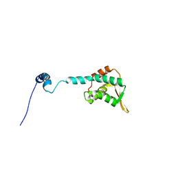
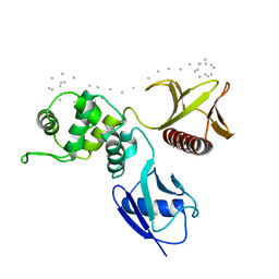
Structure of the Cdt1 C-terminal domain
分子名称: DNA replication factor Cdt1
著者Khayrutdinov, B.I, Bae, W.J, Yun, Y.M, Tsuyama, T, Kim, J.J, Hwang, E, Ryu, K.-S, Cheong, H.-K, Cheong, C, Karplus, P.A, Guntert, P, Tada, S, Jeon, Y.H, Cho, Y.
登録日2009-07-06
公開日2009-10-13
最終更新日2024-05-29
実験手法SOLUTION NMR
主引用文献Structure of the Cdt1 C-terminal domain: Conservation of the winged helix fold in replication licensing factors
Protein Sci., 18, 2009
1SGH
DownloadVisualize
BU of 1sgh by Molmil
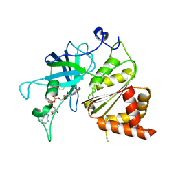
Moesin FERM domain bound to EBP50 C-terminal peptide
分子名称: Ezrin-radixin-moesin binding phosphoprotein 50, Moesin
著者Finnerty, C.M, Chambers, D, Ingraffea, J, Faber, H.R, Karplus, P.A, Bretscher, A.
登録日2004-02-23
公開日2004-06-29
最終更新日2023-08-23
実験手法X-RAY DIFFRACTION (3.5 Å)
主引用文献The EBP50-moesin interaction involves a binding site regulated by direct masking on the FERM domain
J.Cell.Sci., 117, 2004
1TF4
DownloadVisualize
BU of 1tf4 by Molmil
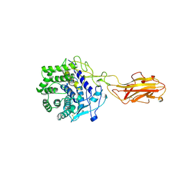
ENDO/EXOCELLULASE FROM THERMOMONOSPORA
分子名称: CALCIUM ION, T. FUSCA ENDO/EXO-CELLULASE E4 CATALYTIC DOMAIN AND CELLULOSE-BINDING DOMAIN
著者Sakon, J, Wilson, D.B, Karplus, P.A.
登録日1997-05-30
公開日1997-09-04
最終更新日2011-07-13
実験手法X-RAY DIFFRACTION (1.9 Å)
主引用文献Structure and mechanism of endo/exocellulase E4 from Thermomonospora fusca.
Nat.Struct.Biol., 4, 1997
1JB9
DownloadVisualize
BU of 1jb9 by Molmil
Crystal Structure of The Ferredoxin:NADP+ Reductase From Maize Root AT 1.7 Angstroms
分子名称: FLAVIN-ADENINE DINUCLEOTIDE, ferredoxin-NADP reductase
著者Faber, H.R, Karplus, P.A, Aliverti, A, Ferioli, C, Spinola, M.
登録日2001-06-03
公開日2001-07-04
最終更新日2023-08-16
実験手法X-RAY DIFFRACTION (1.7 Å)
主引用文献Biochemical and crystallographic characterization of ferredoxin-NADP(+) reductase from nonphotosynthetic tissues.
Biochemistry, 40, 2001

221051

件を2024-06-12に公開中

PDB statisticsPDBj update infoContact PDBjnumon