Loading
PDBj
MenuPDBj@FacebookPDBj@TwitterPDBj@YouTubewwPDB FoundationwwPDB
RCSB PDBPDBeBMRBAdv. SearchSearch help
PDB: 123 results

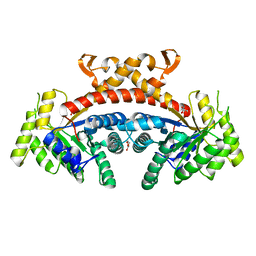
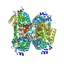
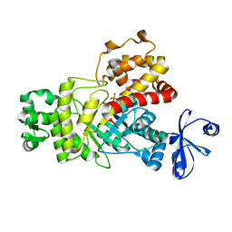
5GK3
DownloadVisualize
BU of 5gk3 by Molmil
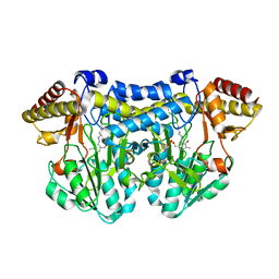
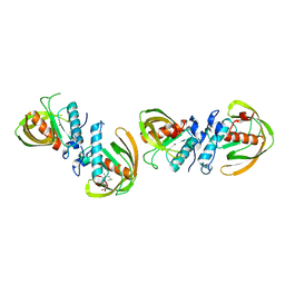
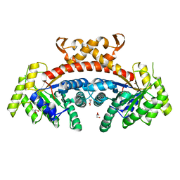
Native structure of fructose 1,6-bisphosphate aldolase from Escherichia coli at 1.8 Angstrom resolution
Descriptor: DI(HYDROXYETHYL)ETHER, Fructose-bisphosphate aldolase class 2, GLYCEROL, ...
Authors:Tran, T.H, Huynh, K.H, Ho, T.H, Kang, L.W.
Deposit date:2016-07-03
Release date:2017-07-05
Last modified:2023-11-08
Method:X-RAY DIFFRACTION (1.8 Å)
Cite:Apo structure of fructose 1,6-bisphosphate aldolase from Escherichia coli at 1.8 Angstrom resolution
To Be Published
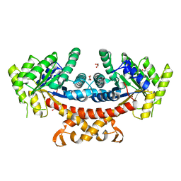
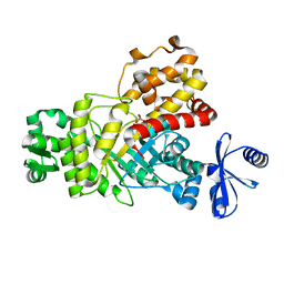
5GK6
DownloadVisualize
BU of 5gk6 by Molmil
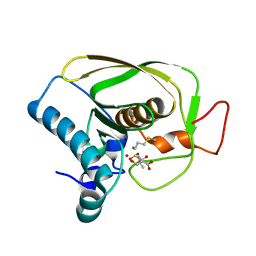
Structure of E.Coli fructose 1,6-bisphosphate aldolase, Citrate bound form
Descriptor: CITRIC ACID, DI(HYDROXYETHYL)ETHER, Fructose-bisphosphate aldolase class 2, ...
Authors:Tran, T.H, Huynh, K.H, Ho, T.H, Kang, L.W.
Deposit date:2016-07-03
Release date:2017-07-05
Last modified:2023-11-08
Method:X-RAY DIFFRACTION (1.8 Å)
Cite:Structure of E.Coli fructose 1,6-bisphosphate aldolase, Citrate bound form
To Be Published
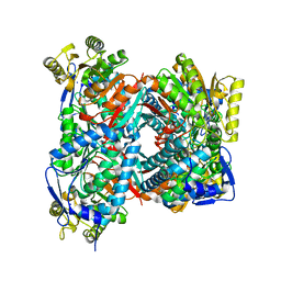
5GK8
DownloadVisualize
BU of 5gk8 by Molmil
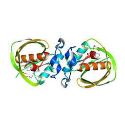
Structure of E.Coli fructose 1,6-bisphosphate aldolase, Acetate bound form
Descriptor: ACETATE ION, DI(HYDROXYETHYL)ETHER, Fructose-bisphosphate aldolase class 2, ...
Authors:Tran, T.H, Huynh, K.H, Ho, T.H, Kang, L.W.
Deposit date:2016-07-03
Release date:2017-07-05
Last modified:2023-11-08
Method:X-RAY DIFFRACTION (2.002 Å)
Cite:Structure of E.Coli fructose 1,6-bisphosphate aldolase, Acetate bound form
To Be Published
5GT6
DownloadVisualize
BU of 5gt6 by Molmil
Apo structure of Aldehyde Dehydrogenase from Bacillus cereus
Descriptor: Betaine-aldehyde dehydrogenase, SODIUM ION
Authors:Ngo, H.P.T, Hong, S.H, Ho, T.H, Oh, D.K, Kang, L.W.
Deposit date:2016-08-18
Release date:2017-09-06
Last modified:2023-11-08
Method:X-RAY DIFFRACTION (2.6 Å)
Cite:crystal structures of aldehyde dehydrogenase from Bacillus cereus having atypical bidirectional oxidizing and reducing activities for all-trans-retinal
To Be Published
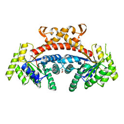
5GK7
DownloadVisualize
BU of 5gk7 by Molmil
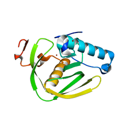
Structure of E.Coli fructose 1,6-bisphosphate aldolase bound to Tris
Descriptor: 2-AMINO-2-HYDROXYMETHYL-PROPANE-1,3-DIOL, DI(HYDROXYETHYL)ETHER, Fructose-bisphosphate aldolase class 2, ...
Authors:Tran, T.H, Huynh, K.H, Ho, T.H, Kang, L.W.
Deposit date:2016-07-03
Release date:2017-07-05
Last modified:2023-11-08
Method:X-RAY DIFFRACTION (1.8 Å)
Cite:Structure of E.Coli fructose 1,6-bisphosphate aldolase, Tris bound form
To Be Published
5GTL
DownloadVisualize
BU of 5gtl by Molmil
NADPH complex structure of Aldehyde Dehydrogenase from Bacillus cereus
Descriptor: Betaine-aldehyde dehydrogenase, NADPH DIHYDRO-NICOTINAMIDE-ADENINE-DINUCLEOTIDE PHOSPHATE, SODIUM ION
Authors:Ngo, H.P.T, Hong, S.H, Ho, T.H, Oh, D.K, Kang, L.W.
Deposit date:2016-08-21
Release date:2017-09-06
Last modified:2023-11-08
Method:X-RAY DIFFRACTION (2 Å)
Cite:Crystal structures of aldehyde dehydrogenase from Bacillus cereus having atypical bidirectional oxidizing and reducing activities for all-trans-retinal
To Be Published
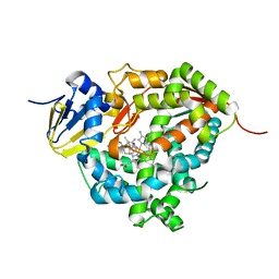
7WZM
DownloadVisualize
BU of 7wzm by Molmil
Crystal structure of Cytochrome P450 184A1 from streptomyces avermitilis in complex with Oleic acid
Descriptor: OLEIC ACID, PROTOPORPHYRIN IX CONTAINING FE, Putative cytochrome P450
Authors:Kim, V.C, Kim, D.G, Lee, S.G, Lee, G.H, Lee, S.A, Kang, L.W.
Deposit date:2022-02-18
Release date:2023-02-22
Last modified:2023-11-29
Method:X-RAY DIFFRACTION (1.68 Å)
Cite:Crystal structure of Cytochrome P450 184A1 from streptomyces avermitilis in complex with Oleic acid
To Be Published
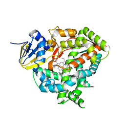
7WZL
DownloadVisualize
BU of 7wzl by Molmil
Crystal structure of Cytochrome P450 184A1 from streptomyces avermitilis
Descriptor: PROTOPORPHYRIN IX CONTAINING FE, Putative cytochrome P450
Authors:Kim, V.C, Kim, D.G, Lee, S.G, Lee, G.H, Lee, S.A, Kang, L.W.
Deposit date:2022-02-18
Release date:2023-02-22
Last modified:2023-11-29
Method:X-RAY DIFFRACTION (2.27 Å)
Cite:Crystal structure of Cytochrome P450 184A1 from streptomyces avermitilis
To Be Published
6IKZ
DownloadVisualize
BU of 6ikz by Molmil
UDP-glucose pyrophosphorylase from acinetobacter baumanii
Descriptor: GLYCEROL, PYROPHOSPHATE, SULFATE ION, ...
Authors:Lee, J.H, Kang, L.W.
Deposit date:2018-10-16
Release date:2019-10-16
Last modified:2023-11-22
Method:X-RAY DIFFRACTION (2.33 Å)
Cite:UTP-bound UGPase from acinetobacter baumanii
To be published
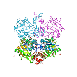
6ILA
DownloadVisualize
BU of 6ila by Molmil
Two Glycerol complexed Crystal structure of fructuronate-tagaturonate epimerase UxaE from Cohnella laeviribosi
Descriptor: Fructuronate-tagaturonate epimerase UxaE, GLYCEROL, PHOSPHATE ION, ...
Authors:Choi, M.Y, Kang, L.W, Ho, T.H, Nguyen, D.Q, Lee, I.H, Lee, J.H, Park, Y.S, Park, H.J.
Deposit date:2018-10-17
Release date:2019-10-23
Last modified:2023-11-22
Method:X-RAY DIFFRACTION (2.69 Å)
Cite:Crystal structure of fructuronate-tagaturonate epimerase UxaE from Cohnella laeviribosi
To be published
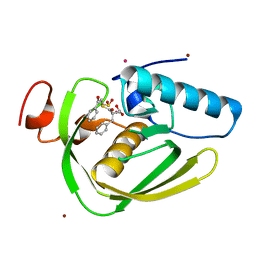
6IKT
DownloadVisualize
BU of 6ikt by Molmil
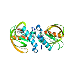
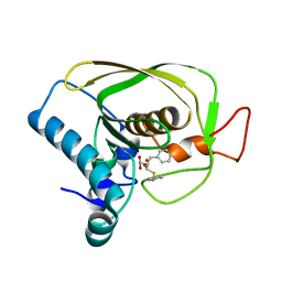
K1U complex structure of peptide deformylase from Xanthomonas oryzae pv. oryzae
Descriptor: (3R)-3-benzyl-4-oxo-4-[(2-oxo-2-phenylethyl)sulfanyl]butanoic acid, CADMIUM ION, NICKEL (II) ION, ...
Authors:Lee, I.H, Kang, L.W.
Deposit date:2018-10-16
Release date:2019-10-23
Last modified:2023-11-22
Method:X-RAY DIFFRACTION (1.9 Å)
Cite:K1U complex structure of peptide deformylase from Xanthomonas oryzae pv. oryzae
To be published
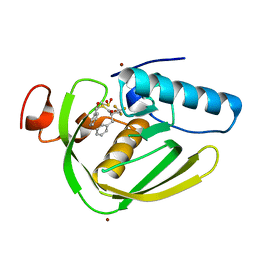
6IL0
DownloadVisualize
BU of 6il0 by Molmil
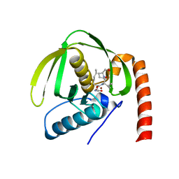
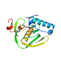
K3U complex structure of peptide deformylase from Xanthomonas oryzae pv. oryzae
Descriptor: NICKEL (II) ION, Peptide deformylase, S-(2-oxo-2-phenylethyl) (2R)-2-benzyl-4,4,4-trifluorobutanethioate
Authors:Lee, I.H, Kang, L.W.
Deposit date:2018-10-16
Release date:2019-10-23
Last modified:2023-11-22
Method:X-RAY DIFFRACTION (1.93 Å)
Cite:FBIs complex structure of peptide deformylase from Xanthomonas oryzae pv. oryzae
To be published
6IL9
DownloadVisualize
BU of 6il9 by Molmil
One Glycerol complexed Crystal structure of fructuronate-tagaturonate epimerase UxaE from Cohnella laeviribosi
Descriptor: Fructuronate-tagaturonate epimerase UxaE from Cohnella laeviribosi in complex with 1 glycerol, GLYCEROL, ZINC ION
Authors:Choi, M.Y, Kang, L.W, Ho, T.H, Nguyen, D.Q, Lee, I.H, Lee, J.H, Park, Y.S, Park, H.J.
Deposit date:2018-10-17
Release date:2019-10-23
Last modified:2023-11-22
Method:X-RAY DIFFRACTION (1.72005355 Å)
Cite:Crystal structure of fructuronate-tagaturonate epimerase UxaE from Cohnella laeviribosi
To Be Published
5B7S
DownloadVisualize
BU of 5b7s by Molmil
Apo structure of Cysteine Desulfurase from Thermococcus onnurineus NA1
Descriptor: Cysteine desulfurase, GLYCEROL
Authors:Ho, T.-H, Kang, L.W.
Deposit date:2016-06-08
Release date:2017-06-14
Last modified:2023-11-15
Method:X-RAY DIFFRACTION (2.58 Å)
Cite:Catalytic Intermediate Crystal Structures of Cysteine Desulfurase from the ArchaeonThermococcus onnurineus NA1.
Archaea, 2017, 2017
6JF8
DownloadVisualize
BU of 6jf8 by Molmil
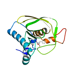
K4U bound crystal structure of class I type b peptide deformylase from Acinetobacter baumannii
Descriptor: L-[(N-HYDROXYAMINO)CARBONYL]PHENYLALANINE, Peptide deformylase, ZINC ION
Authors:Lee, I.H, Ho, T.H, Kang, L.W.
Deposit date:2019-02-08
Release date:2020-02-12
Last modified:2023-11-22
Method:X-RAY DIFFRACTION (1.7 Å)
Cite:K4U bound crystal structure of class I type b peptide deformylase from Acinetobacter baumannii
To be published
6JFS
DownloadVisualize
BU of 6jfs by Molmil
K4U bound crystal structure of class II peptide deformylase from methicillin resistant Staphylococcus aureus
Descriptor: L-[(N-HYDROXYAMINO)CARBONYL]PHENYLALANINE, NICKEL (II) ION, Peptide deformylase
Authors:Lee, I.H, Ho, T.H, Kang, L.W.
Deposit date:2019-02-11
Release date:2020-02-12
Last modified:2023-11-22
Method:X-RAY DIFFRACTION (2.25 Å)
Cite:K4U bound crystal structure of class II peptide deformylase from methicillin resistant Staphylococcus aureus
To be published
6JET
DownloadVisualize
BU of 6jet by Molmil
Actinonin bound crystal structure of class I type a peptide deformylase from Acinetobacter baumannii
Descriptor: ACTINONIN, Peptide deformylase, ZINC ION
Authors:Ho, T.H, Lee, I.H, Kang, L.W.
Deposit date:2019-02-07
Release date:2020-02-12
Last modified:2023-11-22
Method:X-RAY DIFFRACTION (2.6 Å)
Cite:Actinonin bound crystal structure of class I type a peptide deformylase from Acinetobacter baumannii
To be published
6JF6
DownloadVisualize
BU of 6jf6 by Molmil
Met-ala-ser bound crystal structure of class I type b peptide deformylase from Acinetobacter baumannii
Descriptor: MET-ALA-SER, Peptide deformylase, ZINC ION
Authors:Jung, K.H, Ho, T.H, Lee, I.H, Kang, L.W.
Deposit date:2019-02-08
Release date:2020-02-12
Last modified:2023-11-22
Method:X-RAY DIFFRACTION (2.35 Å)
Cite:Met-ala-ser bound crystal structure of class I type b peptide deformylase from Acinetobacter baumannii
To be published
6JFO
DownloadVisualize
BU of 6jfo by Molmil
Formyl-Met-Ala-Ser bound crystal structure of class II peptide deformylase from methicillin resistant Staphylococcus aureus
Descriptor: FME-ALA-SER, MAGNESIUM ION, Peptide deformylase
Authors:Lee, I.H, Ho, T.H, Kang, L.W.
Deposit date:2019-02-11
Release date:2020-02-12
Last modified:2023-11-22
Method:X-RAY DIFFRACTION (1.6 Å)
Cite:Formyl-Met-Ala-Ser bound crystal structure of class II peptide deformylase from methicillin resistant Staphylococcus aureus
To be published
6JF4
DownloadVisualize
BU of 6jf4 by Molmil
K1U bound crystal structure of class I type b peptide deformylase from Acinetobacter baumannii
Descriptor: (3R)-3-benzyl-4-oxo-4-[(2-oxo-2-phenylethyl)sulfanyl]butanoic acid, Peptide deformylase, ZINC ION
Authors:Lee, I.H, Ho, T.H, Kang, L.W.
Deposit date:2019-02-08
Release date:2020-02-12
Last modified:2023-11-22
Method:X-RAY DIFFRACTION (2 Å)
Cite:K1U bound crystal structure of class I type b peptide deformylase from Acinetobacter baumannii
To be published
6JFF
DownloadVisualize
BU of 6jff by Molmil
K3U bound crystal structure of class I type b peptide deformylase from Pseudomonas aeruginosa
Descriptor: NICKEL (II) ION, Peptide deformylase, S-(2-oxo-2-phenylethyl) (2R)-2-benzyl-4,4,4-trifluorobutanethioate
Authors:Lee, I.H, Ho, T.H, Kang, L.W.
Deposit date:2019-02-08
Release date:2020-02-12
Last modified:2023-11-22
Method:X-RAY DIFFRACTION (2.1 Å)
Cite:K3U bound crystal structure of class I type b peptide deformylase from Pseudomonas aeruginosa
To be published
6JFQ
DownloadVisualize
BU of 6jfq by Molmil
K2U bound crystal structure of class II peptide deformylase from methicillin resistant Staphylococcus aureus
Descriptor: (3~{R},4~{R})-4-oxidanyl-3-(phenylmethyl)-4-(phenylmethylsulfanyl)butanoic acid, NICKEL (II) ION, Peptide deformylase
Authors:Lee, I.H, Ho, T.H, Kang, L.W.
Deposit date:2019-02-11
Release date:2020-02-12
Last modified:2023-11-22
Method:X-RAY DIFFRACTION (2.2 Å)
Cite:K2U bound crystal structure of class II peptide deformylase from methicillin resistant Staphylococcus aureus
To be published
6JFD
DownloadVisualize
BU of 6jfd by Molmil
K1U bound crystal structure of class I type b peptide deformylase from Pseudomonas aeruginosa
Descriptor: (3R)-3-benzyl-4-oxo-4-[(2-oxo-2-phenylethyl)sulfanyl]butanoic acid, NICKEL (II) ION, Peptide deformylase
Authors:Lee, I.H, Ho, T.H, Kang, L.W.
Deposit date:2019-02-08
Release date:2020-02-12
Last modified:2023-11-22
Method:X-RAY DIFFRACTION (2.4 Å)
Cite:K1U bound crystal structure of class I type b peptide deformylase from Pseudomonas aeruginosa
To be published
6JFA
DownloadVisualize
BU of 6jfa by Molmil
Met-Ala-Ser bound crystal structure of class I type b peptide deformylase from Pseudomonas aeruginosa
Descriptor: MET-ALA-SER, NICKEL (II) ION, Peptide deformylase
Authors:Lee, I.H, Ho, T.H, Kang, L.W.
Deposit date:2019-02-08
Release date:2020-02-12
Last modified:2023-11-22
Method:X-RAY DIFFRACTION (1.93 Å)
Cite:Met-Ala-Ser bound crystal structure of class I type b peptide deformylase from Pseudomonas aeruginosa
To be published
6JER
DownloadVisualize
BU of 6jer by Molmil
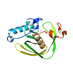
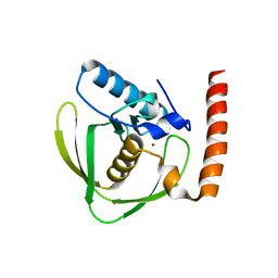
Apo crystal structure of class I type a peptide deformylase from Acinetobacter baumannii
Descriptor: Peptide deformylase, ZINC ION
Authors:Ho, T.H, Lee, I.H, Kang, L.W.
Deposit date:2019-02-07
Release date:2020-02-12
Last modified:2023-11-22
Method:X-RAY DIFFRACTION (2.4 Å)
Cite:Expression, crystallization, and preliminary X-ray crystallographic analysis of peptide deformylase from Acinetobacter baumanii
Biodesign, 5, 2017

222624

PDB entries from 2024-07-17

PDB statisticsPDBj update infoContact PDBjnumon