Loading
PDBj
MenuPDBj@FacebookPDBj@X(formerly Twitter)PDBj@BlueSkyPDBj@YouTubewwPDB FoundationwwPDB
RCSB PDBPDBeBMRBAdv. SearchSearch help
PDB: 72 results

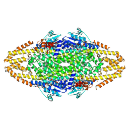
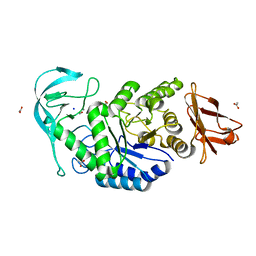
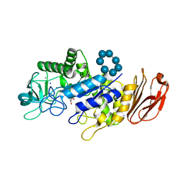
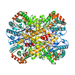
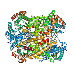
6TP0
DownloadVisualize
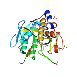
BU of 6tp0 by Molmil
Crystal structure of Bacillus paralicheniformis alpha-amylase in complex with maltose
Descriptor: ACETIC ACID, Amylase, CALCIUM ION, ...
Authors:Rozeboom, H.J, Janssen, D.B.
Deposit date:2019-12-12
Release date:2020-10-14
Last modified:2024-01-24
Method:X-RAY DIFFRACTION (2.04 Å)
Cite:Characterization of the starch surface binding site on Bacillus paralicheniformis alpha-amylase.
Int.J.Biol.Macromol., 165, 2020
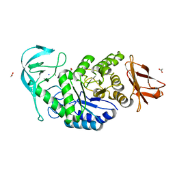
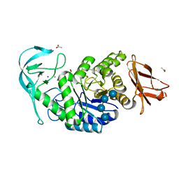
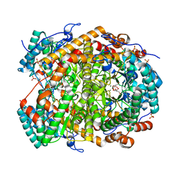
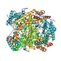
6TP1
DownloadVisualize
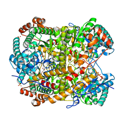
BU of 6tp1 by Molmil
Crystal structure of Bacillus paralicheniformis alpha-amylase in complex with maltotetraose
Descriptor: ACETIC ACID, Amylase, CALCIUM ION, ...
Authors:Rozeboom, H.J, Janssen, D.B.
Deposit date:2019-12-12
Release date:2020-10-14
Last modified:2024-01-24
Method:X-RAY DIFFRACTION (1.94 Å)
Cite:Characterization of the starch surface binding site on Bacillus paralicheniformis alpha-amylase.
Int.J.Biol.Macromol., 165, 2020
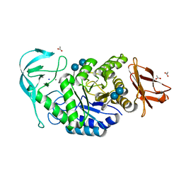
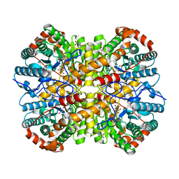
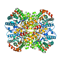
6TOY
DownloadVisualize
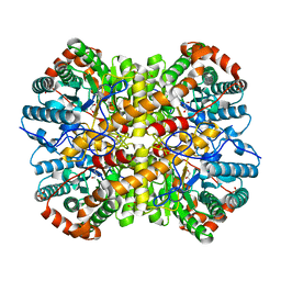
BU of 6toy by Molmil
Crystal structure of Bacillus paralicheniformis wild-type alpha-amylase
Descriptor: ACETIC ACID, Amylase, CALCIUM ION, ...
Authors:Rozeboom, H.J, Janssen, D.B.
Deposit date:2019-12-12
Release date:2020-10-14
Last modified:2024-01-24
Method:X-RAY DIFFRACTION (1.95 Å)
Cite:Characterization of the starch surface binding site on Bacillus paralicheniformis alpha-amylase.
Int.J.Biol.Macromol., 165, 2020
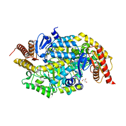
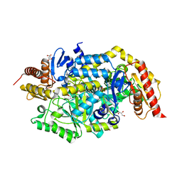
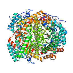
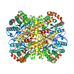
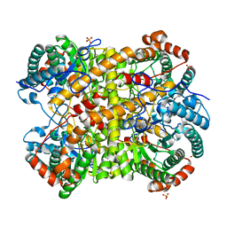
4AO9
DownloadVisualize
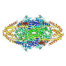
BU of 4ao9 by Molmil
Biochemical properties and crystal structure of a novel beta- phenylalanine aminotransferase from Variovorax paradoxus
Descriptor: BETA-PHENYLALANINE AMINOTRANSFERASE, GLYCEROL, PYRIDOXAL-5'-PHOSPHATE
Authors:Crismaru, C.G, Wybenga, G.G, Szymanski, W, Wijma, H.J, Wu, B, deWildeman, S, Poelarends, G.J, Dijkstra, B.W, Janssen, D.B.
Deposit date:2012-03-25
Release date:2012-10-24
Last modified:2024-05-01
Method:X-RAY DIFFRACTION (1.5 Å)
Cite:Biochemical Properties and Crystal Structure of a Novel Beta-Phenylalanine Aminotransferase from Variovorax Paradoxus
Appl.Environ.Microbiol., 79, 2013
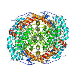
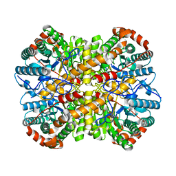
6T8E
DownloadVisualize
BU of 6t8e by Molmil
Crystal structure of native xylose isomerase from Piromyces E2 grown in yeast, in complex with xylose
Descriptor: CALCIUM ION, D-xylose, SULFATE ION, ...
Authors:Rozeboom, H.J, Janssen, D.B.
Deposit date:2019-10-24
Release date:2020-01-29
Last modified:2024-01-24
Method:X-RAY DIFFRACTION (1.86 Å)
Cite:Structure-based directed evolution improves S. cerevisiae growth on xylose by influencing in vivo enzyme performance.
Biotechnol Biofuels, 13, 2020
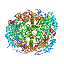
4AOA
DownloadVisualize
BU of 4aoa by Molmil
Biochemical properties and crystal structure of a novel beta- phenylalanine aminotransferase from Variovorax paradoxus
Descriptor: 4'-DEOXY-4'-ACETYLYAMINO-PYRIDOXAL-5'-PHOSPHATE, BETA-PHENYLALANINE AMINOTRANSFERASE, GLYCEROL
Authors:Crismaru, C.G, Wybenga, G.G, Szymanski, W, Wijma, H.J, Wu, B, deWildeman, S, Poelarends, G.J, Dijkstra, B.W, Janssen, D.B.
Deposit date:2012-03-25
Release date:2012-10-24
Last modified:2024-05-01
Method:X-RAY DIFFRACTION (2.28 Å)
Cite:Biochemical Properties and Crystal Structure of a Beta-Phenylalanine Aminotransferase from Variovorax Paradoxus.
Appl.Environ.Microbiol., 79, 2013
6TP2
DownloadVisualize
BU of 6tp2 by Molmil
Crystal structure of Bacillus paralicheniformis alpha-amylase in complex with beta-cyclodextrin
Descriptor: Amylase, CALCIUM ION, Cycloheptakis-(1-4)-(alpha-D-glucopyranose), ...
Authors:Rozeboom, H.J, Janssen, D.B.
Deposit date:2019-12-12
Release date:2020-10-14
Last modified:2024-01-24
Method:X-RAY DIFFRACTION (1.94 Å)
Cite:Characterization of the starch surface binding site on Bacillus paralicheniformis alpha-amylase.
Int.J.Biol.Macromol., 165, 2020
4C5R
DownloadVisualize
BU of 4c5r by Molmil
Structural Investigations into the Stereochemistry and Activity of a Phenylalanine-2,3-Aminomutase from Taxus chinensis
Descriptor: (3S)-3-amino-2,2-difluoro-3-phenylpropanoic acid, GLYCEROL, PHENYLALANINE AMMONIA-LYASE
Authors:Wybenga, G.G, Szymanski, W, Wu, B, Feringa, B.L, Janssen, D.B, Dijkstra, B.W.
Deposit date:2013-09-16
Release date:2014-05-07
Last modified:2024-11-13
Method:X-RAY DIFFRACTION (2.14 Å)
Cite:Structural Investigations Into the Stereochemistry and Activity of a Phenylalanine-2,3-Aminomutase from Taxus Chinensis.
Biochemistry, 53, 2014
4C5S
DownloadVisualize
BU of 4c5s by Molmil
Structural Investigations into the Stereochemistry and Activity of a Phenylalanine-2,3-Aminomutase from Taxus chinensis
Descriptor: (3S)-3-amino-2,2-difluoro-3-phenylpropanoic acid, PHENYLALANINE AMMONIA-LYASE
Authors:Wybenga, G.G, Szymanski, W, Wu, B, Feringa, B.L, Janssen, D.B, Dijkstra, B.W.
Deposit date:2013-09-16
Release date:2014-05-07
Last modified:2024-10-16
Method:X-RAY DIFFRACTION (1.85 Å)
Cite:Structural Investigations Into the Stereochemistry and Activity of a Phenylalanine-2,3-Aminomutase from Taxus Chinensis.
Biochemistry, 53, 2014
5NH6
DownloadVisualize
BU of 5nh6 by Molmil
Crystal structure of xylose isomerase from Piromyces E2 Complexed with one Mg2+ ion and xylitol
Descriptor: MAGNESIUM ION, SULFATE ION, Xylitol, ...
Authors:Rozeboom, H.J, Janssen, D.B.
Deposit date:2017-03-21
Release date:2017-11-01
Last modified:2024-05-01
Method:X-RAY DIFFRACTION (1.75 Å)
Cite:Metal Dependence of the Xylose Isomerase from Piromyces sp. E2 Explored by Activity Profiling and Protein Crystallography.
Biochemistry, 56, 2017
5NHC
DownloadVisualize
BU of 5nhc by Molmil
Crystal structure of xylose isomerase from Piromyces E2 in complex with two Co2+ ions and xylulose
Descriptor: 4-HYDROXYPROLINE, COBALT (II) ION, D-XYLULOSE, ...
Authors:Rozeboom, H.J, Janssen, D.B.
Deposit date:2017-03-21
Release date:2017-11-01
Last modified:2024-05-01
Method:X-RAY DIFFRACTION (1.93 Å)
Cite:Metal Dependence of the Xylose Isomerase from Piromyces sp. E2 Explored by Activity Profiling and Protein Crystallography.
Biochemistry, 56, 2017
5NH7
DownloadVisualize
BU of 5nh7 by Molmil
Crystal structure of xylose isomerase from Piromyces E2 in complex with two Mg2+ ions and xylose
Descriptor: D-xylose, MAGNESIUM ION, SULFATE ION, ...
Authors:Rozeboom, H.J, Janssen, D.B.
Deposit date:2017-03-21
Release date:2017-11-01
Last modified:2024-05-01
Method:X-RAY DIFFRACTION (1.9 Å)
Cite:Metal Dependence of the Xylose Isomerase from Piromyces sp. E2 Explored by Activity Profiling and Protein Crystallography.
Biochemistry, 56, 2017
5NHA
DownloadVisualize
BU of 5nha by Molmil
Crystal structure of xylose isomerase from Piromyces sp. E2 in complex with two Mn2+ ions and sorbitol
Descriptor: MANGANESE (II) ION, SULFATE ION, Xylose isomerase, ...
Authors:Rozeboom, H.J, Janssen, D.B.
Deposit date:2017-03-21
Release date:2017-11-01
Last modified:2024-05-01
Method:X-RAY DIFFRACTION (1.8 Å)
Cite:Metal Dependence of the Xylose Isomerase from Piromyces sp. E2 Explored by Activity Profiling and Protein Crystallography.
Biochemistry, 56, 2017
5NH4
DownloadVisualize
BU of 5nh4 by Molmil
Crystal structure of xylose isomerase from Piromyces E2 in complex with one Mg2+ ions and glycerol
Descriptor: GLYCEROL, MAGNESIUM ION, SULFATE ION, ...
Authors:Rozeboom, H.J, Janssen, D.B.
Deposit date:2017-03-21
Release date:2017-11-01
Last modified:2024-01-17
Method:X-RAY DIFFRACTION (1.8 Å)
Cite:Metal Dependence of the Xylose Isomerase from Piromyces sp. E2 Explored by Activity Profiling and Protein Crystallography.
Biochemistry, 56, 2017
5NHD
DownloadVisualize
BU of 5nhd by Molmil
Crystal structure of xylose isomerase from Piromyces E2 in complex with 2 Ni2+ ions and xylose
Descriptor: D-xylose, NICKEL (II) ION, SULFATE ION, ...
Authors:Rozeboom, H.J, Janssen, D.B.
Deposit date:2017-03-21
Release date:2017-11-01
Last modified:2024-05-01
Method:X-RAY DIFFRACTION (1.8 Å)
Cite:Metal Dependence of the Xylose Isomerase from Piromyces sp. E2 Explored by Activity Profiling and Protein Crystallography.
Biochemistry, 56, 2017
5NHE
DownloadVisualize
BU of 5nhe by Molmil
Crystal structure of xylose isomerase from Piromyces E2 in complex with two Cd2+ ions and xylose
Descriptor: CADMIUM ION, D-xylose, SULFATE ION, ...
Authors:Rozeboom, H.J, Janssen, D.B.
Deposit date:2017-03-21
Release date:2017-11-01
Last modified:2024-05-01
Method:X-RAY DIFFRACTION (1.86 Å)
Cite:Metal Dependence of the Xylose Isomerase from Piromyces sp. E2 Explored by Activity Profiling and Protein Crystallography.
Biochemistry, 56, 2017
5NHB
DownloadVisualize
BU of 5nhb by Molmil
Crystal structure of xylose isomerase from Piromyces E2 in complex with two Fe2+ ions
Descriptor: FE (II) ION, SULFATE ION, Xylose isomerase
Authors:Rozeboom, H.J, Janssen, D.B.
Deposit date:2017-03-21
Release date:2017-11-01
Last modified:2024-05-01
Method:X-RAY DIFFRACTION (2.4 Å)
Cite:Metal Dependence of the Xylose Isomerase from Piromyces sp. E2 Explored by Activity Profiling and Protein Crystallography.
Biochemistry, 56, 2017
5OX2
DownloadVisualize
BU of 5ox2 by Molmil
Crystal structure of thymoligase, a substrate-tailored peptiligase variant
Descriptor: Fragment of prodomain, SULFATE ION, Subtilisin BPN'
Authors:Rozeboom, H.J, Janssen, D.B.
Deposit date:2017-09-05
Release date:2018-01-10
Last modified:2024-11-13
Method:X-RAY DIFFRACTION (2.24 Å)
Cite:Design of a substrate-tailored peptiligase variant for the efficient synthesis of thymosin-alpha1.
Org. Biomol. Chem., 16, 2018
5NH9
DownloadVisualize
BU of 5nh9 by Molmil
Crystal structure of xylose isomerase from Piromyces E2 in complex with two Mn2+ ions and xylose
Descriptor: D-xylose, MANGANESE (II) ION, SULFATE ION, ...
Authors:Rozeboom, H.J, Janssen, D.B.
Deposit date:2017-03-21
Release date:2017-11-01
Last modified:2024-05-01
Method:X-RAY DIFFRACTION (2.08 Å)
Cite:Metal Dependence of the Xylose Isomerase from Piromyces sp. E2 Explored by Activity Profiling and Protein Crystallography.
Biochemistry, 56, 2017
5NH5
DownloadVisualize
BU of 5nh5 by Molmil
Crystal structure of native xylose isomerase from Piromyces E2
Descriptor: CALCIUM ION, FE (II) ION, GLYCEROL, ...
Authors:Rozeboom, H.J, Janssen, D.B.
Deposit date:2017-03-21
Release date:2017-11-01
Last modified:2024-01-17
Method:X-RAY DIFFRACTION (1.8 Å)
Cite:Metal Dependence of the Xylose Isomerase from Piromyces sp. E2 Explored by Activity Profiling and Protein Crystallography.
Biochemistry, 56, 2017
5NH8
DownloadVisualize
BU of 5nh8 by Molmil
Crystal structure of xylose isomerase from Piromyces E2 in complex with two Ca2+ ions and xylose
Descriptor: CALCIUM ION, D-xylose, SULFATE ION, ...
Authors:Rozeboom, H.J, Janssen, D.B.
Deposit date:2017-03-21
Release date:2017-11-01
Last modified:2024-05-01
Method:X-RAY DIFFRACTION (1.86 Å)
Cite:Metal Dependence of the Xylose Isomerase from Piromyces sp. E2 Explored by Activity Profiling and Protein Crystallography.
Biochemistry, 56, 2017
5NHM
DownloadVisualize
BU of 5nhm by Molmil
Crystal structure of apo xylose isomerase from Piromyces E2
Descriptor: ACETIC ACID, GLYCEROL, SULFATE ION, ...
Authors:Rozeboom, H.J, Janssen, D.B.
Deposit date:2017-03-21
Release date:2017-11-01
Last modified:2024-05-01
Method:X-RAY DIFFRACTION (1.67 Å)
Cite:Metal Dependence of the Xylose Isomerase from Piromyces sp. E2 Explored by Activity Profiling and Protein Crystallography.
Biochemistry, 56, 2017
<123


227561

数据于2024-11-20公开中

PDB statisticsPDBj update infoContact PDBjnumon