2BX4
 
 | |
2BX3
 | |
4K02
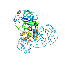
 | | Crystal structure of AtDHNAT1, a 1,4-dihydroxy-2-naphthoyl-CoA thioesterase from Arabidopsis thaliana | | 分子名称: | 1,4-dihydroxy-2-naphthoyl-CoA thioesterase | | 著者 | Furt, F, Allen, W.J, Widhalm, J.R, Madzelan, P, Rizzo, R.C, Basset, G, Wilson, M.A. | | 登録日 | 2013-04-03 | | 公開日 | 2013-04-17 | | 最終更新日 | 2023-09-20 | | 実験手法 | X-RAY DIFFRACTION (1.9 Å) | | 主引用文献 | Functional convergence of structurally distinct thioesterases from cyanobacteria and plants involved in phylloquinone biosynthesis.
Acta Crystallogr.,Sect.D, 69, 2013
|
|
2FL7
 | |
3DB3
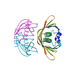
 | | Crystal structure of the tandem tudor domains of the E3 ubiquitin-protein ligase UHRF1 in complex with trimethylated histone H3-K9 peptide | | 分子名称: | E3 ubiquitin-protein ligase UHRF1, Trimethylated histone H3-K9 peptide | | 著者 | Walker, J.R, Avvakumov, G.V, Xue, S, Dong, A, Li, Y, Bountra, C, Weigelt, J, Arrowsmith, C.H, Edwards, A.M, Bochkarev, A, Dhe-Paganon, S, Structural Genomics Consortium (SGC) | | 登録日 | 2008-05-30 | | 公開日 | 2008-09-16 | | 最終更新日 | 2012-04-18 | | 実験手法 | X-RAY DIFFRACTION (2.4 Å) | | 主引用文献 | Recognition of multivalent histone states associated with heterochromatin by UHRF1 protein.
J.Biol.Chem., 286, 2011
|
|
4K7R
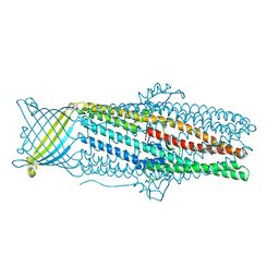 | | Crystal structures of CusC review conformational changes accompanying folding and transmembrane channel formation | | 分子名称: | (2S)-1-(pentanoyloxy)propan-2-yl hexanoate, Cation efflux system protein CusC | | 著者 | Su, C.-C, Lei, H.-T, Bolla, J.R, Yu, E.W. | | 登録日 | 2013-04-17 | | 公開日 | 2013-10-16 | | 最終更新日 | 2024-02-28 | | 実験手法 | X-RAY DIFFRACTION (2.094 Å) | | 主引用文献 | Crystal Structures of CusC Review Conformational Changes Accompanying Folding and Transmembrane Channel Formation.
J.Mol.Biol., 426, 2014
|
|
2BHG
 | |
8E4H
 | |
8E3R
 | |
7NIP
 | |
8E3K
 | |
8E5Y
 | |
8EE9
 | |
3CD9
 | |
3CDC
 | |
5BKC
 | | Crystal structure of AAD-1 in complex with (R)-diclofop, Mn(II), and 2-oxoglutarate | | 分子名称: | (2R)-2-{4-[(3,5-dichloropyridin-2-yl)oxy]phenoxy}propanoic acid, (R)-phenoxypropionate/alpha-ketoglutarate-dioxygenase, 2-OXOGLUTARIC ACID, ... | | 著者 | Chekan, J.R, Nair, S.K. | | 登録日 | 2019-06-02 | | 公開日 | 2019-06-12 | | 最終更新日 | 2023-09-27 | | 実験手法 | X-RAY DIFFRACTION (1.8 Å) | | 主引用文献 | Molecular basis for enantioselective herbicide degradation imparted by aryloxyalkanoate dioxygenases in transgenic plants.
Proc.Natl.Acad.Sci.USA, 116, 2019
|
|
5BK9
 | | AAD-1 Bound to the Vanadyl Ion and Succinate | | 分子名称: | (R)-phenoxypropionate/alpha-ketoglutarate-dioxygenase, PHOSPHATE ION, SUCCINIC ACID, ... | | 著者 | Ongpipattanakul, C, Chekan, J.R. | | 登録日 | 2019-06-01 | | 公開日 | 2019-06-12 | | 最終更新日 | 2023-09-27 | | 実験手法 | X-RAY DIFFRACTION (1.51 Å) | | 主引用文献 | Molecular basis for enantioselective herbicide degradation imparted by aryloxyalkanoate dioxygenases in transgenic plants.
Proc.Natl.Acad.Sci.USA, 116, 2019
|
|
8EBH
 | |
3CDY
 | | AL-09 H87Y, immunoglobulin light chain variable domain | | 分子名称: | IMMUNOGLOBULIN LIGHT CHAIN | | 著者 | Baden, E.M, Randles, E.G, Aboagye, A.K, Thompson, J.R, Ramirez-Alvarado, M. | | 登録日 | 2008-02-27 | | 公開日 | 2008-09-02 | | 最終更新日 | 2023-08-30 | | 実験手法 | X-RAY DIFFRACTION (2.43 Å) | | 主引用文献 | Structural insights into the role of mutations in amyloidogenesis.
J.Biol.Chem., 283, 2008
|
|
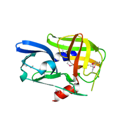
2BRY
 | | Crystal structure of the native monooxygenase domain of MICAL at 1.45 A resolution | | 分子名称: | CHLORIDE ION, FLAVIN-ADENINE DINUCLEOTIDE, GLYCEROL, ... | | 著者 | Siebold, C, Berrow, N, Walter, T.S, Harlos, K, Owens, R.J, Terman, J.R, Stuart, D.I, Kolodkin, A.L, Pasterkamp, R.J, Jones, E.Y. | | 登録日 | 2005-05-13 | | 公開日 | 2005-10-26 | | 最終更新日 | 2024-05-08 | | 実験手法 | X-RAY DIFFRACTION (1.45 Å) | | 主引用文献 | High-Resolution Structure of the Catalytic Region of Mical (Molecule Interacting with Casl), a Multidomain Flavoenzyme-Signaling Molecule.
Proc.Natl.Acad.Sci.USA, 102, 2005
|
|
3CB9
 | |
4K7B
 | | Crystal structure of Extrinsic protein in photosystem II | | 分子名称: | Extrinsic protein in photosystem II, GLYCEROL, SULFATE ION | | 著者 | Nagao, R, Suga, M, Niikura, A, Okumura, A, Koua, F.H.M, Suzuki, T, Tomo, T, Enami, I, Shen, J.R. | | 登録日 | 2013-04-16 | | 公開日 | 2013-09-18 | | 最終更新日 | 2024-03-20 | | 実験手法 | X-RAY DIFFRACTION (1.55 Å) | | 主引用文献 | Crystal Structure of Psb31, a Novel Extrinsic Protein of Photosystem II from a Marine Centric Diatom and Implications for Its Binding and Function
Biochemistry, 52, 2013
|
|
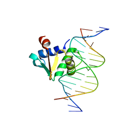
6UHJ
 | | X-ray Structure of C148 mGFP | | 分子名称: | C148 mGFP | | 著者 | Winegar, P.W, Hayes, O.G, McMillan, J.R, Figg, C.A, Focia, P.J, Mirkin, C.A. | | 登録日 | 2019-09-27 | | 公開日 | 2020-03-18 | | 最終更新日 | 2023-11-15 | | 実験手法 | X-RAY DIFFRACTION (1.5 Å) | | 主引用文献 | DNA-Directed Protein Packing within Single Crystals.
Chem, 6, 2020
|
|
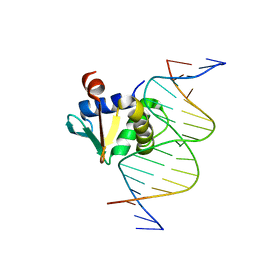
6UHP
 | | Crystal Structure of C148 mGFP-ncDNA-1 | | 分子名称: | C148 mGFP-ncDNA-1 | | 著者 | Winegar, P.W, Hayes, O.G, McMillan, J.R, Figg, C.A, Focia, P.J, Mirkin, C.A. | | 登録日 | 2019-09-27 | | 公開日 | 2020-03-18 | | 最終更新日 | 2023-11-15 | | 実験手法 | X-RAY DIFFRACTION (2.9 Å) | | 主引用文献 | DNA-Directed Protein Packing within Single Crystals.
Chem, 6, 2020
|
|
4GCB
 | |


