Loading
PDBj
メニューPDBj@FacebookPDBj@TwitterPDBj@YouTubewwPDB FoundationwwPDB
RCSB PDBPDBeBMRBAdv. SearchSearch help
PDB: 680 件

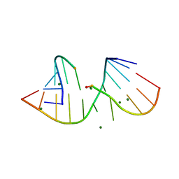
3FS0
DownloadVisualize
BU of 3fs0 by Molmil
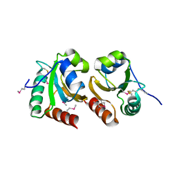
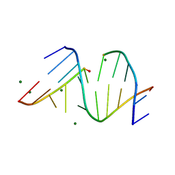
Class II ligase ribozyme product-template duplex, structure 2
分子名称: 5'-R(*CP*CP*AP*GP*UP*CP*GP*GP*AP*AP*C)-3', 5'-R(*GP*GP*UP*GP*AP*GP*GP*CP*UP*G)-3', MAGNESIUM ION
著者Pitt, J.N, Ferre-D'Amare, A.R.
登録日2009-01-08
公開日2009-02-24
最終更新日2023-09-06
実験手法X-RAY DIFFRACTION (2.3 Å)
主引用文献Structure-guided engineering of the regioselectivity of RNA ligase ribozymes.
J.Am.Chem.Soc., 131, 2009
3FTM
DownloadVisualize
BU of 3ftm by Molmil
Class II ligase ribozyme product-template duplex, structure 1
分子名称: 5'-R(*CP*CP*AP*GP*UP*CP*GP*GP*AP*AP*CP*A)-3', 5'-R(*GP*UP*GP*UP*GP*AP*GP*GP*CP*UP*G)-3', MAGNESIUM ION, ...
著者Pitt, J.N, Ferre-D'Amare, A.R.
登録日2009-01-13
公開日2009-02-24
最終更新日2024-02-21
実験手法X-RAY DIFFRACTION (2.7 Å)
主引用文献Structure-guided engineering of the regioselectivity of RNA ligase ribozymes.
J.Am.Chem.Soc., 131, 2009
3GJ1
DownloadVisualize
BU of 3gj1 by Molmil
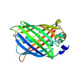
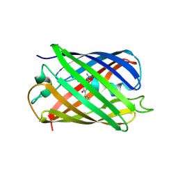
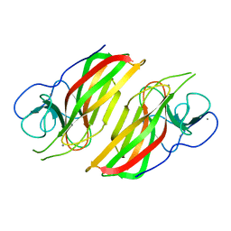
Non photoactivated state of PA-GFP
分子名称: CHLORIDE ION, Green fluorescent protein, SULFATE ION
著者Henderson, J.N, Gepshtein, R, Heenan, J.R, Kallio, K, Huppert, D, Remington, S.J.
登録日2009-03-07
公開日2009-03-24
最終更新日2023-11-22
実験手法X-RAY DIFFRACTION (1.8 Å)
主引用文献Structure and mechanism of the photoactivatable green fluorescent protein.
J.Am.Chem.Soc., 131, 2009
3HZZ
DownloadVisualize
BU of 3hzz by Molmil
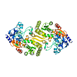
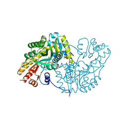
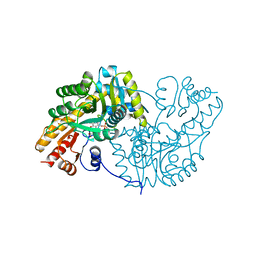
2.4 Angstrom Crystal Structure of Streptomyces collinus crotonyl CoA carboxylase/reductase
分子名称: Crotonyl CoA reductase, SULFATE ION
著者Scarsdale, J.N, Musayev, F.N, Wright, H.T.
登録日2009-06-24
公開日2010-07-21
最終更新日2024-02-21
実験手法X-RAY DIFFRACTION (2.4 Å)
主引用文献Structure of Streptomycs collinus crotonyl COA carboxylase/reductase
To be Published
3HZX
DownloadVisualize
BU of 3hzx by Molmil
Crystal Structure of Staphylococcal nuclease variant D+PHS/V66K at pH 9 determined at 100 K
分子名称: CALCIUM ION, THYMIDINE-3',5'-DIPHOSPHATE, Thermonuclease
著者Schlessman, J.L, De Luca-Westrate, J.N, Garcia-Moreno, B.E.
登録日2009-06-24
公開日2010-05-19
最終更新日2023-09-06
実験手法X-RAY DIFFRACTION (2 Å)
主引用文献Structure of Staphylococcal nuclease D+PHS/V66K reveals internal hydration in protein cavity
To be Published
3GJ2
DownloadVisualize
BU of 3gj2 by Molmil
Photoactivated state of PA-GFP
分子名称: CHLORIDE ION, Green fluorescent protein
著者Henderson, J.N, Gepshtein, R, Heenan, J.R, Kallio, K, Huppert, D, Remington, S.J.
登録日2009-03-07
公開日2009-03-24
最終更新日2023-11-22
実験手法X-RAY DIFFRACTION (1.9 Å)
主引用文献Structure and mechanism of the photoactivatable green fluorescent protein.
J.Am.Chem.Soc., 131, 2009
3GNJ
DownloadVisualize
BU of 3gnj by Molmil
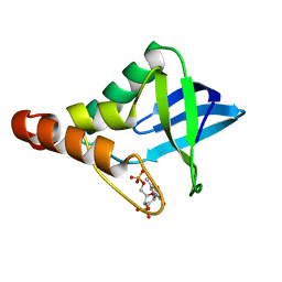
The crystal structure of a thioredoxin-related protein from Desulfitobacterium hafniense DCB
分子名称: Thioredoxin domain protein
著者Tan, K, Volkart, L, Gu, M, Kinney, J.N, Babnigg, G, Kerfeld, C, Joachimiak, A, Midwest Center for Structural Genomics (MCSG)
登録日2009-03-17
公開日2009-05-12
最終更新日2011-07-13
実験手法X-RAY DIFFRACTION (1.99 Å)
主引用文献The crystal structure of a thioredoxin-related protein from Desulfitobacterium hafniense DCB
To be Published
3HH2
DownloadVisualize
BU of 3hh2 by Molmil
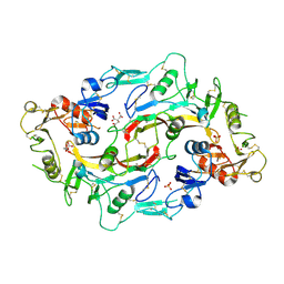
Crystal structure of the myostatin:follistatin 288 complex
分子名称: CITRIC ACID, Follistatin, Growth/differentiation factor 8, ...
著者Cash, J.N, Thompson, T.B.
登録日2009-05-14
公開日2009-08-04
最終更新日2017-11-01
実験手法X-RAY DIFFRACTION (2.15 Å)
主引用文献The structure of myostatin:follistatin 288: insights into receptor utilization and heparin binding
Embo J., 28, 2009
3F8M
DownloadVisualize
BU of 3f8m by Molmil
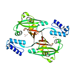
Crystal Structure of PhnF from Mycobacterium smegmatis
分子名称: GLYCEROL, GntR-family protein transcriptional regulator
著者Busby, J.N, Gebhard, S, Cook, G.M, Lott, S.J, Baker, E.N, Money, V.A.
登録日2008-11-12
公開日2009-11-17
最終更新日2023-11-01
実験手法X-RAY DIFFRACTION (1.8 Å)
主引用文献Crystal structure of PhnF, a GntR-family transcription regulator in Mycobacterium smegmatis
To be Published
3FYC
DownloadVisualize
BU of 3fyc by Molmil
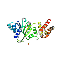
Crystal Structure of Dim1 from the thermophilic archeon, Methanocaldococcus jannaschi
分子名称: PHOSPHATE ION, Probable dimethyladenosine transferase
著者Scarsdale, J.N, Musayev, F.N, Rife, J.P.
登録日2009-01-22
公開日2009-06-30
最終更新日2023-09-06
実験手法X-RAY DIFFRACTION (2.15 Å)
主引用文献Structural and functional divergence within the Dim1/KsgA family of rRNA methyltransferases.
J.Mol.Biol., 391, 2009
3IR8
DownloadVisualize
BU of 3ir8 by Molmil
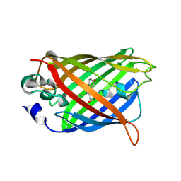
Red fluorescent protein mKeima at pH 7.0
分子名称: Large stokes shift fluorescent protein
著者Henderson, J.N, Osborn, M.F, Koon, N, Gepshtein, R, Huppert, D, Remington, S.J.
登録日2009-08-21
公開日2009-09-08
最終更新日2023-11-15
実験手法X-RAY DIFFRACTION (1.63 Å)
主引用文献Excited state proton transfer in the red fluorescent protein mKeima.
J.Am.Chem.Soc., 131, 2009
1A7W
DownloadVisualize
BU of 1a7w by Molmil
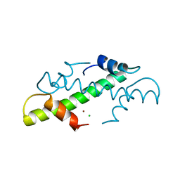
CRYSTAL STRUCTURE OF THE HISTONE HMFB FROM METHANOTHERMUS FERVIDUS
分子名称: CHLORIDE ION, HISTONE HMFB, ZINC ION
著者Decanniere, K, Sandman, K, Reeve, J.N, Heinemann, U.
登録日1998-03-18
公開日1999-03-23
最終更新日2024-04-03
実験手法X-RAY DIFFRACTION (1.55 Å)
主引用文献Crystal structures of recombinant histones HMfA and HMfB from the hyperthermophilic archaeon Methanothermus fervidus.
J.Mol.Biol., 303, 2000
1AKC
DownloadVisualize
BU of 1akc by Molmil
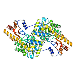
Structural basis for the catalytic activity of aspartate aminotransferase K258H lacking its pyridoxal-5'-phosphate-binding lysine residue
分子名称: 4-[(1,3-DICARBOXY-PROPYLAMINO)-METHYL]-3-HYDROXY-2-METHYL-5-PHOSPHONOOXYMETHYL-PYRIDINIUM, ASPARTATE AMINOTRANSFERASE
著者Malashkevich, V.N, Jansonius, J.N.
登録日1994-02-28
公開日1994-07-31
最終更新日2024-02-07
実験手法X-RAY DIFFRACTION (2.3 Å)
主引用文献Structural basis for the catalytic activity of aspartate aminotransferase K258H lacking the pyridoxal 5'-phosphate-binding lysine residue.
Biochemistry, 34, 1995
1AKA
DownloadVisualize
BU of 1aka by Molmil
STRUCTURAL BASIS FOR THE CATALYTIC ACTIVITY OF ASPARTATE AMINOTRANSFERASE K258H LACKING ITS PYRIDOXAL-5'-PHOSPHATE-BINDING LYSINE RESIDUE
分子名称: ASPARTATE AMINOTRANSFERASE, PHOSPHATE ION, PYRIDOXAL-5'-PHOSPHATE
著者Malashkevich, V.N, Jansonius, J.N.
登録日1994-02-28
公開日1994-07-31
最終更新日2024-02-07
実験手法X-RAY DIFFRACTION (2.1 Å)
主引用文献Structural basis for the catalytic activity of aspartate aminotransferase K258H lacking the pyridoxal 5'-phosphate-binding lysine residue.
Biochemistry, 34, 1995
1AKB
DownloadVisualize
BU of 1akb by Molmil
STRUCTURAL BASIS FOR THE CATALYTIC ACTIVITY OF ASPARTATE AMINOTRANSFERASE K258H LACKING ITS PYRIDOXAL-5'-PHOSPHATE-BINDING LYSINE RESIDUE
分子名称: 2-[(3-HYDROXY-2-METHYL-5-PHOSPHONOOXYMETHYL-PYRIDIN-4-YLMETHYLENE)-AMINO]-SUCCINIC ACID, ASPARTATE AMINOTRANSFERASE
著者Malashkevich, V.N, Jansonius, J.N.
登録日1994-02-28
公開日1994-07-31
最終更新日2024-02-07
実験手法X-RAY DIFFRACTION (2.3 Å)
主引用文献Structural basis for the catalytic activity of aspartate aminotransferase K258H lacking the pyridoxal 5'-phosphate-binding lysine residue.
Biochemistry, 34, 1995
5ZWB
DownloadVisualize
BU of 5zwb by Molmil
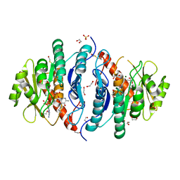
Crystal structure of Pyridoxal kinase (PdxK) from Salmonella typhimurium in complex with ADP, PL-linked to Lys233 via a Schiff base
分子名称: 1,2-ETHANEDIOL, 2-AMINO-2-HYDROXYMETHYL-PROPANE-1,3-DIOL, ADENOSINE-5'-DIPHOSPHATE, ...
著者Deka, G, Benazir, J.F, Kalyani, J.N, Savithri, H.S, Murthy, M.R.N.
登録日2018-05-14
公開日2019-05-29
最終更新日2023-11-22
実験手法X-RAY DIFFRACTION (2.2 Å)
主引用文献Structural and functional studies on Salmonella typhimurium pyridoxal kinase: the first structural evidence for the formation of Schiff base with the substrate.
Febs J., 286, 2019
5ZW9
DownloadVisualize
BU of 5zw9 by Molmil
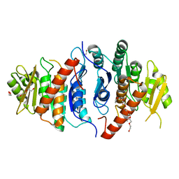
Crystal structure of Pyridoxal kinase (PdxK) from Salmonella typhimurium
分子名称: 1,2-ETHANEDIOL, 2-{2-[2-(2-{2-[2-(2-ETHOXY-ETHOXY)-ETHOXY]-ETHOXY}-ETHOXY)-ETHOXY]-ETHOXY}-ETHANOL, DI(HYDROXYETHYL)ETHER, ...
著者Deka, G, Benazir, J.F, Kalyani, J.N, Savithri, H.S, Murthy, M.R.N.
登録日2018-05-14
公開日2019-05-29
最終更新日2023-11-22
実験手法X-RAY DIFFRACTION (2.6 Å)
主引用文献Structural and functional studies on Salmonella typhimurium pyridoxal kinase: the first structural evidence for the formation of Schiff base with the substrate.
Febs J., 286, 2019
5ZWA
DownloadVisualize
BU of 5zwa by Molmil
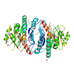
Crystal structure of Pyridoxal kinase (PdxK) from Salmonella typhimurium in complex with ADP, PL-linked to Lys233 via Schiff base in protomer A and the product (PLP) in protomer B
分子名称: 1,2-ETHANEDIOL, ADENOSINE-5'-DIPHOSPHATE, GLYCEROL, ...
著者Deka, G, Benazir, J.F, Kalyani, J.N, Savithri, H.S, Murthy, M.R.N.
登録日2018-05-14
公開日2019-05-29
最終更新日2023-11-22
実験手法X-RAY DIFFRACTION (2.45 Å)
主引用文献Structural and functional studies on Salmonella typhimurium pyridoxal kinase: the first structural evidence for the formation of Schiff base with the substrate.
Febs J., 286, 2019
2QWF
DownloadVisualize
BU of 2qwf by Molmil
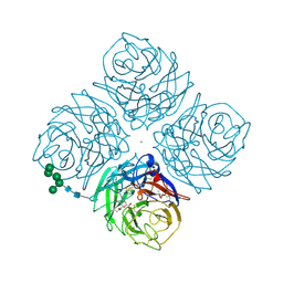
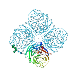
THE X-RAY STRUCTURE OF A COMPLEX OF N-ACETYL-4-GUANIDINO-6-METHYL(PROPYL)CARBOXAMIDE-4,5-DIHYDRO-2H-PYRAN-2-CARBOXYLIC ACID AND A DRUG RESISTANT VARIANT R292K OF TERN N9 INFLUENZA VIRUS NEURAMINIDASE
分子名称: 2-acetamido-2-deoxy-beta-D-glucopyranose, 2-acetamido-2-deoxy-beta-D-glucopyranose-(1-4)-2-acetamido-2-deoxy-beta-D-glucopyranose, 5-N-acetyl-4-guanidino-6-methyl(propyl) carboxamide-4,5-dihydro-2H-pyran-2-carboxylic acid, ...
著者Varghese, J.N.
登録日1998-04-07
公開日1998-11-11
最終更新日2024-04-03
実験手法X-RAY DIFFRACTION (1.9 Å)
主引用文献Drug design against a shifting target: a structural basis for resistance to inhibitors in a variant of influenza virus neuraminidase.
Structure, 6, 1998
6AL7
DownloadVisualize
BU of 6al7 by Molmil
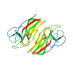
Crystal structure HpiC1 F138S
分子名称: 12-epi-hapalindole C/U synthase, CALCIUM ION
著者Newmister, S.A, Li, S, Garcia-Borras, M, Sanders, J.N, Yang, S, Lowell, A.N, Yu, F, Smith, J.L, Williams, R.M, Houk, K.N, Sherman, D.H.
登録日2017-08-07
公開日2018-03-07
最終更新日2024-03-13
実験手法X-RAY DIFFRACTION (1.687 Å)
主引用文献Structural basis of the Cope rearrangement and cyclization in hapalindole biogenesis.
Nat. Chem. Biol., 14, 2018
2QWK
DownloadVisualize
BU of 2qwk by Molmil
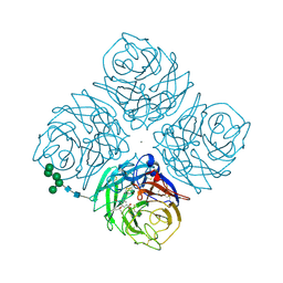
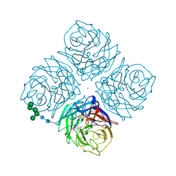
THE X-RAY STRUCTURE OF A COMPLEX OF 5-N-ACETYL-5-AMINO-3-(1-ETHYLPROPOXY)-1-CYCLOHEXENE-1-CARBOXYLIC ACID (GS4071) AND WILDTYPE TERN N9 INFLUENZA VIRUS NEURAMINIDASE
分子名称: (3R,4R,5S)-4-(acetylamino)-5-amino-3-(pentan-3-yloxy)cyclohex-1-ene-1-carboxylic acid, 2-acetamido-2-deoxy-beta-D-glucopyranose, 2-acetamido-2-deoxy-beta-D-glucopyranose-(1-4)-2-acetamido-2-deoxy-beta-D-glucopyranose, ...
著者Varghese, J.N.
登録日1998-04-07
公開日1998-11-11
最終更新日2024-04-03
実験手法X-RAY DIFFRACTION (1.8 Å)
主引用文献Drug design against a shifting target: a structural basis for resistance to inhibitors in a variant of influenza virus neuraminidase.
Structure, 6, 1998
2QWH
DownloadVisualize
BU of 2qwh by Molmil
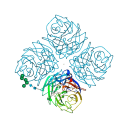
THE X-RAY STRUCTURE OF A COMPLEX OF 5-N-ACETYL-5-AMINO-3-(1-ETHYLPROPOXY)-1-CYCLOHEXENE-1-CARBOXYLIC ACID (GS4071) AND A DRUG RESISTANT VARIANT R292K OF TERN N9 INFLUENZA VIRUS NEURAMINIDASE
分子名称: (3R,4R,5S)-4-(acetylamino)-5-amino-3-(pentan-3-yloxy)cyclohex-1-ene-1-carboxylic acid, 2-acetamido-2-deoxy-beta-D-glucopyranose, 2-acetamido-2-deoxy-beta-D-glucopyranose-(1-4)-2-acetamido-2-deoxy-beta-D-glucopyranose, ...
著者Varghese, J.N.
登録日1998-04-07
公開日1998-11-11
最終更新日2024-04-03
実験手法X-RAY DIFFRACTION (1.8 Å)
主引用文献Drug design against a shifting target: a structural basis for resistance to inhibitors in a variant of influenza virus neuraminidase.
Structure, 6, 1998
2QWA
DownloadVisualize
BU of 2qwa by Molmil
THE X-RAY STRUCTURE OF A DRUG RESISTANT VARIANT R292K OF TERN N9 INFLUENZA VIRUS NEURAMINIDASE
分子名称: 2-acetamido-2-deoxy-beta-D-glucopyranose, 2-acetamido-2-deoxy-beta-D-glucopyranose-(1-4)-2-acetamido-2-deoxy-beta-D-glucopyranose, CALCIUM ION, ...
著者Varghese, J.N.
登録日1998-04-07
公開日1998-11-11
最終更新日2024-04-03
実験手法X-RAY DIFFRACTION (1.7 Å)
主引用文献Drug design against a shifting target: a structural basis for resistance to inhibitors in a variant of influenza virus neuraminidase.
Structure, 6, 1998
2QWJ
DownloadVisualize
BU of 2qwj by Molmil
THE X-RAY STRUCTURE OF A COMPLEX OF 5-N-ACETYL-4-AMINO-6-DIETHYLCARBOXAMIDE-4,5-DIHYDRO-2H-PYRAN-2-CARBOXYLIC ACID AND A DRUG RESISTANT VARIANT R292K OF TERN N9 INFLUENZA VIRUS NEURAMINIDASE
分子名称: 2-acetamido-2-deoxy-beta-D-glucopyranose, 2-acetamido-2-deoxy-beta-D-glucopyranose-(1-4)-2-acetamido-2-deoxy-beta-D-glucopyranose, 5-N-acetyl-4-amino-6-diethyl carboxamide-4,5-dihydro-2H-pyran-2-carboxylic acid, ...
著者Varghese, J.N.
登録日1998-04-07
公開日1998-11-11
最終更新日2024-04-03
実験手法X-RAY DIFFRACTION (2 Å)
主引用文献Drug design against a shifting target: a structural basis for resistance to inhibitors in a variant of influenza virus neuraminidase.
Structure, 6, 1998
6AL6
DownloadVisualize
BU of 6al6 by Molmil
Crystal structure HpiC1 in P42 space group
分子名称: 12-epi-hapalindole C/U synthase, CALCIUM ION
著者Newmister, S.A, Li, S, Garcia-Borras, M, Sanders, J.N, Yang, S, Lowell, A.N, Yu, F, Smith, J.L, Williams, R.M, Houk, K.N, Sherman, D.H.
登録日2017-08-07
公開日2018-03-07
最終更新日2023-10-04
実験手法X-RAY DIFFRACTION (2.088 Å)
主引用文献Structural basis of the Cope rearrangement and cyclization in hapalindole biogenesis.
Nat. Chem. Biol., 14, 2018

224931

件を2024-09-11に公開中

PDB statisticsPDBj update infoContact PDBjnumon