1G1I
 
 | |
1QWY
 | | Latent LytM at 1.3 A resolution | | 分子名称: | ZINC ION, peptidoglycan hydrolase | | 著者 | Odintsov, S.G, Sabala, I, Marcyjaniak, M, Bochtler, M. | | 登録日 | 2003-09-03 | | 公開日 | 2004-01-20 | | 最終更新日 | 2024-02-14 | | 実験手法 | X-RAY DIFFRACTION (1.3 Å) | | 主引用文献 | Latent LytM at 1.3A resolution.
J.Mol.Biol., 335, 2004
|
|
1G3Q
 | |
5B0F
 | | Polyketide cyclase OAC from Cannabis sativa, Y72F mutant | | 分子名称: | GLYCEROL, Olivetolic acid cyclase | | 著者 | Yang, X, Matsui, T, Mori, T, Abe, I, Morita, H. | | 登録日 | 2015-10-28 | | 公開日 | 2016-01-27 | | 最終更新日 | 2023-11-08 | | 実験手法 | X-RAY DIFFRACTION (1.6 Å) | | 主引用文献 | Structural basis for olivetolic acid formation by a polyketide cyclase from Cannabis sativa
Febs J., 283, 2016
|
|
1R12
 | | Native Aplysia ADP ribosyl cyclase | | 分子名称: | ADP-ribosyl cyclase | | 著者 | Love, M.L, Szebenyi, D.M.E, Kriksunov, I.A, Thiel, D.J, Munshi, C, Graeff, R, Lee, H.C, Hao, Q. | | 登録日 | 2003-09-23 | | 公開日 | 2004-03-09 | | 最終更新日 | 2011-07-13 | | 実験手法 | X-RAY DIFFRACTION (1.7 Å) | | 主引用文献 | ADP-ribosyl cyclase; crystal structures reveal a covalent intermediate.
Structure, 12, 2004
|
|
5B6K
 | |
2ONU
 | | Plasmodium falciparum ubiquitin conjugating enzyme PF10_0330, putative homologue of human UBE2H | | 分子名称: | TETRAETHYLENE GLYCOL, Ubiquitin-conjugating enzyme, putative | | 著者 | Dong, A, Lew, J, Lin, L, Hassanali, A, Zhao, Y, Senisterra, G, Wasney, G, Vedadi, M, Kozieradzki, I, Bochkarev, A, Arrowsmith, C.H, Edwards, A.M, Sundstrom, M, Weigelt, J, Hui, R, Brokx, S.J, Structural Genomics Consortium (SGC) | | 登録日 | 2007-01-24 | | 公開日 | 2007-02-06 | | 最終更新日 | 2023-08-30 | | 実験手法 | X-RAY DIFFRACTION (2.38 Å) | | 主引用文献 | Plasmodium falciparum ubiquitin conjugating enzyme PF10_0330, putative homologue of human UBE2H
To be Published
|
|
1Q6O
 | | Structure of 3-keto-L-gulonate 6-phosphate decarboxylase with bound L-gulonaet 6-phosphate | | 分子名称: | 3-keto-L-gulonate 6-phosphate decarboxylase, L-GULURONIC ACID 6-PHOSPHATE, MAGNESIUM ION | | 著者 | Wise, E.L, Yew, W.S, Gerlt, J.A, Rayment, I. | | 登録日 | 2003-08-13 | | 公開日 | 2003-10-28 | | 最終更新日 | 2024-02-14 | | 実験手法 | X-RAY DIFFRACTION (1.202 Å) | | 主引用文献 | Structural Evidence for a 1,2-Enediolate Intermediate in the Reaction Catalyzed by 3-Keto-l-Gulonate 6-Phosphate Decarboxylase, a Member of the Orotidine 5'-Monophosphate Decarboxylase Suprafamily
Biochemistry, 42, 2003
|
|
3TPR
 | | Crystal structure of BACE1 complexed with an inhibitor | | 分子名称: | Beta-secretase 1, CHLORIDE ION, N-[(1S,2R)-1-BENZYL-3-(CYCLOPROPYLAMINO)-2-HYDROXYPROPYL]-5-[METHYL(METHYLSULFONYL)AMINO]-N'-[(1R)-1-PHENYLETHYL]ISOPHTHALAMIDE | | 著者 | Xu, Y.C, Li, M.J, Greenblatt, H, Chen, T.T, Silman, I, Sussman, J.L. | | 登録日 | 2011-09-08 | | 公開日 | 2011-11-23 | | 最終更新日 | 2023-11-01 | | 実験手法 | X-RAY DIFFRACTION (2.55 Å) | | 主引用文献 | Flexibility of the flap in the active site of BACE1 as revealed by crystal structures and molecular dynamics simulations
Acta Crystallogr.,Sect.D, 68, 2012
|
|
2OP2
 | |
1I0S
 | | ARCHAEOGLOBUS FULGIDUS FERRIC REDUCTASE COMPLEX WITH NADP+ | | 分子名称: | CONSERVED HYPOTHETICAL PROTEIN, FLAVIN MONONUCLEOTIDE, NADP NICOTINAMIDE-ADENINE-DINUCLEOTIDE PHOSPHATE | | 著者 | Chiu, H.-J, Johnson, E, Schroder, I, Rees, D.C. | | 登録日 | 2001-01-29 | | 公開日 | 2001-05-02 | | 最終更新日 | 2023-08-09 | | 実験手法 | X-RAY DIFFRACTION (1.65 Å) | | 主引用文献 | Crystal structures of a novel ferric reductase from the hyperthermophilic archaeon Archaeoglobus fulgidus and its complex with NADP+.
Structure, 9, 2001
|
|
1I0R
 | | CRYSTAL STRUCTURE OF FERRIC REDUCTASE FROM ARCHAEOGLOBUS FULGIDUS | | 分子名称: | CONSERVED HYPOTHETICAL PROTEIN, FLAVIN MONONUCLEOTIDE | | 著者 | Chiu, H.-J, Johnson, E, Schroder, I, Rees, D.C. | | 登録日 | 2001-01-29 | | 公開日 | 2001-05-02 | | 最終更新日 | 2017-10-04 | | 実験手法 | X-RAY DIFFRACTION (1.5 Å) | | 主引用文献 | Crystal structures of a novel ferric reductase from the hyperthermophilic archaeon Archaeoglobus fulgidus and its complex with NADP+.
Structure, 9, 2001
|
|
5BMF
 | | Crystal Structure of a Theophylline binding antibody Fab fragment | | 分子名称: | 2-(5-{1-[1-(1,3-dimethyl-2,6-dioxo-2,3,6,7-tetrahydro-1H-purin-8-yl)-4,15-dioxo-8,11-dioxa-5,14-diazaicosan-20-yl]-3,3-dimethyl-6-sulfo-1,3-dihydro-2H-indol-2-ylidene}penta-1,3-dien-1-yl)-1-ethyl-3,3-dimethyl-3H-indolium-5-sulfonate, Fab fragment heavy chain, Fab fragment light chain | | 著者 | Bujotzek, A, Fuchs, A, Changtao, Q, Klostermann, S, Benz, J, Antes, I, Dengl, S, Hoffmann, E, Georges, G. | | 登録日 | 2015-05-22 | | 公開日 | 2015-07-29 | | 最終更新日 | 2024-05-01 | | 実験手法 | X-RAY DIFFRACTION (2.8 Å) | | 主引用文献 | MoFvAb: Modeling the Fv region of antibodies.
Mabs, 7, 2015
|
|
2ORL
 | | Solution structure of the cytochrome c- para-aminophenol adduct | | 分子名称: | 4-AMINOPHENOL, Cytochrome c iso-1, HEME C | | 著者 | Assfalg, M, Bertini, I, Del Conte, R, Giachetti, A, Turano, P. | | 登録日 | 2007-02-03 | | 公開日 | 2007-04-24 | | 最終更新日 | 2021-10-20 | | 実験手法 | SOLUTION NMR | | 主引用文献 | Cytochrome c and organic molecules: solution structure of the p-aminophenol adduct.
Biochemistry, 46, 2007
|
|
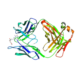
1IC4
 | | CRYSTAL STRUCTURE OF HYHEL-10 FV MUTANT(HD32A)-HEN LYSOZYME COMPLEX | | 分子名称: | IGG1 FAB CHAIN H, LYSOZYME BINDING IG KAPPA CHAIN, LYSOZYME C | | 著者 | Shiroishi, M, Yokota, A, Tsumoto, K, Kondo, H, Nishimiya, Y, Horii, K, Matsushima, M, Ogasahara, K, Yutani, K, Kumagai, I. | | 登録日 | 2001-03-30 | | 公開日 | 2001-07-18 | | 最終更新日 | 2021-11-10 | | 実験手法 | X-RAY DIFFRACTION (2.5 Å) | | 主引用文献 | Structural evidence for entropic contribution of salt bridge formation to a protein antigen-antibody interaction: the case of hen lysozyme-HyHEL-10 Fv complex.
J.Biol.Chem., 276, 2001
|
|
1QCI
 | | LOW TEMPERATURE STRUCTURE OF POKEWEED ANTIVIRAL PROTEIN COMPLEXED WITH ADENINE | | 分子名称: | ADENINE, POKEWEED ANTIVIRAL PROTEIN | | 著者 | Kurinov, I.V, Myers, D.E, Irvin, J.D, Uckun, F.M. | | 登録日 | 1999-05-05 | | 公開日 | 1999-09-15 | | 最終更新日 | 2011-07-13 | | 実験手法 | X-RAY DIFFRACTION (2 Å) | | 主引用文献 | X-ray crystallographic analysis of the structural basis for the interactions of pokeweed antiviral protein with its active site inhibitor and ribosomal RNA substrate analogs.
Protein Sci., 8, 1999
|
|
1I1H
 | | CRYSTAL STRUCTURE ANALYSIS OF PRECORRIN-8X METHYLMUTASE COMPLEX WITH HYDROGENOBYRINIC ACID | | 分子名称: | HYDROGENOBYRINIC ACID, PRECORRIN-8X METHYLMUTASE | | 著者 | Shipman, L.W, Li, D, Roessner, C.A, Scott, A.I, Sacchettini, J.C. | | 登録日 | 2001-02-01 | | 公開日 | 2001-07-18 | | 最終更新日 | 2024-02-07 | | 実験手法 | X-RAY DIFFRACTION (2.6 Å) | | 主引用文献 | Crystal structure of precorrin-8x methyl mutase.
Structure, 9, 2001
|
|
2OWA
 | | Crystal structure of putative GTPase activating protein for ADP ribosylation factor from Cryptosporidium parvum (cgd5_1040) | | 分子名称: | Arfgap-like finger domain containing protein, ZINC ION | | 著者 | Dong, A, Lew, J, Zhao, Y, Hassanali, A, Lin, L, Ravichandran, M, Wasney, G, Vedadi, M, Kozieradzki, I, Bochkarev, A, Edwards, A.M, Arrowsmith, C.H, Weigelt, J, Sundstrom, M, Hui, R, Qiu, W, Structural Genomics Consortium (SGC) | | 登録日 | 2007-02-15 | | 公開日 | 2007-02-27 | | 最終更新日 | 2023-08-30 | | 実験手法 | X-RAY DIFFRACTION (2 Å) | | 主引用文献 | Crystal structure of putative GTPase activating protein for ADP ribosylation factor from Cryptosporidium parvum (cgd5_1040)
To be Published
|
|
1QWG
 | | Crystal structure of Methanococcus jannaschii phosphosulfolactate synthase | | 分子名称: | (2R)-phospho-3-sulfolactate synthase, SULFATE ION | | 著者 | Wise, E.L, Graham, D.E, White, R.H, Rayment, I. | | 登録日 | 2003-09-02 | | 公開日 | 2003-12-09 | | 最終更新日 | 2024-02-14 | | 実験手法 | X-RAY DIFFRACTION (1.6 Å) | | 主引用文献 | The structural determination of phosphosulfolactate synthase from Methanococcus jannaschii at 1.7-A resolution: an enolase that is not an enolase
J.Biol.Chem., 278, 2003
|
|
2P1P
 | | Mechanism of Auxin Perception by the TIR1 ubiquitin ligase | | 分子名称: | 1H-INDOL-3-YLACETIC ACID, INOSITOL HEXAKISPHOSPHATE, SKP1-like protein 1A, ... | | 著者 | Tan, X, Calderon-Villalobos, L.I.A, Sharon, M, Robinson, C.V, Estelle, M, Zheng, N. | | 登録日 | 2007-03-06 | | 公開日 | 2007-04-10 | | 最終更新日 | 2023-08-30 | | 実験手法 | X-RAY DIFFRACTION (2.21 Å) | | 主引用文献 | Mechanism of auxin perception by the TIR1 ubiquitin ligase
Nature, 446, 2007
|
|
5AMX
 | | Crystal Structure of Proteinase K processed with the CrystalDirect automated mounting and cryo-cooling technology | | 分子名称: | PROTEINASE K, SULFATE ION | | 著者 | Zander, U, Hoffmann, G, Cornaciu, I, Cipriani, F, Marquez, J.A. | | 登録日 | 2015-09-02 | | 公開日 | 2016-04-13 | | 最終更新日 | 2016-04-20 | | 実験手法 | X-RAY DIFFRACTION (1.01 Å) | | 主引用文献 | Automated Harvesting and Processing of Protein Crystals Through Laser Photoablation.
Acta Crystallogr.,Sect.D, 72, 2016
|
|
1I5C
 | | STRUCTURE OF CHEA DOMAIN P4 IN COMPLEX WITH ADP | | 分子名称: | ADENOSINE-5'-DIPHOSPHATE, CHEMOTAXIS PROTEIN CHEA | | 著者 | Bilwes, A.M, Quezada, C.M, Croal, L.R, Crane, B.R, Simon, M.I. | | 登録日 | 2001-02-26 | | 公開日 | 2001-08-26 | | 最終更新日 | 2023-08-09 | | 実験手法 | X-RAY DIFFRACTION (1.9 Å) | | 主引用文献 | Nucleotide binding by the histidine kinase CheA.
Nat.Struct.Biol., 8, 2001
|
|
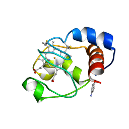
5APC
 | | Hen Egg White Lysozyme illuminated with 0.4THz radiation | | 分子名称: | LYSOZYME C, SODIUM ION | | 著者 | Lundholm, I, Rodilla, H, Wahlgren, W.Y, Duelli, A, Bourenkov, G, Vukusic, J, Friedman, R, Stake, J, Schneider, T, Katona, G. | | 登録日 | 2015-09-15 | | 公開日 | 2016-01-13 | | 最終更新日 | 2018-03-07 | | 実験手法 | X-RAY DIFFRACTION (1.7 Å) | | 主引用文献 | Terahertz Radiation Induces Non-Thermal Structural Changes Associated with Frohlich Condensation in a Protein Crystal
Struct.Dyn., 2, 2015
|
|
3GQ2
 | |
1Q6L
 | | Structure of 3-keto-L-gulonate 6-phosphate decarboxylase with bound L-threonohydroxamate 4-phosphate | | 分子名称: | 3-keto-L-gulonate 6-phosphate decarboxylase, L-THREONOHYDROXAMATE 4-PHOSPHATE, MAGNESIUM ION | | 著者 | Wise, E.L, Yew, W.S, Gerlt, J.A, Rayment, I. | | 登録日 | 2003-08-13 | | 公開日 | 2003-10-28 | | 最終更新日 | 2019-07-24 | | 実験手法 | X-RAY DIFFRACTION (1.8 Å) | | 主引用文献 | Structural Evidence for a 1,2-Enediolate Intermediate in the Reaction Catalyzed by 3-Keto-l-Gulonate 6-Phosphate Decarboxylase, a Member of the Orotidine 5'-Monophosphate Decarboxylase Suprafamily
Biochemistry, 42, 2003
|
|


