4U6U
 
 | |
8CUK
 | |
2HJE
 | |
1ZHH
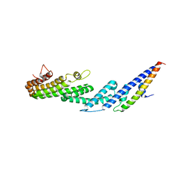
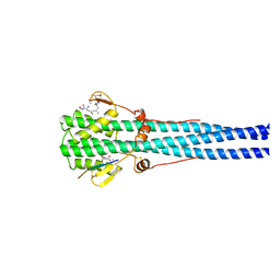
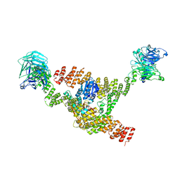
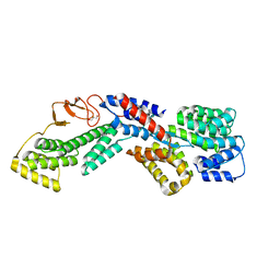
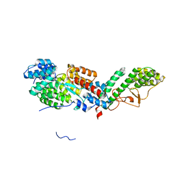
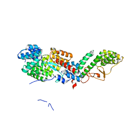
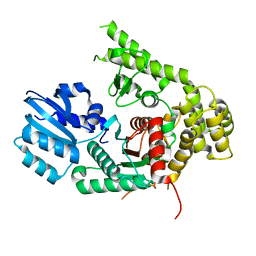
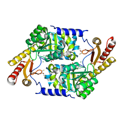
 | | Crystal Structure of the Apo Form of Vibrio Harveyi LUXP Complexed with the Periplasmic Domain of LUXQ | | 分子名称: | 2-[N-CYCLOHEXYLAMINO]ETHANE SULFONIC ACID, Autoinducer 2 sensor kinase/phosphatase luxQ, Autoinducer 2-binding periplasmic protein luxP | | 著者 | Neiditch, M.B, Federle, M.J, Miller, S.T, Bassler, B.L, Hughson, F.M. | | 登録日 | 2005-04-25 | | 公開日 | 2005-05-24 | | 最終更新日 | 2024-02-14 | | 実験手法 | X-RAY DIFFRACTION (1.94 Å) | | 主引用文献 | Regulation of LuxPQ Receptor Activity by the Quorum-Sensing Signal Autoinducer-2.
Mol.Cell, 18, 2005
|
|
1HTM
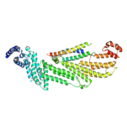
 | | STRUCTURE OF INFLUENZA HAEMAGGLUTININ AT THE PH OF MEMBRANE FUSION | | 分子名称: | HEMAGGLUTININ HA1 CHAIN, HEMAGGLUTININ HA2 CHAIN | | 著者 | Bullough, P.A, Hughson, F.M, Skehel, J.J, Wiley, D.C. | | 登録日 | 1994-11-02 | | 公開日 | 1995-02-14 | | 最終更新日 | 2011-07-13 | | 実験手法 | X-RAY DIFFRACTION (2.5 Å) | | 主引用文献 | Structure of influenza haemagglutinin at the pH of membrane fusion.
Nature, 371, 1994
|
|
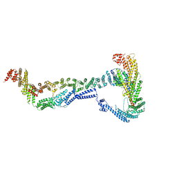
8FTU
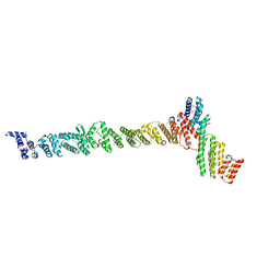
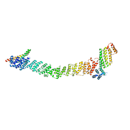 | | Crystal structure of the SNARE Use1 bound to Dsl1 complex subunits Sec39 and Dsl1, Revised Use1 structure | | 分子名称: | Protein transport protein DSL1, Protein transport protein SEC39, Vesicle transport protein USE1 | | 著者 | Travis, S.M, Jeffrey, P.D, Hughson, F.M. | | 登録日 | 2023-01-13 | | 公開日 | 2023-03-01 | | 最終更新日 | 2024-02-28 | | 実験手法 | X-RAY DIFFRACTION (5.73 Å) | | 主引用文献 | Structure of a membrane tethering complex incorporating multiple SNAREs.
Nat.Struct.Mol.Biol., 31, 2024
|
|
5BV0
 | |
5BUZ
 | |
5BV1
 | |
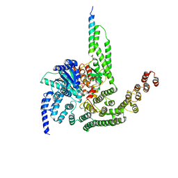
8DIT
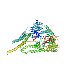
 | | Cryo-EM structure of a HOPS core complex containing Vps33, Vps16, and Vps18 | | 分子名称: | Vacuolar protein sorting-associated protein 16, Vacuolar protein sorting-associated protein 18, Vacuolar protein sorting-associated protein 33 | | 著者 | Port, S.A, Farrell, P.D, Jeffrey, P.D, DiMaio, F, Hughson, F.M. | | 登録日 | 2022-06-29 | | 公開日 | 2022-08-31 | | 最終更新日 | 2024-02-14 | | 実験手法 | ELECTRON MICROSCOPY (5.1 Å) | | 主引用文献 | Cryo-EM structure of the HOPS core complex and its implication for SNARE assembly
To Be Published
|
|
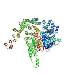
8EKI
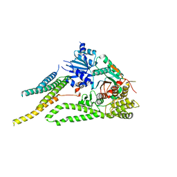
 | | CryoEM structure of the Dsl1 complex bound to SNAREs Sec20 and Use1 | | 分子名称: | Protein transport protein DSL1, Protein transport protein SEC20, Protein transport protein SEC39, ... | | 著者 | DAmico, K.A, Jeffrey, P.D, Hughson, F.M. | | 登録日 | 2022-09-21 | | 公開日 | 2023-10-04 | | 最終更新日 | 2024-02-28 | | 実験手法 | ELECTRON MICROSCOPY (4.5 Å) | | 主引用文献 | Structure of a membrane tethering complex incorporating multiple SNAREs.
Nat.Struct.Mol.Biol., 31, 2024
|
|
1EZ3
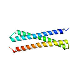 | |
1FIO
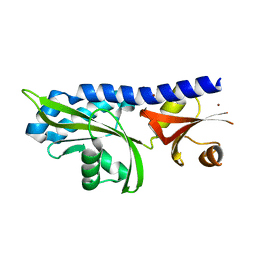
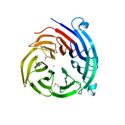
 | | CRYSTAL STRUCTURE OF YEAST T-SNARE PROTEIN SSO1 | | 分子名称: | SSO1 PROTEIN, ZINC ION | | 著者 | Munson, M, Chen, X, Cocina, A.E, Schultz, S.M, Hughson, F.M. | | 登録日 | 2000-08-04 | | 公開日 | 2000-10-11 | | 最終更新日 | 2022-12-21 | | 実験手法 | X-RAY DIFFRACTION (2.1 Å) | | 主引用文献 | Interactions within the yeast t-SNARE Sso1p that control SNARE complex assembly.
Nat.Struct.Biol., 7, 2000
|
|
6U3W
 | |
6U3V
 | |
6TZT
 | |
6WC3
 | |
6XJL
 | |
6XM1
 | |
6XMD
 | |
3K8P
 | |
3KKI
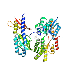
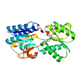
 | | PLP-Dependent Acyl-CoA transferase CqsA | | 分子名称: | 2,3-DIHYDROXY-1,4-DITHIOBUTANE, CAI-1 autoinducer synthase, MAGNESIUM ION, ... | | 著者 | Kelly, R.C, Jeffrey, P.D, Hughson, F.M. | | 登録日 | 2009-11-05 | | 公開日 | 2009-11-24 | | 最終更新日 | 2023-09-06 | | 実験手法 | X-RAY DIFFRACTION (1.8 Å) | | 主引用文献 | The Vibrio cholerae quorum-sensing autoinducer CAI-1: analysis of the biosynthetic enzyme CqsA.
Nat.Chem.Biol., 5, 2009
|
|
1TM2
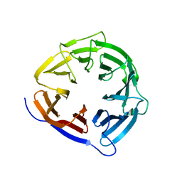
 | | Crystal Structure of the apo form of the Salmonella typhimurium AI-2 receptor LsrB | | 分子名称: | sugar transport protein | | 著者 | Miller, S.T, Xavier, K.B, Campagna, S.R, Taga, M.E, Semmelhack, M.F, Bassler, B.L, Hughson, F.M. | | 登録日 | 2004-06-10 | | 公開日 | 2004-09-28 | | 最終更新日 | 2018-01-31 | | 実験手法 | X-RAY DIFFRACTION (1.9 Å) | | 主引用文献 | Salmonella typhimurium Recognizes a Chemically Distinct Form of the Bacterial Quorum-Sensing Signal AI-2
Mol.Cell, 15, 2004
|
|
1TJY
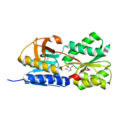 | | Crystal Structure of Salmonella typhimurium AI-2 receptor LsrB in complex with R-THMF | | 分子名称: | (2R,4S)-2-methyl-2,3,3,4-tetrahydroxytetrahydrofuran, sugar transport protein | | 著者 | Miller, S.T, Xavier, K.B, Campagna, S.R, Taga, M.E, Semmelhack, M.F, Bassler, B.L, Hughson, F.M. | | 登録日 | 2004-06-07 | | 公開日 | 2004-09-28 | | 最終更新日 | 2020-07-29 | | 実験手法 | X-RAY DIFFRACTION (1.3 Å) | | 主引用文献 | Salmonella typhimurium Recognizes a Chemically Distinct Form of the Bacterial Quorum-Sensing Signal AI-2
Mol.Cell, 15, 2004
|
|
4H5J
 | |


