Loading
PDBj
MenuPDBj@FacebookPDBj@TwitterPDBj@YouTubewwPDB FoundationwwPDB
RCSB PDBPDBeBMRBAdv. SearchSearch help
PDB: 220 results

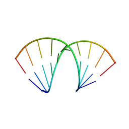
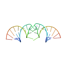
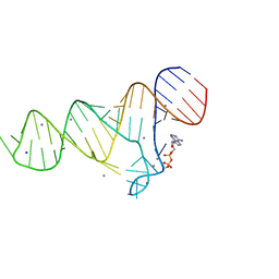
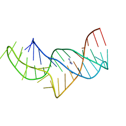
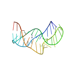
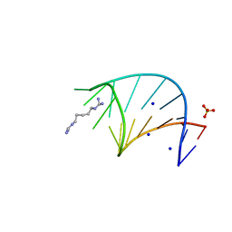
6HC5
DownloadVisualize
BU of 6hc5 by Molmil
The structure of the G. violaceus guanidine II riboswitch P2 stem-loop with audouine
Descriptor: 1-(5-carbamimidamidopentyl)guanidine, RNA (5'-R(*GP*GP*UP*GP*GP*GP*GP*AP*CP*GP*AP*CP*CP*CP*CP*AP*(CBV)P*C)-3'), SODIUM ION, ...
Authors:Huang, L, Lilley, D.M.J.
Deposit date:2018-08-14
Release date:2019-02-20
Last modified:2024-05-15
Method:X-RAY DIFFRACTION (1.413 Å)
Cite:Structure-guided design of a high-affinity ligand for a riboswitch.
Rna, 25, 2019
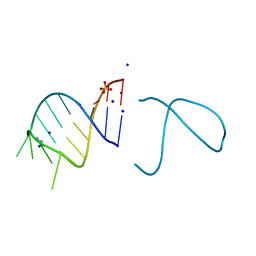
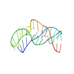
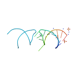
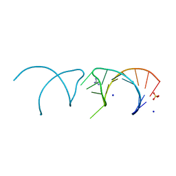
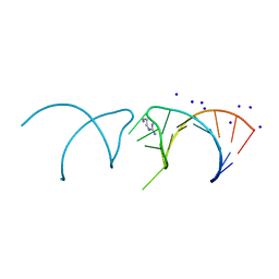
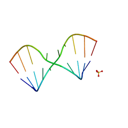
5LR3
DownloadVisualize
BU of 5lr3 by Molmil
RNA duplex has central consecutive GA pairs flanked by G-U basepairs
Descriptor: RNA (5'-R(*GP*(CBV)P*CP*GP*GP*AP*UP*GP*GP*C)-3'), SULFATE ION
Authors:Huang, L, Lilley, D.M.J.
Deposit date:2016-08-18
Release date:2017-06-21
Last modified:2024-05-08
Method:X-RAY DIFFRACTION (1.65 Å)
Cite:Control of box C/D snoRNP assembly by N(6)-methylation of adenine.
EMBO Rep., 18, 2017
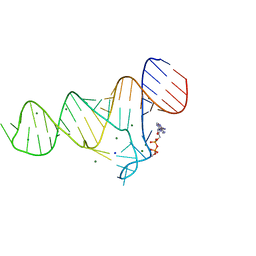
5LR5
DownloadVisualize
BU of 5lr5 by Molmil
N6-methyladenine is accommodated in a conventional A-U basepair
Descriptor: RNA (5'-R(*GP*GP*(6MZ)P*CP*UP*AP*GP*UP*(CBV)P*C)-3'), SULFATE ION
Authors:Huang, L, Lilley, D.M.J.
Deposit date:2016-08-18
Release date:2017-06-21
Last modified:2024-05-08
Method:X-RAY DIFFRACTION (2.4 Å)
Cite:Control of box C/D snoRNP assembly by N(6)-methylation of adenine.
EMBO Rep., 18, 2017
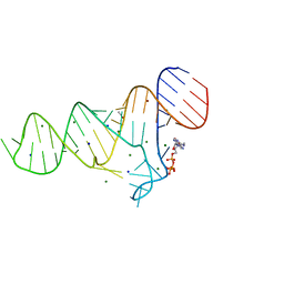
5NEF
DownloadVisualize
BU of 5nef by Molmil
The structure of the G. violaceus guanidine II riboswitch P1 stem-loop with guanidine
Descriptor: GUANIDINE, RNA (5'-R(*GP*GP*UP*GP*GP*GP*GP*AP*CP*GP*AP*CP*CP*CP*CP*AP*(CBV)P*C)-3'), SODIUM ION, ...
Authors:Huang, L, Wang, J, Lilley, D.M.J.
Deposit date:2017-03-10
Release date:2017-06-07
Last modified:2024-05-08
Method:X-RAY DIFFRACTION (1.91 Å)
Cite:The Structure of the Guanidine-II Riboswitch.
Cell Chem Biol, 24, 2017
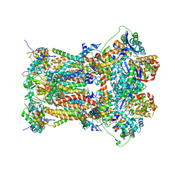
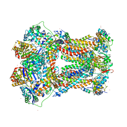
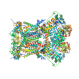
3L74
DownloadVisualize
BU of 3l74 by Molmil
Cytochrome BC1 complex from chicken with famoxadone bound
Descriptor: 1,2-dioleoyl-sn-glycero-3-phosphoethanolamine, AZIDE ION, CARDIOLIPIN, ...
Authors:Huang, L, Berry, E.A.
Deposit date:2009-12-28
Release date:2010-02-02
Last modified:2023-09-06
Method:X-RAY DIFFRACTION (2.76 Å)
Cite:Famoxadone and related inhibitors bind like methoxy acrylate inhibitors in the Qo site of the BC1 compl and fix the rieske iron-sulfur protein in a positio close to but distinct from that seen with stigmatellin and other "DISTAL" Qo inhibitors.
To be Published
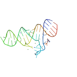
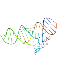
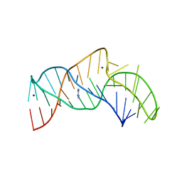
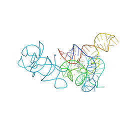
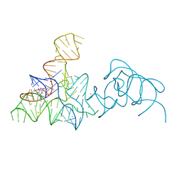
6TB7
DownloadVisualize
BU of 6tb7 by Molmil
Crystal structure of the ADP-binding domain of the NAD+ riboswitch with Adenosine monophosphate (AMP)
Descriptor: ADENOSINE MONOPHOSPHATE, BROMIDE ION, MAGNESIUM ION, ...
Authors:Huang, L, Lilley, D.M.J.
Deposit date:2019-11-01
Release date:2020-09-30
Last modified:2024-05-15
Method:X-RAY DIFFRACTION (2.53 Å)
Cite:Structure and ligand binding of the ADP-binding domain of the NAD + riboswitch.
Rna, 26, 2020
5NDI
DownloadVisualize
BU of 5ndi by Molmil
The structure of the E.coli guanidine II riboswitch P1 stem-loop
Descriptor: GUANIDINE, RNA (5'-R(*UP*UP*UP*GP*CP*AP*GP*GP*AP*CP*GP*AP*CP*CP*UP*GP*(CBV)P*AP*AP*A)-3'), SODIUM ION
Authors:Huang, L, Wang, J, Lilley, D.M.J.
Deposit date:2017-03-08
Release date:2017-05-31
Last modified:2024-05-08
Method:X-RAY DIFFRACTION (2.57 Å)
Cite:The Structure of the Guanidine-II Riboswitch.
Cell Chem Biol, 24, 2017
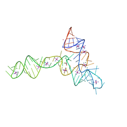
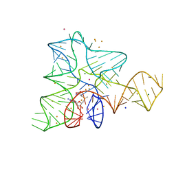
5O69
DownloadVisualize
BU of 5o69 by Molmil
The structure of the thermobifida fusca guanidine III riboswitch with agmatine.
Descriptor: AGMATINE, MAGNESIUM ION, RNA (37-MER), ...
Authors:Huang, L, Wang, J, Lilley, D.M.J.
Deposit date:2017-06-06
Release date:2017-10-18
Last modified:2024-05-08
Method:X-RAY DIFFRACTION (2.319 Å)
Cite:Structure of the Guanidine III Riboswitch.
Cell Chem Biol, 24, 2017
2A06
DownloadVisualize
BU of 2a06 by Molmil
Bovine cytochrome bc1 complex with stigmatellin bound
Descriptor: 1,2-dioleoyl-sn-glycero-3-phosphoethanolamine, AZIDE ION, CARDIOLIPIN, ...
Authors:Huang, L.S, Cobessi, D, Tung, E.Y, Berry, E.A.
Deposit date:2005-06-16
Release date:2005-06-21
Last modified:2023-08-23
Method:X-RAY DIFFRACTION (2.1 Å)
Cite:Binding of the Respiratory Chain Inhibitor Antimycin to the Mitochondrial bc(1) Complex: A New Crystal Structure Reveals an Altered Intramolecular Hydrogen-bonding Pattern.
J.Mol.Biol., 351, 2005
5NEO
DownloadVisualize
BU of 5neo by Molmil
The structure of the G. violaceus guanidine II riboswitch P1 stem-loop
Descriptor: AMMONIUM ION, RNA (5'-R(*GP*GP*UP*GP*GP*GP*GP*AP*CP*GP*AP*CP*CP*CP*CP*AP*(CBV)P*C)-3'), SODIUM ION, ...
Authors:Huang, L, Wang, J, Lilley, D.M.J.
Deposit date:2017-03-11
Release date:2017-05-31
Last modified:2024-05-08
Method:X-RAY DIFFRACTION (1.69 Å)
Cite:The Structure of the Guanidine-II Riboswitch.
Cell Chem Biol, 24, 2017
6TFG
DownloadVisualize
BU of 6tfg by Molmil
Crystal structure of the ADP-binding domain of the NAD+ riboswitch with Adenosine 3-phosphate 5-phosphosulfate (APPS)
Descriptor: 3'-PHOSPHATE-ADENOSINE-5'-PHOSPHATE SULFATE, Chains: A, MAGNESIUM ION, ...
Authors:Huang, L, Lilley, D.M.J.
Deposit date:2019-11-14
Release date:2020-09-23
Last modified:2024-01-24
Method:X-RAY DIFFRACTION (2.45 Å)
Cite:Structure and ligand binding of the ADP-binding domain of the NAD+ riboswitch.
Rna, 26, 2020
6TFH
DownloadVisualize
BU of 6tfh by Molmil
Crystal structure of the ADP-binding domain of the NAD+ riboswitch with Nicotinamide adenine dinucleotide, reduced (NADH); soaking with Manganese(II) (Mn2+)
Descriptor: Chains: A, MANGANESE (II) ION, NICOTINAMIDE-ADENINE-DINUCLEOTIDE, ...
Authors:Huang, L, Lilley, D.M.J.
Deposit date:2019-11-14
Release date:2020-09-23
Last modified:2024-01-24
Method:X-RAY DIFFRACTION (2.95 Å)
Cite:Structure and ligand binding of the ADP-binding domain of the NAD+ riboswitch.
Rna, 26, 2020
5NEP
DownloadVisualize
BU of 5nep by Molmil
The structure of the G. violaceus guanidine II riboswitch P1 stem-loop with methylguanidine
Descriptor: 1-METHYLGUANIDINE, RNA (5'-R(*GP*GP*UP*GP*GP*GP*GP*AP*CP*GP*AP*CP*CP*CP*CP*AP*(CBV)P*C)-3'), SODIUM ION, ...
Authors:Huang, L, Wang, J, Lilley, D.M.J.
Deposit date:2017-03-11
Release date:2017-05-31
Last modified:2024-05-08
Method:X-RAY DIFFRACTION (1.6 Å)
Cite:The Structure of the Guanidine-II Riboswitch.
Cell Chem Biol, 24, 2017
3TGU
DownloadVisualize
BU of 3tgu by Molmil
Cytochrome bc1 complex from chicken with pfvs-designed moa inhibitor bound
Descriptor: 1,2-dioleoyl-sn-glycero-3-phosphoethanolamine, CARDIOLIPIN, Coenzyme Q10, ...
Authors:Huang, L.-S, Yang, G.-F, Berry, E.A.
Deposit date:2011-08-17
Release date:2012-07-04
Last modified:2023-09-13
Method:X-RAY DIFFRACTION (2.7 Å)
Cite:Computational discovery of picomolar Q(o) site inhibitors of cytochrome bc1 complex.
J.Am.Chem.Soc., 134, 2012
5NY8
DownloadVisualize
BU of 5ny8 by Molmil
The structure of the thermobifida fusca guanidine III riboswitch with aminoguanidine
Descriptor: AMINOGUANIDINE, MAGNESIUM ION, RNA (41-MER), ...
Authors:Huang, L, Wang, J, Lilley, D.M.J.
Deposit date:2017-05-11
Release date:2017-10-18
Last modified:2024-05-08
Method:X-RAY DIFFRACTION (2.04 Å)
Cite:Structure of the Guanidine III Riboswitch.
Cell Chem Biol, 24, 2017
5NZ6
DownloadVisualize
BU of 5nz6 by Molmil
The structure of the thermobifida fusca guanidine III riboswitch with guanidine in space group P3212.
Descriptor: GUANIDINE, RNA (41-MER)
Authors:Huang, L, Wang, J, Lilley, D.M.J.
Deposit date:2017-05-12
Release date:2017-10-18
Last modified:2024-01-17
Method:X-RAY DIFFRACTION (2.94 Å)
Cite:Structure of the Guanidine III Riboswitch.
Cell Chem Biol, 24, 2017
5NOM
DownloadVisualize
BU of 5nom by Molmil
The structure of the G. violaceus guanidine II riboswitch P1 stem-loop with guanidine
Descriptor: GUANIDINE, RNA (5'-R(*GP*GP*UP*GP*GP*GP*GP*AP*CP*GP*AP*CP*CP*CP*CP*AP*(CBV)P*C)-3'), SODIUM ION, ...
Authors:Huang, L, Wang, J, Lilley, D.M.J.
Deposit date:2017-04-12
Release date:2017-05-31
Last modified:2024-01-17
Method:X-RAY DIFFRACTION (1.93 Å)
Cite:The Structure of the Guanidine-II Riboswitch.
Cell Chem Biol, 24, 2017
6TF0
DownloadVisualize
BU of 6tf0 by Molmil
Crystal structure of the ADP-binding domain of the NAD+ riboswitch with Nicotinamide adenine dinucleotide, reduced (NADH)
Descriptor: 1,4-DIHYDRONICOTINAMIDE ADENINE DINUCLEOTIDE, Chains: A, MAGNESIUM ION, ...
Authors:Huang, L, Lilley, D.M.J.
Deposit date:2019-11-12
Release date:2020-09-23
Last modified:2024-05-15
Method:X-RAY DIFFRACTION (2.1 Å)
Cite:Structure and ligand binding of the ADP-binding domain of the NAD+ riboswitch.
Rna, 26, 2020
6TFE
DownloadVisualize
BU of 6tfe by Molmil
Crystal structure of the ADP-binding domain of the NAD+ riboswitch with N6-Methyl-adenosine-5'-triphosphate (m6ATP)
Descriptor: BROMIDE ION, Chains: A, MAGNESIUM ION, ...
Authors:Huang, L, Lilley, D.M.J.
Deposit date:2019-11-14
Release date:2020-09-23
Last modified:2024-05-15
Method:X-RAY DIFFRACTION (2.3 Å)
Cite:Structure and ligand binding of the ADP-binding domain of the NAD+ riboswitch.
Rna, 26, 2020
5O62
DownloadVisualize
BU of 5o62 by Molmil
The structure of the thermobifida fusca guanidine III riboswitch with 1-Ethylguanidine.
Descriptor: MAGNESIUM ION, N-ETHYLGUANIDINE, RNA (41-MER)
Authors:Huang, L, Wang, J, Lilley, D.M.J.
Deposit date:2017-06-04
Release date:2017-10-18
Last modified:2024-05-08
Method:X-RAY DIFFRACTION (2.119 Å)
Cite:Structure of the Guanidine III Riboswitch.
Cell Chem Biol, 24, 2017
5NEX
DownloadVisualize
BU of 5nex by Molmil
The structure of the G. violaceus guanidine II riboswitch P1 stem-loop with agmatine
Descriptor: AGMATINE, RNA (5'-R(*GP*GP*UP*GP*GP*GP*GP*AP*CP*GP*AP*CP*CP*CP*CP*AP*(CBV)P*C)-3'), SODIUM ION
Authors:Huang, L, Wang, J, Lilley, D.M.J.
Deposit date:2017-03-13
Release date:2017-05-31
Last modified:2024-05-08
Method:X-RAY DIFFRACTION (1.72 Å)
Cite:The Structure of the Guanidine-II Riboswitch.
Cell Chem Biol, 24, 2017
3OWZ
DownloadVisualize
BU of 3owz by Molmil
Crystal structure of glycine riboswitch, soaked in Iridium
Descriptor: Domain II of glycine riboswitch, GLYCINE, IRIDIUM HEXAMMINE ION, ...
Authors:Huang, L, Serganov, A, Patel, D.J.
Deposit date:2010-09-20
Release date:2010-12-29
Last modified:2024-02-21
Method:X-RAY DIFFRACTION (2.949 Å)
Cite:Structural insights into ligand recognition by a sensing domain of the cooperative glycine riboswitch.
Mol.Cell, 40, 2010
5FKF
DownloadVisualize
BU of 5fkf by Molmil
SAM-I riboswitch bearing the H. marismortui Kt-7 variant 3bn is UC
Descriptor: BARIUM ION, POTASSIUM ION, S-ADENOSYLMETHIONINE, ...
Authors:Huang, L, Lilley, D.M.J.
Deposit date:2015-10-15
Release date:2016-05-25
Last modified:2024-01-10
Method:X-RAY DIFFRACTION (2.8 Å)
Cite:A Critical Base Pair in K-Turns Determines the Conformational Class Adopted, and Correlates with Biological Function.
Nucleic Acids Res., 44, 2016
5FKD
DownloadVisualize
BU of 5fkd by Molmil
SAM-I riboswitch bearing the H. marismortui Kt-7 variant 3bn is UA
Descriptor: BARIUM ION, POTASSIUM ION, S-ADENOSYLMETHIONINE, ...
Authors:Huang, L, Lilley, D.M.J.
Deposit date:2015-10-15
Release date:2016-05-25
Last modified:2024-01-10
Method:X-RAY DIFFRACTION (3 Å)
Cite:A Critical Base Pair in K-Turns Determines the Conformational Class Adopted, and Correlates with Biological Function.
Nucleic Acids Res., 44, 2016
5FJC
DownloadVisualize
BU of 5fjc by Molmil
SAM-I riboswitch bearing the H. marismortui Kt-7 variant C-2bU
Descriptor: BARIUM ION, MAGNESIUM ION, POTASSIUM ION, ...
Authors:Huang, L, Lilley, D.M.J.
Deposit date:2015-10-07
Release date:2016-05-25
Last modified:2024-01-10
Method:X-RAY DIFFRACTION (1.71 Å)
Cite:A Critical Base Pair in K-Turns Determines the Conformational Class Adopted, and Correlates with Biological Function.
Nucleic Acids Res., 44, 2016

222926

건을2024-07-24부터공개중

PDB statisticsPDBj update infoContact PDBjnumon