6Q8U
 
 | |
5G4U
 | |
6YML
 | |
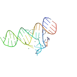
5NDH
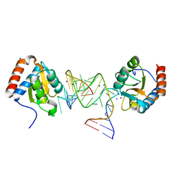
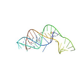
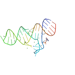
 | | The structure of the G. violaceus guanidine II riboswitch P2 stem-loop | | 分子名称: | GUANIDINE, MAGNESIUM ION, RNA (5'-R(*GP*(CBV)P*GP*GP*GP*GP*AP*CP*GP*AP*CP*CP*CP*CP*GP*C)-3'), ... | | 著者 | Huang, L, Wang, J, Lilley, D.M.J. | | 登録日 | 2017-03-08 | | 公開日 | 2017-05-31 | | 最終更新日 | 2024-05-08 | | 実験手法 | X-RAY DIFFRACTION (1.81 Å) | | 主引用文献 | The Structure of the Guanidine-II Riboswitch.
Cell Chem Biol, 24, 2017
|
|
6YMM
 | |
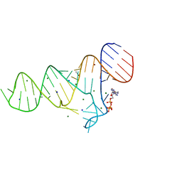
5NEQ
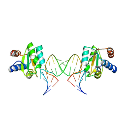
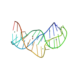
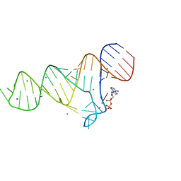
 | | The structure of the G. violaceus guanidine II riboswitch P1 stem-loop with aminoguanidine | | 分子名称: | AMINOGUANIDINE, RNA (5'-R(*GP*GP*UP*GP*GP*GP*GP*AP*CP*GP*AP*CP*CP*CP*CP*AP*(CBV)P*C)-3'), SODIUM ION, ... | | 著者 | Huang, L, Wang, J, Lilley, D.M.J. | | 登録日 | 2017-03-11 | | 公開日 | 2017-05-31 | | 最終更新日 | 2024-01-17 | | 実験手法 | X-RAY DIFFRACTION (1.69 Å) | | 主引用文献 | The Structure of the Guanidine-II Riboswitch.
Cell Chem Biol, 24, 2017
|
|
6FZ0
 | |
5NWQ
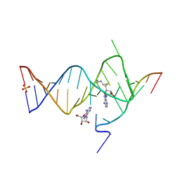
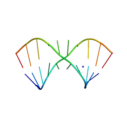
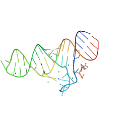
 | | The structure of the thermobifida fusca guanidine III riboswitch with guanidine. | | 分子名称: | GUANIDINE, Guanidine III riboswitch, MAGNESIUM ION, ... | | 著者 | Huang, L, Wang, J, Lilley, D.M.J. | | 登録日 | 2017-05-08 | | 公開日 | 2017-10-18 | | 最終更新日 | 2024-05-08 | | 実験手法 | X-RAY DIFFRACTION (1.91 Å) | | 主引用文献 | Structure of the Guanidine III Riboswitch.
Cell Chem Biol, 24, 2017
|
|
5NZD
 | | The structure of the thermobifida fusca guanidine III riboswitch in space group P212121. | | 分子名称: | ACETATE ION, MAGNESIUM ION, SODIUM ION, ... | | 著者 | Huang, L, Wang, J, Lilley, D.M.J. | | 登録日 | 2017-05-13 | | 公開日 | 2017-10-18 | | 最終更新日 | 2024-05-08 | | 実験手法 | X-RAY DIFFRACTION (2.007 Å) | | 主引用文献 | Structure of the Guanidine III Riboswitch.
Cell Chem Biol, 24, 2017
|
|
5NZ3
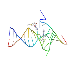
 | | The structure of the thermobifida fusca guanidine III riboswitch with methylguanidine | | 分子名称: | 1-METHYLGUANIDINE, MAGNESIUM ION, RNA (41-MER), ... | | 著者 | Huang, L, Wang, J, Lilley, D.M.J. | | 登録日 | 2017-05-12 | | 公開日 | 2017-10-18 | | 最終更新日 | 2024-05-08 | | 実験手法 | X-RAY DIFFRACTION (2.059 Å) | | 主引用文献 | Structure of the Guanidine III Riboswitch.
Cell Chem Biol, 24, 2017
|
|
5LR4
 | |
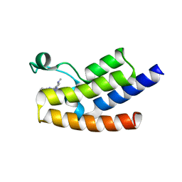
1LXD
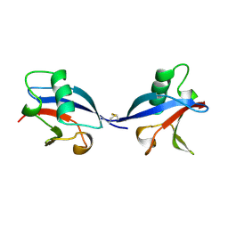 | | CRYSTAL STRUCTURE OF THE RAS INTERACTING DOMAIN OF RALGDS, A GUANINE NUCLEOTIDE DISSOCIATION STIMULATOR OF RAL PROTEIN | | 分子名称: | RALGDSB | | 著者 | Huang, L, Weng, X.W, Hofer, F, Martin, G.S, Kim, S.H. | | 登録日 | 1997-03-05 | | 公開日 | 1998-03-11 | | 最終更新日 | 2011-07-13 | | 実験手法 | X-RAY DIFFRACTION (2.4 Å) | | 主引用文献 | Three-dimensional structure of the Ras-interacting domain of RalGDS.
Nat.Struct.Biol., 4, 1997
|
|
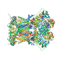
3L70
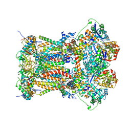
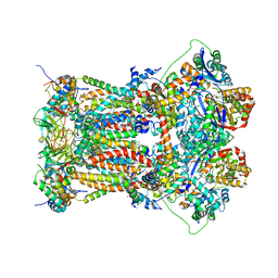 | | Cytochrome BC1 complex from chicken with trifloxystrobin bound | | 分子名称: | 1,2-dioleoyl-sn-glycero-3-phosphoethanolamine, CARDIOLIPIN, Coenzyme Q10, ... | | 著者 | Huang, L, Berry, E.A. | | 登録日 | 2009-12-27 | | 公開日 | 2010-02-02 | | 最終更新日 | 2023-09-06 | | 実験手法 | X-RAY DIFFRACTION (2.75 Å) | | 主引用文献 | Famoxadone and related inhibitors bind like methoxy acrylate inhibitors in the Qo site of the BC1 compl and fix the rieske iron-sulfur protein in a positio close to but distinct from that seen with stigmatellin and other "distal" Qo inhibitors.
To be Published
|
|
3L75
 | | Cytochrome BC1 complex from chicken with fenamidone bound | | 分子名称: | (5S)-5-methyl-2-(methylsulfanyl)-5-phenyl-3-(phenylamino)-3,5-dihydro-4H-imidazol-4-one, 1,2-dioleoyl-sn-glycero-3-phosphoethanolamine, AZIDE ION, ... | | 著者 | Huang, L, Berry, E.A. | | 登録日 | 2009-12-28 | | 公開日 | 2010-02-02 | | 最終更新日 | 2023-09-06 | | 実験手法 | X-RAY DIFFRACTION (2.79 Å) | | 主引用文献 | FAMOXADONE AND RELATED INHIBITORS BIND LIKE METHOXY ACRYLATE INHIBITORS IN THE Qo SITE OF THE BC1 COMPL AND FIX THE RIESKE IRON-SULFUR PROTEIN IN A POSITIO CLOSE TO BUT DISTINCT FROM THAT SEEN WITH STIGMATELLIN AND OTHER "DISTAL" Qo INHIBITORS.
To be Published
|
|
6J3P
 | | Crystal structure of the human GCN5 bromodomain in complex with compound (R,R)-36n | | 分子名称: | 2-{[(3R,5R)-5-(2,3-dihydro-1,4-benzodioxin-6-yl)-1-methylpiperidin-3-yl]amino}-3-methyl-3,5-dihydro-4H-pyrrolo[3,2-d]pyrimidin-4-one, Histone acetyltransferase KAT2A | | 著者 | Huang, L.Y, Li, H, Niu, L, Wu, C.Y, Yu, Y.M, Li, L.L, Yang, S.Y. | | 登録日 | 2019-01-05 | | 公開日 | 2019-05-01 | | 最終更新日 | 2023-11-22 | | 実験手法 | X-RAY DIFFRACTION (1.598 Å) | | 主引用文献 | Discovery of Pyrrolo[3,2- d]pyrimidin-4-one Derivatives as a New Class of Potent and Cell-Active Inhibitors of P300/CBP-Associated Factor Bromodomain.
J.Med.Chem., 62, 2019
|
|
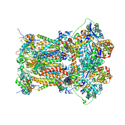
2A06
 | | Bovine cytochrome bc1 complex with stigmatellin bound | | 分子名称: | 1,2-dioleoyl-sn-glycero-3-phosphoethanolamine, AZIDE ION, CARDIOLIPIN, ... | | 著者 | Huang, L.S, Cobessi, D, Tung, E.Y, Berry, E.A. | | 登録日 | 2005-06-16 | | 公開日 | 2005-06-21 | | 最終更新日 | 2023-08-23 | | 実験手法 | X-RAY DIFFRACTION (2.1 Å) | | 主引用文献 | Binding of the Respiratory Chain Inhibitor Antimycin to the Mitochondrial bc(1) Complex: A New Crystal Structure Reveals an Altered Intramolecular Hydrogen-bonding Pattern.
J.Mol.Biol., 351, 2005
|
|
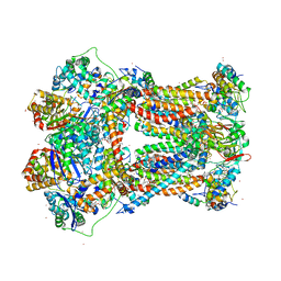
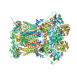
3L71
 | | Cytochrome BC1 complex from chicken with azoxystrobin bound | | 分子名称: | 1,2-dioleoyl-sn-glycero-3-phosphoethanolamine, CARDIOLIPIN, Coenzyme Q10, ... | | 著者 | Huang, L, Berry, E.A. | | 登録日 | 2009-12-27 | | 公開日 | 2010-02-02 | | 最終更新日 | 2023-09-06 | | 実験手法 | X-RAY DIFFRACTION (2.84 Å) | | 主引用文献 | Famoxadone and related inhibitors bind like methoxy acrylate inhibitors in the Qo site of the BC1 compl and fix the rieske iron-sulfur protein in a positio close to but distinct from that seen with stigmatellin and other "DISTAL" Qo inhibitors.
To be Published
|
|
6TF0
 | | Crystal structure of the ADP-binding domain of the NAD+ riboswitch with Nicotinamide adenine dinucleotide, reduced (NADH) | | 分子名称: | 1,4-DIHYDRONICOTINAMIDE ADENINE DINUCLEOTIDE, Chains: A, MAGNESIUM ION, ... | | 著者 | Huang, L, Lilley, D.M.J. | | 登録日 | 2019-11-12 | | 公開日 | 2020-09-23 | | 最終更新日 | 2024-05-15 | | 実験手法 | X-RAY DIFFRACTION (2.1 Å) | | 主引用文献 | Structure and ligand binding of the ADP-binding domain of the NAD+ riboswitch.
Rna, 26, 2020
|
|
6TFE
 | |
3L74
 | | Cytochrome BC1 complex from chicken with famoxadone bound | | 分子名称: | 1,2-dioleoyl-sn-glycero-3-phosphoethanolamine, AZIDE ION, CARDIOLIPIN, ... | | 著者 | Huang, L, Berry, E.A. | | 登録日 | 2009-12-28 | | 公開日 | 2010-02-02 | | 最終更新日 | 2023-09-06 | | 実験手法 | X-RAY DIFFRACTION (2.76 Å) | | 主引用文献 | Famoxadone and related inhibitors bind like methoxy acrylate inhibitors in the Qo site of the BC1 compl and fix the rieske iron-sulfur protein in a positio close to but distinct from that seen with stigmatellin and other "DISTAL" Qo inhibitors.
To be Published
|
|
6TB7
 | |
6TFH
 | |
3L73
 | | Cytochrome BC1 complex from chicken with triazolone inhibitor | | 分子名称: | 1,2-dioleoyl-sn-glycero-3-phosphoethanolamine, 4-[7-(3,3-dimethylbut-1-yn-1-yl)naphthalen-1-yl]-5-methoxy-2-methyl-2,4-dihydro-3H-1,2,4-triazol-3-one, CARDIOLIPIN, ... | | 著者 | Huang, L, Berry, E.A. | | 登録日 | 2009-12-27 | | 公開日 | 2010-02-02 | | 最終更新日 | 2023-09-06 | | 実験手法 | X-RAY DIFFRACTION (3.04 Å) | | 主引用文献 | Famoxadone and related inhibitors bind like methoxy acrylate inhibitors in the Qo site of the BC1 compl and fix the rieske iron-sulfur protein in a positio close to but distinct from that seen with stigmatellin and other "DISTAL" Qo inhibitors.
To be Published
|
|
6TFG
 | |
3TGU
 | | Cytochrome bc1 complex from chicken with pfvs-designed moa inhibitor bound | | 分子名称: | 1,2-dioleoyl-sn-glycero-3-phosphoethanolamine, CARDIOLIPIN, Coenzyme Q10, ... | | 著者 | Huang, L.-S, Yang, G.-F, Berry, E.A. | | 登録日 | 2011-08-17 | | 公開日 | 2012-07-04 | | 最終更新日 | 2023-09-13 | | 実験手法 | X-RAY DIFFRACTION (2.7 Å) | | 主引用文献 | Computational discovery of picomolar Q(o) site inhibitors of cytochrome bc1 complex.
J.Am.Chem.Soc., 134, 2012
|
|


