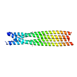
8HBR
 
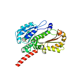
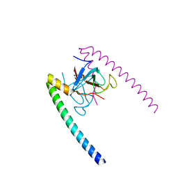
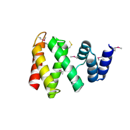 | | The C-terminal domain of Spiral2 | | 分子名称: | TOG domain-containing protein | | 著者 | Hayashi, I. | | 登録日 | 2022-10-30 | | 公開日 | 2023-01-18 | | 実験手法 | X-RAY DIFFRACTION (2.2 Å) | | 主引用文献 | Crystal structure of the C-terminal domain of the plant-specific microtubule-associated protein Spiral2.
Acta Crystallogr.,Sect.F, 79, 2023
|
|
1PA7
 | |
1K40
 | |
4EI9
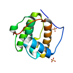
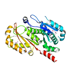
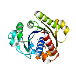
 | | Crystal structure of Bacillus cereus TubZ, GTP-form | | 分子名称: | 5'-GUANOSINE-DIPHOSPHATE-MONOTHIOPHOSPHATE, GUANOSINE-5'-DIPHOSPHATE, Plasmid replication protein RepX | | 著者 | Hayashi, I, Hoshino, S. | | 登録日 | 2012-04-05 | | 公開日 | 2012-08-15 | | 最終更新日 | 2024-03-20 | | 実験手法 | X-RAY DIFFRACTION (3.3 Å) | | 主引用文献 | Filament formation of the FtsZ/tubulin-like protein TubZ from the Bacillus cereus pXO1 plasmid.
J.Biol.Chem., 287, 2012
|
|
4EI7
 | |
4EI8
 | |
3WOY
 | |
3WOZ
 | |
7C7Y
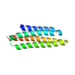
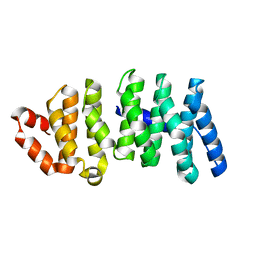
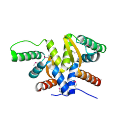
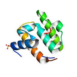
 | | C-terminal domain of B. cereus TubY | | 分子名称: | Uncharacterized protein | | 著者 | Hayashi, I. | | 登録日 | 2020-05-27 | | 公開日 | 2020-10-28 | | 最終更新日 | 2021-03-17 | | 実験手法 | X-RAY DIFFRACTION (2.6 Å) | | 主引用文献 | The C-terminal region of the plasmid partitioning protein TubY is a tetramer that can bind membranes and DNA.
J.Biol.Chem., 295, 2020
|
|
1G3Q
 | |
1G3R
 | |
6AHT
 | |
1TXQ
 | |
1UEG
 | |
2GW4
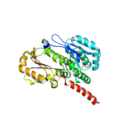
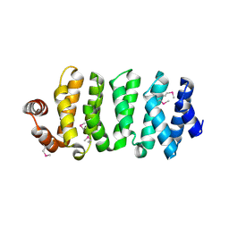
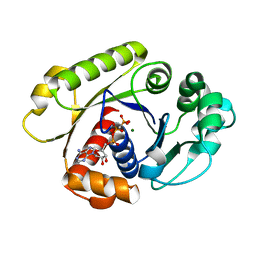
 | | Crystal structure of stony coral fluorescent protein Kaede, red form | | 分子名称: | Kaede, NICKEL (II) ION | | 著者 | Hayashi, I, Mizuno, H, Miyawako, A, Ikura, M. | | 登録日 | 2006-05-03 | | 公開日 | 2007-05-08 | | 最終更新日 | 2023-11-15 | | 実験手法 | X-RAY DIFFRACTION (1.6 Å) | | 主引用文献 | Crystallographic evidence for water-assisted photo-induced peptide cleavage in the stony coral fluorescent protein Kaede.
J.Mol.Biol., 372, 2007
|
|
2GW3
 | | Crystal structure of stony coral fluorescent protein Kaede, green form | | 分子名称: | Kaede, NICKEL (II) ION | | 著者 | Hayashi, I, Mizuno, H, Miyawaki, A, Ikura, M. | | 登録日 | 2006-05-03 | | 公開日 | 2007-05-08 | | 最終更新日 | 2023-11-15 | | 実験手法 | X-RAY DIFFRACTION (1.4 Å) | | 主引用文献 | Crystallographic evidence for water-assisted photo-induced peptide cleavage in the stony coral fluorescent protein Kaede.
J.Mol.Biol., 372, 2007
|
|
2HQH
 | |


