Loading
PDBj
MenuPDBj@FacebookPDBj@TwitterPDBj@YouTubewwPDB FoundationwwPDB
RCSB PDBPDBeBMRBAdv. SearchSearch help
PDB: 319 results

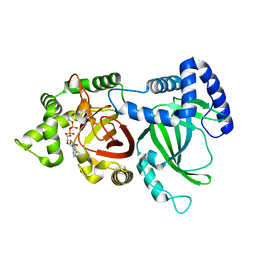
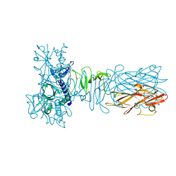
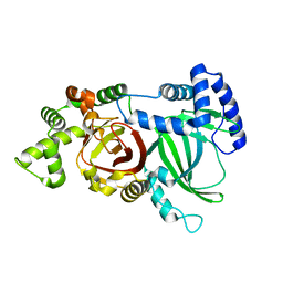
5H03
DownloadVisualize
BU of 5h03 by Molmil
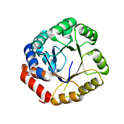
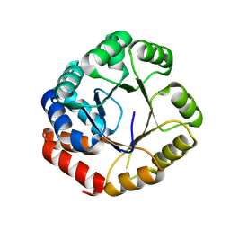
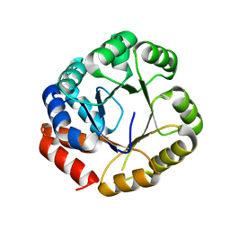
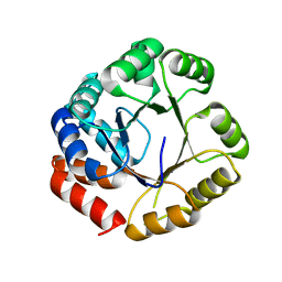
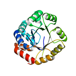
Crystal structure of an ADP-ribosylating toxin BECa from C. perfringens
Descriptor: Binary enterotoxin of Clostridium perfringens component a
Authors:Kawahara, K, Yonogi, S, Munetomo, R, Oki, H, Yoshida, T, Ohkubo, T, Kumeda, Y, Matsuda, S, Kodama, T, Iida, T, Nakamura, S.
Deposit date:2016-10-03
Release date:2016-11-02
Last modified:2023-11-08
Method:X-RAY DIFFRACTION (1.89 Å)
Cite:Crystal structure of the ADP-ribosylating component of BEC, the binary enterotoxin of Clostridium perfringens.
Biochem.Biophys.Res.Commun., 480, 2016
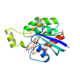
1A2B
DownloadVisualize
BU of 1a2b by Molmil
HUMAN RHOA COMPLEXED WITH GTP ANALOGUE
Descriptor: 5'-GUANOSINE-DIPHOSPHATE-MONOTHIOPHOSPHATE, MAGNESIUM ION, TRANSFORMING PROTEIN RHOA
Authors:Ihara, K, Muraguchi, S, Kato, M, Shimizu, T, Shirakawa, M, Kuroda, S, Kaibuchi, K, Hakoshima, T.
Deposit date:1997-12-26
Release date:1998-06-17
Last modified:2024-05-22
Method:X-RAY DIFFRACTION (2.4 Å)
Cite:Crystal structure of human RhoA in a dominantly active form complexed with a GTP analogue.
J.Biol.Chem., 273, 1998
5H04
DownloadVisualize
BU of 5h04 by Molmil
Crystal structure of an ADP-ribosylating toxin BECa of a novel binary enterotoxin of C. perfringens with NADH
Descriptor: 1,4-DIHYDRONICOTINAMIDE ADENINE DINUCLEOTIDE, Binary enterotoxin of Clostridium perfringens component a
Authors:Kawahara, K, Yonogi, S, Munetomo, R, Oki, H, Yoshida, T, Ohkubo, T, Kumeda, Y, Matsuda, S, Kodama, T, Iida, T, Nakamura, S.
Deposit date:2016-10-03
Release date:2016-11-02
Last modified:2023-11-08
Method:X-RAY DIFFRACTION (1.825 Å)
Cite:Crystal structure of the ADP-ribosylating component of BEC, the binary enterotoxin of Clostridium perfringens.
Biochem.Biophys.Res.Commun., 480, 2016
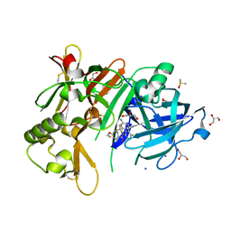
5YGX
DownloadVisualize
BU of 5ygx by Molmil
Structure of BACE1 in complex with N-(3-((4R,5R,6S)-2-amino-6-(1,1-difluoroethyl)-5-fluoro-4-methyl-5,6-dihydro-4H-1,3-oxazin-4-yl)-4-fluorophenyl)-5-(fluoromethoxy)pyrazine-2-carboxamide
Descriptor: Beta-secretase 1, DIMETHYL SULFOXIDE, GLYCEROL, ...
Authors:Nakahara, K, Fuchino, K, Komano, K, Asada, N, Tadano, G, Hasegawa, T, Yamamoto, T, Sako, Y, Ogawa, M, Unemura, C, Hosono, M, Sakaguchi, G, Ando, S, Ohnishi, S, Kido, Y, Fukushima, T, Dhuyvetter, D, Borghys, H, Gijsen, H, Yamano, Y, Iso, Y, Kusakabe, K.
Deposit date:2017-09-27
Release date:2018-08-08
Last modified:2023-11-22
Method:X-RAY DIFFRACTION (2.2 Å)
Cite:Discovery of Potent and Centrally Active 6-Substituted 5-Fluoro-1,3-dihydro-oxazine beta-Secretase (BACE1) Inhibitors via Active Conformation Stabilization
J. Med. Chem., 61, 2018
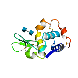
1LZY
DownloadVisualize
BU of 1lzy by Molmil
X-RAY STRUCTURE OF TURKEY EGG LYSOZYME COMPLEX WITH DI-N-ACETYLCHITOBIOSE. RECOGNITION AND BINDING OF ALPHA-ANOMERIC FORM
Descriptor: 2-acetamido-2-deoxy-beta-D-glucopyranose-(1-4)-2-acetamido-2-deoxy-alpha-D-glucopyranose, TURKEY EGG WHITE LYSOZYME
Authors:Harata, K.
Deposit date:1995-01-09
Release date:1995-02-27
Last modified:2024-10-23
Method:X-RAY DIFFRACTION (1.55 Å)
Cite:X-ray structure of monoclinic turkey egg lysozyme at 1.3 A resolution.
Acta Crystallogr.,Sect.D, 49, 1993
5AX6
DownloadVisualize
BU of 5ax6 by Molmil
The crystal structure of CofB, the minor pilin subunit of CFA/III from human enterotoxigenic Escherichia coli.
Descriptor: ACETATE ION, CofB
Authors:Kawahara, K, Oki, K, Fukaksua, F, Maruno, T, Kobayashi, Y, Daisuke, M, Taniguchi, T, Honda, T, Iida, T, Nakamura, S, Ohkubo, T.
Deposit date:2015-07-16
Release date:2016-03-09
Last modified:2020-02-26
Method:X-RAY DIFFRACTION (1.88 Å)
Cite:Homo-trimeric Structure of the Type IVb Minor Pilin CofB Suggests Mechanism of CFA/III Pilus Assembly in Human Enterotoxigenic Escherichia coli
J.Mol.Biol., 428, 2016
1EHD
DownloadVisualize
BU of 1ehd by Molmil
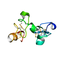
CRYSTAL STRUCTURE OF URTICA DIOICA AGGLUTININ ISOLECTIN VI
Descriptor: AGGLUTININ ISOLECTIN VI
Authors:Harata, K, Muraki, M.
Deposit date:2000-02-20
Release date:2000-04-05
Last modified:2019-12-25
Method:X-RAY DIFFRACTION (1.5 Å)
Cite:Crystal structures of Urtica dioica agglutinin and its complex with tri-N-acetylchitotriose.
J.Mol.Biol., 297, 2000
1EHH
DownloadVisualize
BU of 1ehh by Molmil
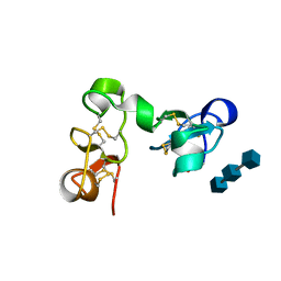
CRYSTAL STRUCTURE OF URTICA DIOICA AGGLUTININ ISOLECTIN VI COMPLEX WITH TRI-N-ACETYLCHITOTRIOSE
Descriptor: 2-acetamido-2-deoxy-beta-D-glucopyranose-(1-4)-2-acetamido-2-deoxy-beta-D-glucopyranose-(1-4)-2-acetamido-2-deoxy-beta-D-glucopyranose, AGGLUTININ ISOLECTIN VI
Authors:Harata, K, Muraki, M.
Deposit date:2000-02-21
Release date:2000-04-05
Last modified:2024-10-16
Method:X-RAY DIFFRACTION (1.9 Å)
Cite:Crystal structures of Urtica dioica agglutinin and its complex with tri-N-acetylchitotriose.
J.Mol.Biol., 297, 2000
133L
DownloadVisualize
BU of 133l by Molmil
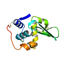
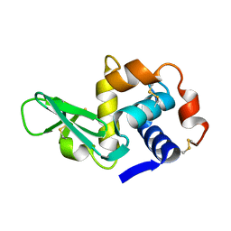
ROLE OF ARG 115 IN THE CATALYTIC ACTION OF HUMAN LYSOZYME. X-RAY STRUCTURE OF HIS 115 AND GLU 115 MUTANTS
Descriptor: HUMAN LYSOZYME
Authors:Harata, K, Muraki, M, Jigami, Y.
Deposit date:1993-06-01
Release date:1993-10-31
Last modified:2024-10-30
Method:X-RAY DIFFRACTION (1.77 Å)
Cite:Role of Arg115 in the catalytic action of human lysozyme. X-ray structure of His115 and Glu115 mutants.
J.Mol.Biol., 233, 1993
134L
DownloadVisualize
BU of 134l by Molmil
ROLE OF ARG 115 IN THE CATALYTIC ACTION OF HUMAN LYSOZYME. X-RAY STRUCTURE OF HIS 115 AND GLU 115 MUTANTS
Descriptor: HUMAN LYSOZYME
Authors:Harata, K, Muraki, M, Jigami, Y.
Deposit date:1993-06-01
Release date:1993-10-31
Last modified:2024-10-30
Method:X-RAY DIFFRACTION (1.77 Å)
Cite:Role of Arg115 in the catalytic action of human lysozyme. X-ray structure of His115 and Glu115 mutants.
J.Mol.Biol., 233, 1993
135L
DownloadVisualize
BU of 135l by Molmil
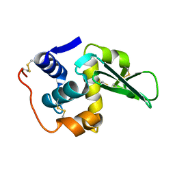
X-RAY STRUCTURE OF MONOCLINIC TURKEY EGG LYSOZYME AT 1.3 ANGSTROMS RESOLUTION
Descriptor: TURKEY EGG WHITE LYSOZYME
Authors:Harata, K.
Deposit date:1993-06-10
Release date:1993-10-31
Last modified:2024-10-30
Method:X-RAY DIFFRACTION (1.3 Å)
Cite:X-ray structure of monoclinic turkey egg lysozyme at 1.3 A resolution.
Acta Crystallogr.,Sect.D, 49, 1993
1B9O
DownloadVisualize
BU of 1b9o by Molmil
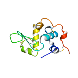
HUMAN ALPHA-LACTALBUMIN, LOW TEMPERATURE FORM
Descriptor: CALCIUM ION, PROTEIN (ALPHA-LACTALBUMIN)
Authors:Harata, K, Abe, Y, Muraki, M.
Deposit date:1999-02-14
Release date:1999-03-31
Last modified:2024-10-30
Method:X-RAY DIFFRACTION (1.15 Å)
Cite:Crystallographic evaluation of internal motion of human alpha-lactalbumin refined by full-matrix least-squares method.
J.Mol.Biol., 287, 1999
1JEF
DownloadVisualize
BU of 1jef by Molmil
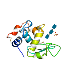
TURKEY LYSOZYME COMPLEX WITH (GLCNAC)3
Descriptor: 2-acetamido-2-deoxy-beta-D-glucopyranose-(1-4)-2-acetamido-2-deoxy-beta-D-glucopyranose-(1-4)-2-acetamido-2-deoxy-beta-D-glucopyranose, LYSOZYME, SULFATE ION
Authors:Harata, K, Muraki, M.
Deposit date:1997-04-23
Release date:1997-10-15
Last modified:2024-10-16
Method:X-RAY DIFFRACTION (1.77 Å)
Cite:X-ray structure of turkey-egg lysozyme complex with tri-N-acetylchitotriose. Lack of binding ability at subsite A.
Acta Crystallogr.,Sect.D, 53, 1997
2DZV
DownloadVisualize
BU of 2dzv by Molmil
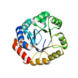
Structure of mutant tryptophan synthase alpha-subunit (D146A) from a hyperthermophile, Pyrococcus furiosus
Descriptor: Tryptophan synthase alpha chain
Authors:Ogasahara, K, Yamagata, Y, Yutani, K, RIKEN Structural Genomics/Proteomics Initiative (RSGI)
Deposit date:2006-09-30
Release date:2007-08-28
Last modified:2023-10-25
Method:X-RAY DIFFRACTION (2.4 Å)
Cite:Structures of mutant tryptophan synthase alpha-subunits from a hyperthermophile, Pyrococcus furiosus
To be Published
2DZT
DownloadVisualize
BU of 2dzt by Molmil
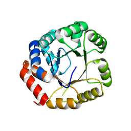
Structure of mutant tryptophan synthase alpha-subunit (D110A) from a hyperthermophile, Pyrococcus furiosus
Descriptor: Tryptophan synthase alpha chain
Authors:Ogasahara, K, Yamagata, Y, Yutani, K, RIKEN Structural Genomics/Proteomics Initiative (RSGI)
Deposit date:2006-09-30
Release date:2007-08-28
Last modified:2023-10-25
Method:X-RAY DIFFRACTION (2.4 Å)
Cite:Structures of mutant tryptophan synthase alpha-subunits from a hyperthermophile, Pyrococcus furiosus
To be Published
2DZU
DownloadVisualize
BU of 2dzu by Molmil
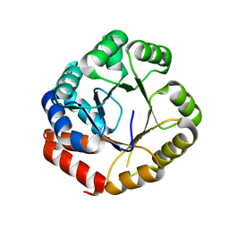
Structure of mutant tryptophan synthase alpha-subunit (D110N) from a hyperthermophile, Pyrococcus furiosus
Descriptor: Tryptophan synthase alpha chain
Authors:Ogasahara, K, Yamagata, Y, Yutani, K, RIKEN Structural Genomics/Proteomics Initiative (RSGI)
Deposit date:2006-09-30
Release date:2007-08-28
Last modified:2023-10-25
Method:X-RAY DIFFRACTION (2.46 Å)
Cite:Structures of mutant tryptophan synthase alpha-subunits from a hyperthermophile, Pyrococcus furiosus
To be Published
2DZS
DownloadVisualize
BU of 2dzs by Molmil
Structure of mutant tryptophan synthase alpha-subunit (E103A) from a hyperthermophile, Pyrococcus furiosus
Descriptor: Tryptophan synthase alpha chain
Authors:Ogasahara, K, Yamagata, Y, Yutani, K, RIKEN Structural Genomics/Proteomics Initiative (RSGI)
Deposit date:2006-09-30
Release date:2007-08-28
Last modified:2023-10-25
Method:X-RAY DIFFRACTION (2.4 Å)
Cite:Structures of mutant tryptophan synthase alpha-subunits from a hyperthermophile, Pyrococcus furiosus
To be Published
2DZW
DownloadVisualize
BU of 2dzw by Molmil
Structure of mutant tryptophan synthase alpha-subunit (E244A) from a hyperthermophile, Pyrococcus furiosus
Descriptor: Tryptophan synthase alpha chain
Authors:Ogasahara, K, Yamagata, Y, Yutani, K, RIKEN Structural Genomics/Proteomics Initiative (RSGI)
Deposit date:2006-09-30
Release date:2007-08-28
Last modified:2023-10-25
Method:X-RAY DIFFRACTION (2.4 Å)
Cite:Structures of mutant tryptophan synthase alpha-subunits from a hyperthermophile, Pyrococcus furiosus
To be Published
2E09
DownloadVisualize
BU of 2e09 by Molmil
Structure of mutant tryptophan synthase alpha-subunit (E74A) from a hyperthermophile, Pyrococcus furiosus
Descriptor: Tryptophan synthase alpha chain
Authors:Ogasahara, K, Yamagata, Y, Yutani, K, RIKEN Structural Genomics/Proteomics Initiative (RSGI)
Deposit date:2006-10-02
Release date:2007-08-28
Last modified:2023-10-25
Method:X-RAY DIFFRACTION (2.4 Å)
Cite:Structures of mutant tryptophan synthase alpha-subunits from a hyperthermophile, Pyrococcus furiosus
To be Published
2DZP
DownloadVisualize
BU of 2dzp by Molmil
Structure of mutant tryptophan synthase alpha-subunit (D17N) from a hyperthermophile, Pyrococcus furiosus
Descriptor: Tryptophan synthase alpha chain
Authors:Ogasahara, K, Yamagata, Y, Yutani, K, RIKEN Structural Genomics/Proteomics Initiative (RSGI)
Deposit date:2006-09-30
Release date:2007-08-28
Last modified:2023-10-25
Method:X-RAY DIFFRACTION (2.4 Å)
Cite:Structures of mutant tryptophan synthase alpha-subunits from a hyperthermophile, Pyrococcus furiosus
To be Published
2DZX
DownloadVisualize
BU of 2dzx by Molmil
Structure of mutant tryptophan synthase alpha-subunit (E131-132A) from a hyperthermophile, Pyrococcus furiosus
Descriptor: Tryptophan synthase alpha chain
Authors:Ogasahara, K, Yamagata, Y, Yutani, K, RIKEN Structural Genomics/Proteomics Initiative (RSGI)
Deposit date:2006-09-30
Release date:2007-08-28
Last modified:2023-10-25
Method:X-RAY DIFFRACTION (2.4 Å)
Cite:Structures of mutant tryptophan synthase alpha-subunits from a hyperthermophile, Pyrococcus furiosus
To be Published
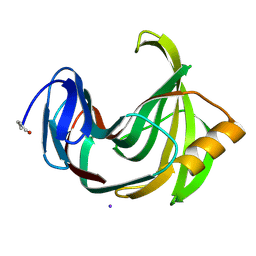
2DFC
DownloadVisualize
BU of 2dfc by Molmil
Xylanase II from Tricoderma reesei at 293K
Descriptor: Endo-1,4-beta-xylanase 2, IODIDE ION
Authors:Harata, K, Akiba, T.
Deposit date:2006-02-28
Release date:2006-07-11
Last modified:2024-10-23
Method:X-RAY DIFFRACTION (1.19 Å)
Cite:Structure of an orthorhombic form of xylanase II from Trichoderma reesei and analysis of thermal displacement.
ACTA CRYSTALLOGR.,SECT.D, 62, 2006
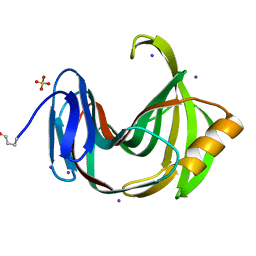
2DFB
DownloadVisualize
BU of 2dfb by Molmil
Xylanase II from Tricoderma reesei at 100K
Descriptor: Endo-1,4-beta-xylanase 2, IODIDE ION, SULFATE ION
Authors:Harata, K, Akiba, T.
Deposit date:2006-02-28
Release date:2006-07-11
Last modified:2024-10-09
Method:X-RAY DIFFRACTION (1.11 Å)
Cite:Structure of an orthorhombic form of xylanase II from Trichoderma reesei and analysis of thermal displacement.
ACTA CRYSTALLOGR.,SECT.D, 62, 2006
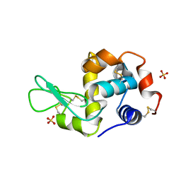
1LJN
DownloadVisualize
BU of 1ljn by Molmil
Crystal Structure of Turkey Egg Lysozyme Complex with Di-N-acetylchitobiose at 1.19A Resolution
Descriptor: 2-acetamido-2-deoxy-beta-D-glucopyranose-(1-4)-2-acetamido-2-deoxy-beta-D-glucopyranose, SULFATE ION, lysozyme C
Authors:Harata, K, Kanai, R.
Deposit date:2002-04-22
Release date:2002-06-05
Last modified:2024-10-23
Method:X-RAY DIFFRACTION (1.19 Å)
Cite:Crystallographic dissection of the thermal motion of protein-sugar complex.
Proteins, 48, 2002
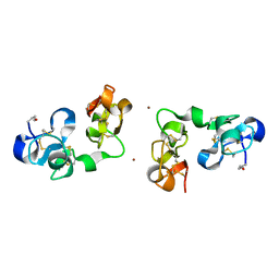
1IQB
DownloadVisualize
BU of 1iqb by Molmil
Crystal Structure of Urtica dioica Agglutinin Isolectin I
Descriptor: AGGLUTININ ISOLECTIN I, ZINC ION
Authors:Harata, K, Schubert, W.D, Muraki, M.
Deposit date:2001-07-15
Release date:2001-11-07
Last modified:2023-10-25
Method:X-RAY DIFFRACTION (1.9 Å)
Cite:Structure of Urtica dioica agglutinin isolectin I: dimer formation mediated by two zinc ions bound at the sugar-binding site.
Acta Crystallogr.,Sect.D, 57, 2001

226707

PDB entries from 2024-10-30

PDB statisticsPDBj update infoContact PDBjnumon