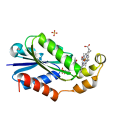
Loading
PDBj
MenuPDBj@FacebookPDBj@TwitterPDBj@YouTubewwPDB FoundationwwPDB
RCSB PDBPDBeBMRBAdv. SearchSearch help
PDB: 1351 results

6MQY
DownloadVisualize
BU of 6mqy by Molmil
Crystal Structure of the 13-cis Product of All-trans Retinal-Bound R111K:Y134F:T54V:R132Q:P39Y:R59Y:L121E Mutant of Human Cellular Retinoic Acid Binding Protein II Irradiated with 400 nm Laser (30 seconds) at 1.9 Angstrom
Descriptor: Cellular retinoic acid-binding protein 2, GLYCEROL, RETINAL
Authors:Ghanbarpour, A, Geiger, J.
Deposit date:2018-10-11
Release date:2019-01-09
Last modified:2023-10-11
Method:X-RAY DIFFRACTION (1.9 Å)
Cite:Mimicking Microbial Rhodopsin Isomerization in a Single Crystal.
J. Am. Chem. Soc., 141, 2019
8G64
DownloadVisualize
BU of 8g64 by Molmil
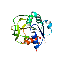
Heme-bound flavodoxin FldH from Fusobacterium nucleatum
Descriptor: FLAVIN MONONUCLEOTIDE, Flavodoxin, PROTOPORPHYRIN IX CONTAINING FE, ...
Authors:Chan, A.C, Wolthers, K.R, Murphy, M.E.
Deposit date:2023-02-14
Release date:2023-08-09
Last modified:2024-05-22
Method:X-RAY DIFFRACTION (1.6 Å)
Cite:A new member of the flavodoxin superfamily from Fusobacterium nucleatum that functions in heme trafficking and reduction of anaerobilin.
J.Biol.Chem., 299, 2023
3OTG
DownloadVisualize
BU of 3otg by Molmil
Crystal Structure of CalG1, Calicheamicin Glycostyltransferase, TDP bound form
Descriptor: CHLORIDE ION, CalG1, THYMIDINE-5'-DIPHOSPHATE
Authors:Chang, A, Singh, S, Bingman, C.A, Thorson, J.S, Phillips Jr, G.N, Center for Eukaryotic Structural Genomics (CESG), Enzyme Discovery for Natural Product Biosynthesis (NatPro)
Deposit date:2010-09-11
Release date:2010-12-15
Last modified:2017-11-08
Method:X-RAY DIFFRACTION (2.08 Å)
Cite:Complete set of glycosyltransferase structures in the calicheamicin biosynthetic pathway reveals the origin of regiospecificity.
Proc.Natl.Acad.Sci.USA, 108, 2011
3OTI
DownloadVisualize
BU of 3oti by Molmil
Crystal Structure of CalG3, Calicheamicin Glycostyltransferase, TDP and calicheamicin T0 bound form
Descriptor: CHLORIDE ION, CalG3, Calicheamicin T0, ...
Authors:Chang, A, Singh, S, Bingman, C.A, Thorson, J.S, Phillips Jr, G.N, Center for Eukaryotic Structural Genomics (CESG), Enzyme Discovery for Natural Product Biosynthesis (NatPro)
Deposit date:2010-09-11
Release date:2010-12-15
Last modified:2024-02-21
Method:X-RAY DIFFRACTION (1.597 Å)
Cite:Complete set of glycosyltransferase structures in the calicheamicin biosynthetic pathway reveals the origin of regiospecificity.
Proc.Natl.Acad.Sci.USA, 108, 2011
3OTH
DownloadVisualize
BU of 3oth by Molmil
Crystal Structure of CalG1, Calicheamicin Glycostyltransferase, TDP and calicheamicin alpha3I bound form
Descriptor: CalG1, Calicheamicin alpha3I, THYMIDINE-5'-DIPHOSPHATE
Authors:Chang, A, Singh, S, Bingman, C.A, Thorson, J.S, Phillips Jr, G.N, Center for Eukaryotic Structural Genomics (CESG), Enzyme Discovery for Natural Product Biosynthesis (NatPro)
Deposit date:2010-09-11
Release date:2010-12-15
Last modified:2017-11-08
Method:X-RAY DIFFRACTION (2.301 Å)
Cite:Complete set of glycosyltransferase structures in the calicheamicin biosynthetic pathway reveals the origin of regiospecificity.
Proc.Natl.Acad.Sci.USA, 108, 2011
3PKP
DownloadVisualize
BU of 3pkp by Molmil
Q83S Variant of S. Enterica RmlA with dATP
Descriptor: 2'-DEOXYADENOSINE 5'-TRIPHOSPHATE, Glucose-1-phosphate thymidylyltransferase, MAGNESIUM ION
Authors:Chang, A, Moretti, R, Bingman, C.A, Thorson, J.S, Phillips Jr, G.N, Center for Eukaryotic Structural Genomics (CESG)
Deposit date:2010-11-11
Release date:2011-01-12
Last modified:2024-02-21
Method:X-RAY DIFFRACTION (2.6 Å)
Cite:Expanding the Nucleotide and Sugar 1-Phosphate Promiscuity of Nucleotidyltransferase RmlA via Directed Evolution.
J.Biol.Chem., 286, 2011
3PKQ
DownloadVisualize
BU of 3pkq by Molmil
Q83D Variant of S. Enterica RmlA with dGTP
Descriptor: 2'-DEOXYGUANOSINE-5'-TRIPHOSPHATE, DI(HYDROXYETHYL)ETHER, Glucose-1-phosphate thymidylyltransferase, ...
Authors:Chang, A, Moretti, R, Bingman, C.A, Thorson, J.S, Phillips Jr, G.N, Center for Eukaryotic Structural Genomics (CESG)
Deposit date:2010-11-11
Release date:2011-01-12
Last modified:2023-09-06
Method:X-RAY DIFFRACTION (2.4 Å)
Cite:Expanding the Nucleotide and Sugar 1-Phosphate Promiscuity of Nucleotidyltransferase RmlA via Directed Evolution.
J.Biol.Chem., 286, 2011
6E5R
DownloadVisualize
BU of 6e5r by Molmil
Crystal structure of the apo domain-swapped dimer Q108K:T51D:A28C mutant of human Cellular Retinol Binding Protein II
Descriptor: ACETATE ION, GLYCEROL, Retinol-binding protein 2
Authors:Ghanbarpour, A, Geiger, J.
Deposit date:2018-07-22
Release date:2019-08-07
Last modified:2023-10-11
Method:X-RAY DIFFRACTION (2.592 Å)
Cite:Engineering the hCRBPII Domain-Swapped Dimer into a New Class of Protein Switches.
J.Am.Chem.Soc., 141, 2019
6ON7
DownloadVisualize
BU of 6on7 by Molmil
Crystal Structure of Apo Domain-Swapped Dimer Q108K:T51D:A28C:L36C Mutant of Human Cellular Retinol Binding Protein II
Descriptor: Retinol-binding protein 2
Authors:Ghanbarpour, A, Geiger, J.
Deposit date:2019-04-20
Release date:2019-10-16
Last modified:2023-10-11
Method:X-RAY DIFFRACTION (1.981 Å)
Cite:Engineering the hCRBPII Domain-Swapped Dimer into a New Class of Protein Switches.
J.Am.Chem.Soc., 141, 2019
6MCV
DownloadVisualize
BU of 6mcv by Molmil
Crystal Structure of Holo Retinal-Bound Domain-Swapped Dimer of Wild Type Human Cellular Retinol Binding Protein II
Descriptor: RETINAL, Retinol-binding protein 2
Authors:Ghanbarpour, A, Geiger, J.
Deposit date:2018-09-02
Release date:2019-10-16
Last modified:2023-10-11
Method:X-RAY DIFFRACTION (3.3 Å)
Cite:Engineering the hCRBPII Domain-Swapped Dimer into a New Class of Protein Switches.
J.Am.Chem.Soc., 141, 2019
6MQJ
DownloadVisualize
BU of 6mqj by Molmil
Crystal Structure of the All-Trans Retinal bound R111K:Y134F:T54V:R132Q:P39Y:R59Y:L121Q mutant of Human Cellular Retinoic Acid Binding Protein II Irradiated with 532 nm Laser for 10 minutes at 2.1 Angstrom Resolution
Descriptor: Cellular retinoic acid-binding protein 2, RETINAL
Authors:Ghanbarpour, A, Geiger, J.
Deposit date:2018-10-09
Release date:2019-01-09
Last modified:2023-10-11
Method:X-RAY DIFFRACTION (2.125 Å)
Cite:Mimicking Microbial Rhodopsin Isomerization in a Single Crystal.
J. Am. Chem. Soc., 141, 2019
6MOQ
DownloadVisualize
BU of 6moq by Molmil
Crystal Structure of the All-trans Retinal-Bound R111K:Y134F:T54V:R132Q:P39Y:R59Y:L121W Mutant of Human Cellular Retinoic Acid Binding Protein II in the Dark at 2.2 Angstrom Resolution
Descriptor: Cellular retinoic acid-binding protein 2, RETINAL
Authors:Ghanbarpour, A, Geiger, J.
Deposit date:2018-10-04
Release date:2019-01-09
Last modified:2023-10-11
Method:X-RAY DIFFRACTION (2.198 Å)
Cite:Mimicking Microbial Rhodopsin Isomerization in a Single Crystal.
J. Am. Chem. Soc., 141, 2019
8GR3
DownloadVisualize
BU of 8gr3 by Molmil
Crystal structure of K151L/Y158F mutant of GATase subunit of Methanocaldococcus jannaschii GMP synthetase
Descriptor: GMP synthase [glutamine-hydrolyzing] subunit A, SULFATE ION
Authors:Chandrashekarmath, A, Bellur, A.
Deposit date:2022-08-31
Release date:2023-07-12
Last modified:2023-11-29
Method:X-RAY DIFFRACTION (2.4 Å)
Cite:Crystal structure of K151L/Y158F mutant of GATase subunit of Methanocaldococcus jannaschii GMP synthetase
To Be Published
8GR1
DownloadVisualize
BU of 8gr1 by Molmil
Crystal structure of D110V/K151L mutant of GATase subunit of Methanocaldococcus jannaschii GMP synthetase
Descriptor: GMP synthase [glutamine-hydrolyzing] subunit A, SULFATE ION
Authors:Chandrashekarmath, A, Bellur, A.
Deposit date:2022-08-31
Release date:2023-07-12
Last modified:2023-11-29
Method:X-RAY DIFFRACTION (2.5 Å)
Cite:Crystal structure of D110V/K151L mutant of GATase subunit of Methanocaldococcus jannaschii GMP synthetase
To Be Published
6MOP
DownloadVisualize
BU of 6mop by Molmil
Crystal Structure of the All-trans Retinal-Bound R111K:Y134F:T54V:R132Q:P39Y:R59Y:L121E Mutant of Human Cellular Retinoic Acid Binding Protein II in the Dark at 1.9 Angstrom Resolution
Descriptor: Cellular retinoic acid-binding protein 2, RETINAL
Authors:Ghanbarpour, A, Geiger, J.
Deposit date:2018-10-04
Release date:2019-01-09
Last modified:2023-10-11
Method:X-RAY DIFFRACTION (1.9 Å)
Cite:Mimicking Microbial Rhodopsin Isomerization in a Single Crystal.
J. Am. Chem. Soc., 141, 2019
6MPK
DownloadVisualize
BU of 6mpk by Molmil
Crystal Structure of the 13-cis Retinal-Bound R111K:Y134F:T54V:R132Q:P39Y:R59Y:L121E:A32Y Mutant of Human Cellular Retinoic Acid Binding Protein II in the Dark at 1.58 Angstrom Resolution
Descriptor: Cellular retinoic acid-binding protein 2, GLYCEROL, RETINAL
Authors:Ghanbarpour, A, Geiger, J.
Deposit date:2018-10-07
Release date:2019-01-09
Last modified:2023-10-11
Method:X-RAY DIFFRACTION (1.58 Å)
Cite:Mimicking Microbial Rhodopsin Isomerization in a Single Crystal.
J. Am. Chem. Soc., 141, 2019
6MOV
DownloadVisualize
BU of 6mov by Molmil
Crystal Structure of the All-trans Retinal bound R111K:Y134F:T54V:R132Q:P39Y:R59Y:L121Q Human Cellular Retinoic Acid Binding Protein II in the Dark at 1.75 Angstrom Resolution
Descriptor: Cellular retinoic acid-binding protein 2, RETINAL
Authors:Ghanbarpour, A, Geiger, J.
Deposit date:2018-10-04
Release date:2019-01-09
Last modified:2023-10-11
Method:X-RAY DIFFRACTION (1.752 Å)
Cite:Mimicking Microbial Rhodopsin Isomerization in a Single Crystal.
J. Am. Chem. Soc., 141, 2019
6MOX
DownloadVisualize
BU of 6mox by Molmil
Crystal Structure of the All-trans Retinal-Bound R111K:Y134F:T54V:R132Q:P39Q:R59Y:L121E Human Cellular Retinoic Acid Binding Protein II Mutant in the Dark at 2.18 Angstrom Resolution
Descriptor: Cellular retinoic acid-binding protein 2, RETINAL
Authors:Ghanbarpour, A, Geiger, J.
Deposit date:2018-10-04
Release date:2019-01-09
Last modified:2023-10-11
Method:X-RAY DIFFRACTION (2.18 Å)
Cite:Mimicking Microbial Rhodopsin Isomerization in a Single Crystal.
J. Am. Chem. Soc., 141, 2019
6MLB
DownloadVisualize
BU of 6mlb by Molmil
Crystal structure of the holo retinal-bound domain-swapped dimer Q108K:K40L:T51F mutant of human cellular retinol binding protein II
Descriptor: ACETATE ION, GLYCEROL, RETINAL, ...
Authors:Ghanbarpour, A, Geiger, J.
Deposit date:2018-09-27
Release date:2019-10-16
Last modified:2023-10-11
Method:X-RAY DIFFRACTION (2.15 Å)
Cite:Engineering the hCRBPII Domain-Swapped Dimer into a New Class of Protein Switches.
J.Am.Chem.Soc., 141, 2019
6MR0
DownloadVisualize
BU of 6mr0 by Molmil
Crystal Structure of the All-trans Retinal Bound R111K:Y134F:T54V:R132Q:P39Q:R59Y:L121E Human Cellular Retinoic Acid Binding Protein II Mutant After 5 Minutes UV irradiation at 2.6 Angstrom Resolution
Descriptor: Cellular retinoic acid-binding protein 2, RETINAL
Authors:Ghanbarpour, A, Geiger, J.
Deposit date:2018-10-11
Release date:2019-01-09
Last modified:2023-10-11
Method:X-RAY DIFFRACTION (2.649 Å)
Cite:Mimicking Microbial Rhodopsin Isomerization in a Single Crystal.
J. Am. Chem. Soc., 141, 2019
6MKV
DownloadVisualize
BU of 6mkv by Molmil
Crystal structure of Retinal-bound holo Q108K:K40L:T51W domain-swapped dimer of human cellular retinol binding protein 2
Descriptor: ACETATE ION, GLYCEROL, RETINAL, ...
Authors:Ghanbarpour, A, Geiger, J.
Deposit date:2018-09-26
Release date:2019-10-16
Last modified:2023-10-11
Method:X-RAY DIFFRACTION (2.107 Å)
Cite:Engineering the hCRBPII Domain-Swapped Dimer into a New Class of Protein Switches.
J.Am.Chem.Soc., 141, 2019
6N81
DownloadVisualize
BU of 6n81 by Molmil
Crystal structure of GII.4 2002 norovirus P domain in complex with cross-reactive human antibody A1227
Descriptor: A1227 Fab heavy chain, A1227 Fab light chain, Major capsid protein
Authors:Changela, A, Verardi, R, Kwong, P.D.
Deposit date:2018-11-28
Release date:2019-06-19
Last modified:2023-10-11
Method:X-RAY DIFFRACTION (2.579 Å)
Cite:Sera Antibody Repertoire Analyses Reveal Mechanisms of Broad and Pandemic Strain Neutralizing Responses after Human Norovirus Vaccination.
Immunity, 50, 2019
6MQI
DownloadVisualize
BU of 6mqi by Molmil
Crystal Structure of the All-trans Retinal bound R111K:Y134F:T54V:R132Q:P39Y:R59Y:L121Q mutant of Human Cellular Retinoic Acid Binding Protein II Irradiated with 400 nm Laser for 5 minutes at 1.87 Angstrom Resolution
Descriptor: Cellular retinoic acid-binding protein 2, RETINAL
Authors:Ghanbarpour, A, Geiger, J.
Deposit date:2018-10-09
Release date:2019-01-09
Last modified:2023-10-11
Method:X-RAY DIFFRACTION (1.87 Å)
Cite:Mimicking Microbial Rhodopsin Isomerization in a Single Crystal.
J. Am. Chem. Soc., 141, 2019
6MQZ
DownloadVisualize
BU of 6mqz by Molmil
Crystal Structure of the 13-cis Product of the All-trans Retinal-Bound R111K:Y134F:T54V:R132Q:P39Y:R59Y:L121E Mutant of Human Cellular Retinoic Acid Binding Protein II Irradiated with 400 nm Laser (5 minutes) at 2.07 Angstrom
Descriptor: Cellular retinoic acid-binding protein 2, GLYCEROL, RETINAL
Authors:Ghanbarpour, A, Geiger, J.
Deposit date:2018-10-11
Release date:2019-01-09
Last modified:2023-10-11
Method:X-RAY DIFFRACTION (2.07 Å)
Cite:Mimicking Microbial Rhodopsin Isomerization in a Single Crystal.
J. Am. Chem. Soc., 141, 2019
6MQX
DownloadVisualize
BU of 6mqx by Molmil
Crystal Structure of All-trans Retinal-Bound R111K:Y134F:T54V:R132Q:P39Y:R59Y:L121E Human Cellular Retinoic Acid Binding Protein II Irradiated with 400 nm Laser (30 seconds) and Subsequently Dark Adapted (25 minutes) at 2.0 Angstrom Resolution
Descriptor: Cellular retinoic acid-binding protein 2, GLYCEROL, RETINAL
Authors:Ghanbarpour, A, Geiger, J.
Deposit date:2018-10-11
Release date:2019-01-09
Last modified:2023-10-11
Method:X-RAY DIFFRACTION (2.01 Å)
Cite:Mimicking Microbial Rhodopsin Isomerization in a Single Crystal.
J. Am. Chem. Soc., 141, 2019

222624

건을2024-07-17부터공개중

PDB statisticsPDBj update infoContact PDBjnumon