1SZB
 
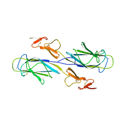 | | Crystal structure of the human MBL-associated protein 19 (MAp19) | | 分子名称: | CALCIUM ION, mannose binding lectin-associated serine protease-2 related protein, MAp19 (19kDa) | | 著者 | Gregory, L.A, Thielens, N.M, Arlaud, G.J, Fontecilla-Camps, J.C, Gaboriaud, C. | | 登録日 | 2004-04-05 | | 公開日 | 2004-06-22 | | 最終更新日 | 2023-08-23 | | 実験手法 | X-RAY DIFFRACTION (2.5 Å) | | 主引用文献 | The X-ray structure of human MBL-associated protein 19 (MAp19) and its interaction site with mannan-binding lectin and L-ficolin
J.Biol.Chem., 279, 2004
|
|
2V7F
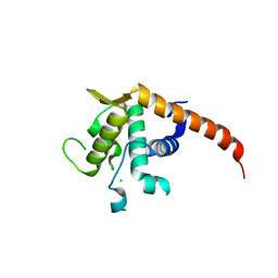 | | Structure of P. abyssi RPS19 protein | | 分子名称: | CHLORIDE ION, RPS19E SSU RIBOSOMAL PROTEIN S19E | | 著者 | Gregory, L.A, Aguissa-Toure, A.H, Pinaud, N, Legrand, P, Gleizes, P.E, Fribourg, S. | | 登録日 | 2007-07-30 | | 公開日 | 2007-09-11 | | 最終更新日 | 2024-05-08 | | 実験手法 | X-RAY DIFFRACTION (1.15 Å) | | 主引用文献 | Molecular Basis of Diamond Blackfan Anemia: Structure and Function Analysis of Rps19.
Nucleic Acids Res., 35, 2007
|
|
1NZI
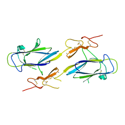 | | Crystal Structure of the CUB1-EGF Interaction Domain of Complement Protease C1s | | 分子名称: | CALCIUM ION, Complement C1s component, MAGNESIUM ION | | 著者 | Gregory, L.A, Thielens, N.M, Arlaud, G.J, Fontecilla-Camps, J.C, Gaboriaud, C. | | 登録日 | 2003-02-18 | | 公開日 | 2003-06-10 | | 最終更新日 | 2011-07-13 | | 実験手法 | X-RAY DIFFRACTION (1.5 Å) | | 主引用文献 | X-ray structure of the Ca2+-binding interaction domain of C1s. Insights into the assembly of the C1 complex of complement
J.Biol.Chem., 278, 2003
|
|


