Loading
PDBj
MenuPDBj@FacebookPDBj@TwitterPDBj@YouTubewwPDB FoundationwwPDB
RCSB PDBPDBeBMRBAdv. SearchSearch help
PDB: 6 results

3QY9
DownloadVisualize
BU of 3qy9 by Molmil
The Crystal Structure of Dihydrodipicolinate reductase from Staphylococcus aureus
Descriptor: ACETATE ION, Dihydrodipicolinate reductase, GLYCEROL, ...
Authors:Girish, T.S, Gopal, B.
Deposit date:2011-03-03
Release date:2011-08-03
Last modified:2023-11-01
Method:X-RAY DIFFRACTION (1.8 Å)
Cite:Structure and nucleotide specificity of Staphylococcus aureus dihydrodipicolinate reductase (DapB)
Febs Lett., 585, 2011
2PI7
DownloadVisualize
BU of 2pi7 by Molmil
Structure of the catalytic domain of the chick retinal neurite inhibitor-Receptor Protein Tyrosine Phosphatase CRYP-2/cPTPRO
Descriptor: NITRATE ION, Protein tyrosine phosphatase CRYP-2
Authors:Girish, T.S, Gopal, B.
Deposit date:2007-04-13
Release date:2007-05-01
Last modified:2023-10-25
Method:X-RAY DIFFRACTION (2.59 Å)
Cite:The crystal structure of the catalytic domain of the chick retinal neurite inhibitor-receptor protein tyrosine phosphatase CRYP-2/cPTPRO
Proteins, 68, 2007
4EWT
DownloadVisualize
BU of 4ewt by Molmil
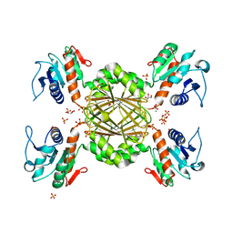
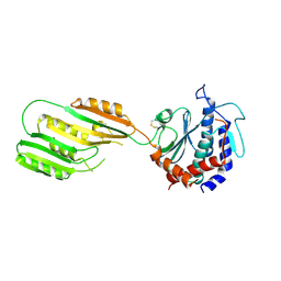
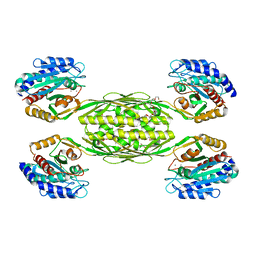
The crystal structure of a putative aminohydrolase from methicillin resistant Staphylococcus aureus
Descriptor: 1-DEOXY-1-THIO-HEPTAETHYLENE GLYCOL, DI(HYDROXYETHYL)ETHER, MANGANESE (II) ION, ...
Authors:Girish, T.S, Vivek, B, Colaco, M, Misquith, S, Gopal, B.
Deposit date:2012-04-27
Release date:2013-02-20
Last modified:2023-11-08
Method:X-RAY DIFFRACTION (2.1 Å)
Cite:Structure of an amidohydrolase, SACOL0085, from methicillin-resistant Staphylococcus aureus COL
Acta Crystallogr.,Sect.F, 69, 2013
3KI9
DownloadVisualize
BU of 3ki9 by Molmil
Crystal structure of Staphylococcus aureus metallopeptidase (Sapep/DapE) in the Mn2+ bound form
Descriptor: MANGANESE (II) ION, PHOSPHATE ION, Putative dipeptidase SACOL1801
Authors:Girish, T.S, Gopal, B.
Deposit date:2009-11-01
Release date:2010-07-07
Last modified:2023-11-01
Method:X-RAY DIFFRACTION (2.9 Å)
Cite:Crystal structure of Staphylococcus aureus metallopeptidase (Sapep) reveals large domain motions between the manganese-bound and apo-states
J.Biol.Chem., 285, 2010
3KHX
DownloadVisualize
BU of 3khx by Molmil
Crystal structure of Staphylococcus aureus metallopeptidase (Sapep/DapE) in the apo-form
Descriptor: Putative dipeptidase SACOL1801
Authors:Girish, T.S, Gopal, B.
Deposit date:2009-10-31
Release date:2010-07-07
Last modified:2023-11-01
Method:X-RAY DIFFRACTION (2.3 Å)
Cite:Crystal structure of Staphylococcus aureus metallopeptidase (Sapep) reveals large domain motions between the manganese-bound and apo-states
J.Biol.Chem., 285, 2010
3KHZ
DownloadVisualize
BU of 3khz by Molmil
Crystal Structure of R350A mutant of Staphylococcus aureus metallopeptidase (Sapep/DapE) in the apo-form
Descriptor: Putative dipeptidase SACOL1801
Authors:Girish, T.S, Gopal, B.
Deposit date:2009-10-31
Release date:2010-07-07
Last modified:2023-11-01
Method:X-RAY DIFFRACTION (2.5 Å)
Cite:Crystal structure of Staphylococcus aureus metallopeptidase (Sapep) reveals large domain motions between the manganese-bound and apo-states
J.Biol.Chem., 285, 2010

223532

PDB entries from 2024-08-07

PDB statisticsPDBj update infoContact PDBjnumon