Loading
PDBj
MenuPDBj@FacebookPDBj@TwitterPDBj@YouTubewwPDB FoundationwwPDB
RCSB PDBPDBeBMRBAdv. SearchSearch help
PDB: 121 results

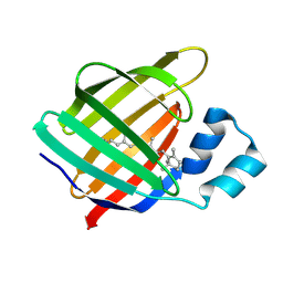
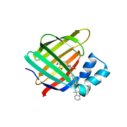
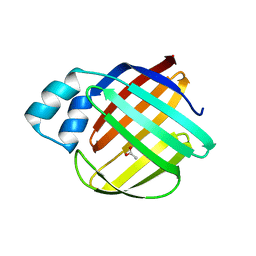
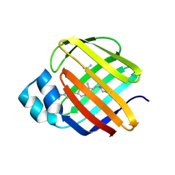
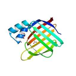
5U6G
DownloadVisualize
BU of 5u6g by Molmil
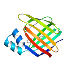
Crystal Structure of the holo Domain-Swapped Dimer mutant Q108K:K40D Human Cellular Retinol Binding Protein II bound with all trans retinal
Descriptor: RETINAL, Retinol-binding protein 2
Authors:Assar, Z, Geiger, J.H.
Deposit date:2016-12-08
Release date:2017-10-18
Last modified:2023-10-04
Method:X-RAY DIFFRACTION (2.6 Å)
Cite:Crystal Structure of the holo Domain-Swapped Dimer mutant Q108K:K40D Human Cellular Retinol Binding Protein II bound with all trans retinal
To Be Published
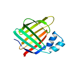
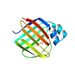
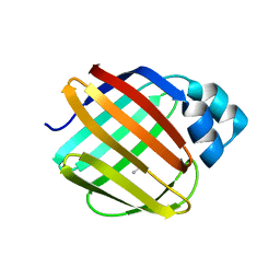
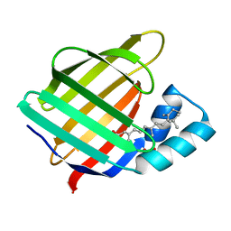
6C7Z
DownloadVisualize
BU of 6c7z by Molmil
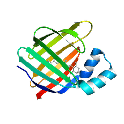
Crystal structure of the Q108K:K40L:T51V:R58F mutant of human Cellular Retinol Binding Protein II in complex with synthetic Ligand Julolidine
Descriptor: (2E,4E)-3-methyl-5-(2,3,6,7-tetrahydro-1H,5H-pyrido[3,2,1-ij]quinolin-9-yl)penta-2,4-dienal, ACETATE ION, Retinol-binding protein 2
Authors:Nosrati, M, Geiger, J.H.
Deposit date:2018-01-23
Release date:2018-04-25
Last modified:2023-10-04
Method:X-RAY DIFFRACTION (1.42 Å)
Cite:A Genetically Encoded Ratiometric pH Probe: Wavelength Regulation-Inspired Design of pH Indicators.
Chembiochem, 19, 2018
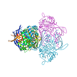
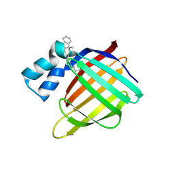
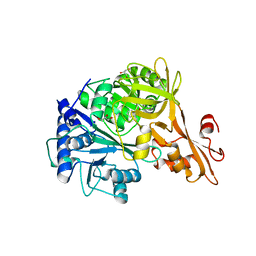
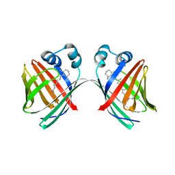
1JKI
DownloadVisualize
BU of 1jki by Molmil
myo-Inositol-1-phosphate Synthase Complexed with an Inhibitor, 2-deoxy-glucitol-6-phosphate
Descriptor: 1,4-DIHYDRONICOTINAMIDE ADENINE DINUCLEOTIDE, 2-DEOXY-GLUCITOL-6-PHOSPHATE, AMMONIUM ION, ...
Authors:Stein, A.J, Geiger, J.H.
Deposit date:2001-07-12
Release date:2002-04-10
Last modified:2023-08-16
Method:X-RAY DIFFRACTION (2.2 Å)
Cite:The crystal structure and mechanism of 1-L-myo-inositol- 1-phosphate synthase
J.Biol.Chem., 277, 2002
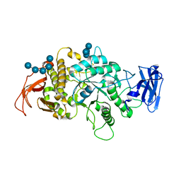
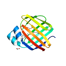
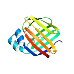
8SDB
DownloadVisualize
BU of 8sdb by Molmil
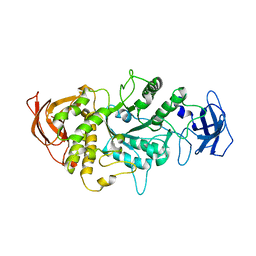
Crystal Structure of E.Coli Branching Enzyme in complex with malto-octose
Descriptor: 1,4-alpha-glucan branching enzyme GlgB, alpha-D-glucopyranose, alpha-D-glucopyranose-(1-4)-alpha-D-glucopyranose, ...
Authors:Bingham, C.R, Nayebi, H, Fawaz, R, Geiger, J.H.
Deposit date:2023-04-06
Release date:2023-07-12
Last modified:2023-08-16
Method:X-RAY DIFFRACTION (3 Å)
Cite:The Structure of Maltooctaose-Bound Escherichia coli Branching Enzyme Suggests a Mechanism for Donor Chain Specificity.
Molecules, 28, 2023
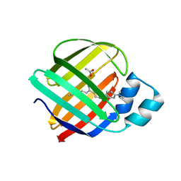
4I9R
DownloadVisualize
BU of 4i9r by Molmil
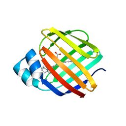
Crystal Structure of the R111K:R132L:Y134F:T54V:R59W:A32W Mutant of the Cellular Retinoic Acid Binding Protein Type II in Complex with All-Trans Retinal at 2.6 Angstrom Resolution
Descriptor: Cellular retinoic acid-binding protein 2, RETINAL
Authors:Nosrati, M, Geiger, J.H.
Deposit date:2012-12-05
Release date:2013-10-09
Last modified:2023-09-20
Method:X-RAY DIFFRACTION (2.6 Å)
Cite:Rational Design of a Colorimetric pH Sensor from a Soluble Retinoic Acid Chaperone.
J.Am.Chem.Soc., 135, 2013
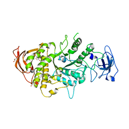
4LPC
DownloadVisualize
BU of 4lpc by Molmil
Crystal Structure of E.Coli Branching Enzyme in complex with maltoheptaose
Descriptor: 1,4-alpha-glucan branching enzyme GlgB, GLYCEROL, alpha-D-glucopyranose-(1-4)-alpha-D-glucopyranose, ...
Authors:Feng, L, Geiger, J.H.
Deposit date:2013-07-16
Release date:2015-04-01
Last modified:2024-02-28
Method:X-RAY DIFFRACTION (2.39 Å)
Cite:Crystal Structures of Escherichia coli Branching Enzyme in Complex with Linear Oligosaccharides.
Biochemistry, 54, 2015
4LQ1
DownloadVisualize
BU of 4lq1 by Molmil
Crystal Structure of E.Coli Branching Enzyme in complex with maltohexaose
Descriptor: 1,4-alpha-glucan branching enzyme GlgB, GLYCEROL, alpha-D-glucopyranose-(1-4)-alpha-D-glucopyranose, ...
Authors:Feng, L, Geiger, J.H.
Deposit date:2013-07-17
Release date:2015-04-01
Last modified:2024-02-28
Method:X-RAY DIFFRACTION (2.55 Å)
Cite:Crystal Structures of Escherichia coli Branching Enzyme in Complex with Linear Oligosaccharides.
Biochemistry, 54, 2015
4M6S
DownloadVisualize
BU of 4m6s by Molmil
Crystal structure of the R111K:R132Y:Y134F:T54V:R59W:A32W mutant of the Cellular Retinoic Acid Binding Protein Type II in complex with All-Trans Retinal at 2.38 Angstrom Resolution
Descriptor: Cellular retinoic acid-binding protein 2, RETINAL
Authors:Nosrati, M, Geiger, J.H.
Deposit date:2013-08-10
Release date:2013-10-09
Last modified:2023-09-20
Method:X-RAY DIFFRACTION (2.47 Å)
Cite:Rational Design of a Colorimetric pH Sensor from a Soluble Retinoic Acid Chaperone.
J.Am.Chem.Soc., 135, 2013
4M7M
DownloadVisualize
BU of 4m7m by Molmil
Crystal structure of the R111K:R132Q:Y134F:T54V:R59W:A32W mutant of the Cellular Retinoic Acid Binding Protein Type II in complex with All-Trans Retinal at 2.57 Angstrom Resolution
Descriptor: Cellular retinoic acid-binding protein 2, RETINAL
Authors:Nosrati, M, Geiger, J.H.
Deposit date:2013-08-12
Release date:2013-10-09
Last modified:2014-02-19
Method:X-RAY DIFFRACTION (2.571 Å)
Cite:Rational Design of a Colorimetric pH Sensor from a Soluble Retinoic Acid Chaperone.
J.Am.Chem.Soc., 135, 2013
4QZT
DownloadVisualize
BU of 4qzt by Molmil
Crystal Structure of wild type Human Cellular Retinol Binding Protein II (hCRBPII) bound to retinol at 7 KeV beam energy
Descriptor: ACETATE ION, RETINOL, Retinol-binding protein 2
Authors:Assar, Z, Geiger, J.H.
Deposit date:2014-07-28
Release date:2014-12-10
Last modified:2023-09-20
Method:X-RAY DIFFRACTION (1.9 Å)
Cite:Structures of holo wild-type human cellular retinol-binding protein II (hCRBPII) bound to retinol and retinal.
Acta Crystallogr.,Sect.D, 70, 2014
4QGW
DownloadVisualize
BU of 4qgw by Molmil
Crystal sturcture of the R132K:R111L:L121D mutant of Cellular Retinoic Acid Binding ProteinII complexed with a synthetic ligand (Merocyanine) at 1.77 angstrom resolution
Descriptor: (2E,4E,6E)-3-methyl-6-(1,3,3-trimethyl-1,3-dihydro-2H-indol-2-ylidene)hexa-2,4-dienal, 2-[3-(2-HYDROXY-1,1-DIHYDROXYMETHYL-ETHYLAMINO)-PROPYLAMINO]-2-HYDROXYMETHYL-PROPANE-1,3-DIOL, Cellular retinoic acid-binding protein 2
Authors:Nosrati, M, Yapici, I, Geiger, J.H.
Deposit date:2014-05-26
Release date:2015-01-28
Last modified:2015-02-11
Method:X-RAY DIFFRACTION (1.77 Å)
Cite:"Turn-on" protein fluorescence: in situ formation of cyanine dyes.
J.Am.Chem.Soc., 137, 2015
4QGV
DownloadVisualize
BU of 4qgv by Molmil
Crystal structure of the R132K:R111L mutant of Cellular Retinoic Acid Binding ProteinII complexed with a synthetic ligand (Merocyanine) at 1.73 angstrom resolution.
Descriptor: (2E,4E,6E)-3-methyl-6-(1,3,3-trimethyl-1,3-dihydro-2H-indol-2-ylidene)hexa-2,4-dienal, Cellular retinoic acid-binding protein 2
Authors:Nosrati, M, Yapici, I, Geiger, J.H.
Deposit date:2014-05-25
Release date:2015-01-28
Last modified:2023-09-20
Method:X-RAY DIFFRACTION (1.73 Å)
Cite:"Turn-on" protein fluorescence: in situ formation of cyanine dyes.
J.Am.Chem.Soc., 137, 2015
4QGX
DownloadVisualize
BU of 4qgx by Molmil
Crystal structure of the R132K:R111L:L121E mutant of Cellular Retinoic Acid Binding ProteinII complexed with a synthetic ligand (Merocyanine) at 1.47 angstrom resolution
Descriptor: (2E,4E,6E)-3-methyl-6-(1,3,3-trimethyl-1,3-dihydro-2H-indol-2-ylidene)hexa-2,4-dienal, Cellular retinoic acid-binding protein 2
Authors:Nosrati, M, Yapici, I, Geiger, J.H.
Deposit date:2014-05-26
Release date:2015-01-28
Last modified:2023-09-20
Method:X-RAY DIFFRACTION (1.471 Å)
Cite:"Turn-on" protein fluorescence: in situ formation of cyanine dyes.
J.Am.Chem.Soc., 137, 2015
4QZU
DownloadVisualize
BU of 4qzu by Molmil
Crystal Structure of wild type Human Cellular Retinol Binding Protein II (hCRBPII) bound to retinol at 11 KeV beam energy
Descriptor: ACETATE ION, GLYCEROL, RETINOL, ...
Authors:Assar, Z, Geiger, J.H.
Deposit date:2014-07-28
Release date:2014-12-10
Last modified:2023-09-20
Method:X-RAY DIFFRACTION (1.5 Å)
Cite:Structures of holo wild-type human cellular retinol-binding protein II (hCRBPII) bound to retinol and retinal.
Acta Crystallogr.,Sect.D, 70, 2014
4RLQ
DownloadVisualize
BU of 4rlq by Molmil
Crystal structure of a benzoate coenzyme A ligase with o-Toluic acid
Descriptor: 2-methylbenzoic acid, Benzoate-coenzyme A ligase, GLYCEROL
Authors:Strom, S, Nosrati, M, Thornburg, C, Walker, K.D, Geiger, J.H.
Deposit date:2014-10-17
Release date:2015-09-30
Last modified:2024-02-28
Method:X-RAY DIFFRACTION (1.63 Å)
Cite:Kinetically and Crystallographically Guided Mutations of a Benzoate CoA Ligase (BadA) Elucidate Mechanism and Expand Substrate Permissivity.
Biochemistry, 54, 2015
8VZZ
DownloadVisualize
BU of 8vzz by Molmil
Q108K:K40L:T51V:T53S:Y19W:R58W mutant of hCRBPII bound to synthetic fluorophore TD-1V
Descriptor: (2E)-3-{5-[4-(dimethylamino)phenyl]thiophen-2-yl}but-2-enal, ACETATE ION, Retinol-binding protein 2
Authors:Nosssoni, Z, Bingham, C.R, Geiger, J.H, Borhan, B.
Deposit date:2024-02-13
Release date:2024-04-03
Method:X-RAY DIFFRACTION (1.22 Å)
Cite:Regulation of Emission via a Protein-Bound Fluorophore
To Be Published
8W02
DownloadVisualize
BU of 8w02 by Molmil
Q108K:K40L:T51V:T53S:R58W mutant of hCRBPII bound to synthetic fluorophore TD-1V
Descriptor: (2E)-3-{5-[4-(dimethylamino)phenyl]thiophen-2-yl}but-2-enal, ACETATE ION, Retinol-binding protein 2
Authors:Ghanbarpour, A, Geiger, J.H, Borhan, B.
Deposit date:2024-02-13
Release date:2024-04-03
Method:X-RAY DIFFRACTION (1.499 Å)
Cite:Regulation of Emission via a Protein-Bound Fluorophore
To Be Published
8W00
DownloadVisualize
BU of 8w00 by Molmil
Q108K:K40L:T51V:T53S:Y19W:R58W:L117E mutant of hCRBPII bound to synthetic fluorophore TD-1V
Descriptor: (2E)-3-{5-[4-(dimethylamino)phenyl]thiophen-2-yl}but-2-enal, ACETATE ION, Retinol-binding protein 2
Authors:Nossoni, Z, Bingham, C.R, Geiger, J.H.
Deposit date:2024-02-13
Release date:2024-04-03
Method:X-RAY DIFFRACTION (1.23 Å)
Cite:Regulation of Emission via a Protein-Bound Fluorophore
To Be Published
8VZY
DownloadVisualize
BU of 8vzy by Molmil
Q108K:K40L:T51V:T53S:R58Y mutant of hCRBPII bound to synthetic fluorophore TD-1V
Descriptor: (2E)-3-{5-[4-(dimethylamino)phenyl]thiophen-2-yl}but-2-enal, ACETATE ION, Retinol-binding protein 2
Authors:Nossoni, Z, Bingham, C.R, Geiger, J.H, Borhan, B.
Deposit date:2024-02-13
Release date:2024-04-03
Method:X-RAY DIFFRACTION (1.34 Å)
Cite:Regulation of Emission via a Protein-Bound Fluorophore
To Be Published
8VZX
DownloadVisualize
BU of 8vzx by Molmil
Q108K:K40L:T53A:R58F mutant of hCRBPII bound to synthetic fluorophore TD-1V
Descriptor: (2E)-3-{5-[4-(dimethylamino)phenyl]thiophen-2-yl}but-2-enal, ACETATE ION, Retinol-binding protein 2
Authors:Nossoni, Z, Bingham, C.R, Geiger, J.H.
Deposit date:2024-02-13
Release date:2024-04-03
Method:X-RAY DIFFRACTION (1.47 Å)
Cite:Regulation of Emission via a Protein-Bound Fluorophore
To Be Published
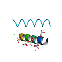
2DPQ
DownloadVisualize
BU of 2dpq by Molmil
The crystal structures of the calcium-bound con-G and con-T(K7gamma) dimeric peptides demonstrate a novel metal-dependent helix-forming motif
Descriptor: CALCIUM ION, CHLORIDE ION, Conantokin-G
Authors:Cnudde, S.E, Prorok, M, Dai, Q, Castellino, F.J, Geiger, J.H.
Deposit date:2006-05-13
Release date:2007-04-24
Last modified:2024-04-03
Method:X-RAY DIFFRACTION (1.25 Å)
Cite:The crystal structures of the calcium-bound con-G and con-T[K7gamma] dimeric peptides demonstrate a metal-dependent helix-forming motif
J.Am.Chem.Soc., 129, 2007
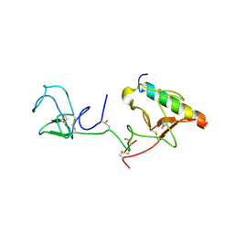
2DOI
DownloadVisualize
BU of 2doi by Molmil
The X-ray crystallographic structure of the angiogenesis inhibitor, angiostatin, bound to a peptide from the group A streptococcus protein PAM
Descriptor: Angiostatin, Plasminogen-binding group A streptococcal M-like protein PAM
Authors:Cnudde, S.E, Prorok, M, Castellino, F.J, Geiger, J.H.
Deposit date:2006-04-29
Release date:2006-12-05
Last modified:2023-10-25
Method:X-RAY DIFFRACTION (3.1 Å)
Cite:X-ray crystallographic structure of the angiogenesis inhibitor, angiostatin, bound to a peptide from the group A streptococcal surface protein PAM
Biochemistry, 45, 2006
2FRS
DownloadVisualize
BU of 2frs by Molmil
Crystal structure of the f15w mutant of apo-cellular retinoic acid binding protein type ii at 1.51 angstroms resolution
Descriptor: Cellular retinoic acid-binding protein 2, SODIUM ION
Authors:Vaezeslami, S, Geiger, J.H.
Deposit date:2006-01-20
Release date:2006-09-19
Last modified:2024-02-14
Method:X-RAY DIFFRACTION (1.51 Å)
Cite:The structure of Apo-wild-type cellular retinoic acid binding protein II at 1.4 A and its relationship to ligand binding and nuclear translocation.
J.Mol.Biol., 363, 2006
2FS6
DownloadVisualize
BU of 2fs6 by Molmil
Crystal Structure of Apo-Cellular Retinoic Acid Binding Protein Type II At 1.35 Angstroms Resolution
Descriptor: ACETATE ION, CHLORIDE ION, Cellular retinoic acid-binding protein 2, ...
Authors:Vaezeslami, S, Geiger, J.H.
Deposit date:2006-01-21
Release date:2006-09-19
Last modified:2024-02-14
Method:X-RAY DIFFRACTION (1.35 Å)
Cite:The structure of Apo-wild-type cellular retinoic acid binding protein II at 1.4 A and its relationship to ligand binding and nuclear translocation.
J.Mol.Biol., 363, 2006
2FR3
DownloadVisualize
BU of 2fr3 by Molmil
Crystal Structure of Cellular Retinoic Acid Binding Protein Type II in Complex with All-Trans-Retinoic Acid at 1.48 Angstroms Resolution
Descriptor: ACETATE ION, Cellular retinoic acid binding protein 2, RETINOIC ACID
Authors:Vaezeslami, S, Geiger, J.H.
Deposit date:2006-01-18
Release date:2006-09-19
Last modified:2024-02-14
Method:X-RAY DIFFRACTION (1.48 Å)
Cite:The structure of Apo-wild-type cellular retinoic acid binding protein II at 1.4 A and its relationship to ligand binding and nuclear translocation.
J.Mol.Biol., 363, 2006

222926

PDB entries from 2024-07-24

PDB statisticsPDBj update infoContact PDBjnumon