Loading
PDBj
MenuPDBj@FacebookPDBj@TwitterPDBj@YouTubewwPDB FoundationwwPDB
RCSB PDBPDBeBMRBAdv. SearchSearch help
PDB: 187 results

1P1F
DownloadVisualize
BU of 1p1f by Molmil
Crystal structure of apo 1L-myo-inositol 1-phosphate synthase
Descriptor: Inositol-3-phosphate synthase
Authors:Jin, X, Geiger, J.H.
Deposit date:2003-04-12
Release date:2003-07-08
Last modified:2024-02-14
Method:X-RAY DIFFRACTION (2.6 Å)
Cite:Structures of NAD(+)- and NADH-bound 1-l-myo-inositol 1-phosphate synthase.
Acta Crystallogr.,Sect.D, 59, 2003
4ZGU
DownloadVisualize
BU of 4zgu by Molmil
Crystal structure of monomer Y60W hCRBPII
Descriptor: ACETATE ION, Retinol-binding protein 2
Authors:Nossoni, Z, Geiger, J.
Deposit date:2015-04-24
Release date:2016-06-08
Last modified:2024-03-06
Method:X-RAY DIFFRACTION (1.49 Å)
Cite:Domain-Swapped Dimers of Intracellular Lipid-Binding Proteins: Evidence for Ordered Folding Intermediates.
Structure, 24, 2016
5DPQ
DownloadVisualize
BU of 5dpq by Molmil
Crystal Structure of E72A mutant of domain swapped dimer Human Cellular Retinol Binding Protein
Descriptor: ACETATE ION, Retinol-binding protein 2
Authors:Assar, Z, Nossoni, Z, Wang, W, Geiger, J.H, Borhan, B.
Deposit date:2015-09-14
Release date:2016-09-14
Last modified:2024-03-06
Method:X-RAY DIFFRACTION (1.775 Å)
Cite:Domain-Swapped Dimers of Intracellular Lipid-Binding Proteins: Evidence for Ordered Folding Intermediates.
Structure, 24, 2016
5E6Z
DownloadVisualize
BU of 5e6z by Molmil
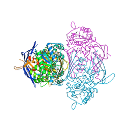
Crystal structure of Ecoli Branching Enzyme with beta cyclodextrin
Descriptor: 1,4-alpha-glucan branching enzyme GlgB, Cycloheptakis-(1-4)-(alpha-D-glucopyranose), GLYCEROL
Authors:Feng, L, Nosrati, M, Geiger, J.H.
Deposit date:2015-10-11
Release date:2015-12-16
Last modified:2023-09-27
Method:X-RAY DIFFRACTION (1.878 Å)
Cite:Crystal structures of Escherichia coli branching enzyme in complex with cyclodextrins.
Acta Crystallogr D Struct Biol, 72, 2016
5E6Y
DownloadVisualize
BU of 5e6y by Molmil
Crystal structure of E.Coli branching enzyme in complex with alpha cyclodextrin
Descriptor: 1,4-alpha-glucan branching enzyme GlgB, Cyclohexakis-(1-4)-(alpha-D-glucopyranose), GLYCEROL
Authors:Feng, L, Nosrati, M, Geiger, J.H.
Deposit date:2015-10-11
Release date:2015-12-16
Last modified:2023-09-27
Method:X-RAY DIFFRACTION (2.6 Å)
Cite:Crystal structures of Escherichia coli branching enzyme in complex with cyclodextrins.
Acta Crystallogr D Struct Biol, 72, 2016
6MCU
DownloadVisualize
BU of 6mcu by Molmil
Crystal structure of the holo retinal-bound domain-swapped dimer Q108K:T51D:A28H mutant of human Cellular Retinol Binding Protein II
Descriptor: GLYCEROL, RETINAL, Retinol-binding protein 2
Authors:Ghanbarpour, A, Geiger, J.
Deposit date:2018-09-02
Release date:2019-10-16
Last modified:2023-10-11
Method:X-RAY DIFFRACTION (2.572 Å)
Cite:Engineering the hCRBPII Domain-Swapped Dimer into a New Class of Protein Switches.
J.Am.Chem.Soc., 141, 2019
6MCV
DownloadVisualize
BU of 6mcv by Molmil
Crystal Structure of Holo Retinal-Bound Domain-Swapped Dimer of Wild Type Human Cellular Retinol Binding Protein II
Descriptor: RETINAL, Retinol-binding protein 2
Authors:Ghanbarpour, A, Geiger, J.
Deposit date:2018-09-02
Release date:2019-10-16
Last modified:2023-10-11
Method:X-RAY DIFFRACTION (3.3 Å)
Cite:Engineering the hCRBPII Domain-Swapped Dimer into a New Class of Protein Switches.
J.Am.Chem.Soc., 141, 2019
6MLB
DownloadVisualize
BU of 6mlb by Molmil
Crystal structure of the holo retinal-bound domain-swapped dimer Q108K:K40L:T51F mutant of human cellular retinol binding protein II
Descriptor: ACETATE ION, GLYCEROL, RETINAL, ...
Authors:Ghanbarpour, A, Geiger, J.
Deposit date:2018-09-27
Release date:2019-10-16
Last modified:2023-10-11
Method:X-RAY DIFFRACTION (2.15 Å)
Cite:Engineering the hCRBPII Domain-Swapped Dimer into a New Class of Protein Switches.
J.Am.Chem.Soc., 141, 2019
6MKV
DownloadVisualize
BU of 6mkv by Molmil
Crystal structure of Retinal-bound holo Q108K:K40L:T51W domain-swapped dimer of human cellular retinol binding protein 2
Descriptor: ACETATE ION, GLYCEROL, RETINAL, ...
Authors:Ghanbarpour, A, Geiger, J.
Deposit date:2018-09-26
Release date:2019-10-16
Last modified:2023-10-11
Method:X-RAY DIFFRACTION (2.107 Å)
Cite:Engineering the hCRBPII Domain-Swapped Dimer into a New Class of Protein Switches.
J.Am.Chem.Soc., 141, 2019
2G7B
DownloadVisualize
BU of 2g7b by Molmil
Crystal Structure of the R132K:R111L:L121E mutant of Cellular Retinoic Acid Binding Protein Type II In Complex With All-Trans-Retinal At 1.18 Angstroms Resolution
Descriptor: ALL-TRANS AXEROPHTHENE, Cellular retinoic acid-binding protein 2, SODIUM ION
Authors:Vaezeslami, S, Geiger, J.H.
Deposit date:2006-02-27
Release date:2007-04-17
Last modified:2021-10-20
Method:X-RAY DIFFRACTION (1.18 Å)
Cite:Protein design: reengineering cellular retinoic acid binding protein II into a rhodopsin protein mimic.
J.Am.Chem.Soc., 129, 2007
2G79
DownloadVisualize
BU of 2g79 by Molmil
Crystal Structure of the R132K:Y134F Mutant of Cellular Retinoic Acid Binding Protein Type II in Complex with All-Trans-Retinal at 1.69 Angstroms Resolution
Descriptor: Cellular retinoic acid-binding protein 2, RETINAL, SODIUM ION, ...
Authors:Vaezeslami, S, Geiger, J.H.
Deposit date:2006-02-27
Release date:2007-04-17
Last modified:2024-02-14
Method:X-RAY DIFFRACTION (1.69 Å)
Cite:Protein design: reengineering cellular retinoic acid binding protein II into a rhodopsin protein mimic.
J.Am.Chem.Soc., 129, 2007
4ZCB
DownloadVisualize
BU of 4zcb by Molmil
Human CRBPII mutant - Y60W dimer
Descriptor: Retinol-binding protein 2
Authors:Nossoni, Z, Assar, Z, Wang, W, Geiger, J, Borhan, B.
Deposit date:2015-04-15
Release date:2016-04-20
Last modified:2023-09-27
Method:X-RAY DIFFRACTION (1.7 Å)
Cite:Domain-Swapped Dimers of Intracellular Lipid-Binding Proteins: Evidence for Ordered Folding Intermediates.
Structure, 24, 2016
7LHM
DownloadVisualize
BU of 7lhm by Molmil
Crystal structure of Q108K:K40L:T51V:T53C:R58W:T29L:Y19W:Q4A:Q38L mutant of cellular retinol binding protein II complex with all-trans-retinal
Descriptor: ACETATE ION, GLYCEROL, RETINAL, ...
Authors:Ehyaei, N, Geiger, J.H, Borhan, B.
Deposit date:2021-01-25
Release date:2021-04-14
Last modified:2023-10-18
Method:X-RAY DIFFRACTION (1.5 Å)
Cite:Crystal structure of Q108K:K40L:T51V:T53C:R58W:T29L:Y19W:Q4A:Q38L mutant of cellular retinol binding protein II complex with all-trans-retinal
To Be Published
4M6S
DownloadVisualize
BU of 4m6s by Molmil
Crystal structure of the R111K:R132Y:Y134F:T54V:R59W:A32W mutant of the Cellular Retinoic Acid Binding Protein Type II in complex with All-Trans Retinal at 2.38 Angstrom Resolution
Descriptor: Cellular retinoic acid-binding protein 2, RETINAL
Authors:Nosrati, M, Geiger, J.H.
Deposit date:2013-08-10
Release date:2013-10-09
Last modified:2023-09-20
Method:X-RAY DIFFRACTION (2.47 Å)
Cite:Rational Design of a Colorimetric pH Sensor from a Soluble Retinoic Acid Chaperone.
J.Am.Chem.Soc., 135, 2013
4M7M
DownloadVisualize
BU of 4m7m by Molmil
Crystal structure of the R111K:R132Q:Y134F:T54V:R59W:A32W mutant of the Cellular Retinoic Acid Binding Protein Type II in complex with All-Trans Retinal at 2.57 Angstrom Resolution
Descriptor: Cellular retinoic acid-binding protein 2, RETINAL
Authors:Nosrati, M, Geiger, J.H.
Deposit date:2013-08-12
Release date:2013-10-09
Last modified:2014-02-19
Method:X-RAY DIFFRACTION (2.571 Å)
Cite:Rational Design of a Colorimetric pH Sensor from a Soluble Retinoic Acid Chaperone.
J.Am.Chem.Soc., 135, 2013
6MQJ
DownloadVisualize
BU of 6mqj by Molmil
Crystal Structure of the All-Trans Retinal bound R111K:Y134F:T54V:R132Q:P39Y:R59Y:L121Q mutant of Human Cellular Retinoic Acid Binding Protein II Irradiated with 532 nm Laser for 10 minutes at 2.1 Angstrom Resolution
Descriptor: Cellular retinoic acid-binding protein 2, RETINAL
Authors:Ghanbarpour, A, Geiger, J.
Deposit date:2018-10-09
Release date:2019-01-09
Last modified:2023-10-11
Method:X-RAY DIFFRACTION (2.125 Å)
Cite:Mimicking Microbial Rhodopsin Isomerization in a Single Crystal.
J. Am. Chem. Soc., 141, 2019
6MOQ
DownloadVisualize
BU of 6moq by Molmil
Crystal Structure of the All-trans Retinal-Bound R111K:Y134F:T54V:R132Q:P39Y:R59Y:L121W Mutant of Human Cellular Retinoic Acid Binding Protein II in the Dark at 2.2 Angstrom Resolution
Descriptor: Cellular retinoic acid-binding protein 2, RETINAL
Authors:Ghanbarpour, A, Geiger, J.
Deposit date:2018-10-04
Release date:2019-01-09
Last modified:2023-10-11
Method:X-RAY DIFFRACTION (2.198 Å)
Cite:Mimicking Microbial Rhodopsin Isomerization in a Single Crystal.
J. Am. Chem. Soc., 141, 2019
6MOP
DownloadVisualize
BU of 6mop by Molmil
Crystal Structure of the All-trans Retinal-Bound R111K:Y134F:T54V:R132Q:P39Y:R59Y:L121E Mutant of Human Cellular Retinoic Acid Binding Protein II in the Dark at 1.9 Angstrom Resolution
Descriptor: Cellular retinoic acid-binding protein 2, RETINAL
Authors:Ghanbarpour, A, Geiger, J.
Deposit date:2018-10-04
Release date:2019-01-09
Last modified:2023-10-11
Method:X-RAY DIFFRACTION (1.9 Å)
Cite:Mimicking Microbial Rhodopsin Isomerization in a Single Crystal.
J. Am. Chem. Soc., 141, 2019
6MPK
DownloadVisualize
BU of 6mpk by Molmil
Crystal Structure of the 13-cis Retinal-Bound R111K:Y134F:T54V:R132Q:P39Y:R59Y:L121E:A32Y Mutant of Human Cellular Retinoic Acid Binding Protein II in the Dark at 1.58 Angstrom Resolution
Descriptor: Cellular retinoic acid-binding protein 2, GLYCEROL, RETINAL
Authors:Ghanbarpour, A, Geiger, J.
Deposit date:2018-10-07
Release date:2019-01-09
Last modified:2023-10-11
Method:X-RAY DIFFRACTION (1.58 Å)
Cite:Mimicking Microbial Rhodopsin Isomerization in a Single Crystal.
J. Am. Chem. Soc., 141, 2019
6MQY
DownloadVisualize
BU of 6mqy by Molmil
Crystal Structure of the 13-cis Product of All-trans Retinal-Bound R111K:Y134F:T54V:R132Q:P39Y:R59Y:L121E Mutant of Human Cellular Retinoic Acid Binding Protein II Irradiated with 400 nm Laser (30 seconds) at 1.9 Angstrom
Descriptor: Cellular retinoic acid-binding protein 2, GLYCEROL, RETINAL
Authors:Ghanbarpour, A, Geiger, J.
Deposit date:2018-10-11
Release date:2019-01-09
Last modified:2023-10-11
Method:X-RAY DIFFRACTION (1.9 Å)
Cite:Mimicking Microbial Rhodopsin Isomerization in a Single Crystal.
J. Am. Chem. Soc., 141, 2019
6MOV
DownloadVisualize
BU of 6mov by Molmil
Crystal Structure of the All-trans Retinal bound R111K:Y134F:T54V:R132Q:P39Y:R59Y:L121Q Human Cellular Retinoic Acid Binding Protein II in the Dark at 1.75 Angstrom Resolution
Descriptor: Cellular retinoic acid-binding protein 2, RETINAL
Authors:Ghanbarpour, A, Geiger, J.
Deposit date:2018-10-04
Release date:2019-01-09
Last modified:2023-10-11
Method:X-RAY DIFFRACTION (1.752 Å)
Cite:Mimicking Microbial Rhodopsin Isomerization in a Single Crystal.
J. Am. Chem. Soc., 141, 2019
6MR0
DownloadVisualize
BU of 6mr0 by Molmil
Crystal Structure of the All-trans Retinal Bound R111K:Y134F:T54V:R132Q:P39Q:R59Y:L121E Human Cellular Retinoic Acid Binding Protein II Mutant After 5 Minutes UV irradiation at 2.6 Angstrom Resolution
Descriptor: Cellular retinoic acid-binding protein 2, RETINAL
Authors:Ghanbarpour, A, Geiger, J.
Deposit date:2018-10-11
Release date:2019-01-09
Last modified:2023-10-11
Method:X-RAY DIFFRACTION (2.649 Å)
Cite:Mimicking Microbial Rhodopsin Isomerization in a Single Crystal.
J. Am. Chem. Soc., 141, 2019
6MOX
DownloadVisualize
BU of 6mox by Molmil
Crystal Structure of the All-trans Retinal-Bound R111K:Y134F:T54V:R132Q:P39Q:R59Y:L121E Human Cellular Retinoic Acid Binding Protein II Mutant in the Dark at 2.18 Angstrom Resolution
Descriptor: Cellular retinoic acid-binding protein 2, RETINAL
Authors:Ghanbarpour, A, Geiger, J.
Deposit date:2018-10-04
Release date:2019-01-09
Last modified:2023-10-11
Method:X-RAY DIFFRACTION (2.18 Å)
Cite:Mimicking Microbial Rhodopsin Isomerization in a Single Crystal.
J. Am. Chem. Soc., 141, 2019
6MQX
DownloadVisualize
BU of 6mqx by Molmil
Crystal Structure of All-trans Retinal-Bound R111K:Y134F:T54V:R132Q:P39Y:R59Y:L121E Human Cellular Retinoic Acid Binding Protein II Irradiated with 400 nm Laser (30 seconds) and Subsequently Dark Adapted (25 minutes) at 2.0 Angstrom Resolution
Descriptor: Cellular retinoic acid-binding protein 2, GLYCEROL, RETINAL
Authors:Ghanbarpour, A, Geiger, J.
Deposit date:2018-10-11
Release date:2019-01-09
Last modified:2023-10-11
Method:X-RAY DIFFRACTION (2.01 Å)
Cite:Mimicking Microbial Rhodopsin Isomerization in a Single Crystal.
J. Am. Chem. Soc., 141, 2019
6MQI
DownloadVisualize
BU of 6mqi by Molmil
Crystal Structure of the All-trans Retinal bound R111K:Y134F:T54V:R132Q:P39Y:R59Y:L121Q mutant of Human Cellular Retinoic Acid Binding Protein II Irradiated with 400 nm Laser for 5 minutes at 1.87 Angstrom Resolution
Descriptor: Cellular retinoic acid-binding protein 2, RETINAL
Authors:Ghanbarpour, A, Geiger, J.
Deposit date:2018-10-09
Release date:2019-01-09
Last modified:2023-10-11
Method:X-RAY DIFFRACTION (1.87 Å)
Cite:Mimicking Microbial Rhodopsin Isomerization in a Single Crystal.
J. Am. Chem. Soc., 141, 2019

220472

건을2024-05-29부터공개중

PDB statisticsPDBj update infoContact PDBjnumon