Loading
PDBj
MenuPDBj@FacebookPDBj@TwitterPDBj@YouTubewwPDB FoundationwwPDB
RCSB PDBPDBeBMRBAdv. SearchSearch help
PDB: 163 results

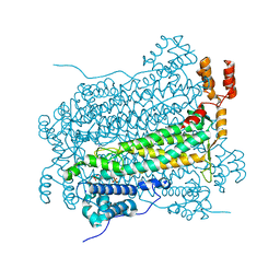
4F26
DownloadVisualize
BU of 4f26 by Molmil
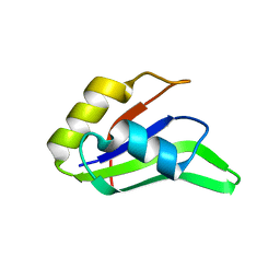
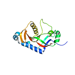
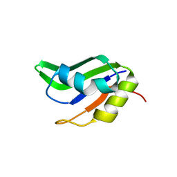
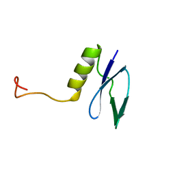
Crystal structure of the second RRM domain of human PABPC1 a pH 9.0
Descriptor: Polyadenylate-binding protein 1
Authors:Kozlov, G, Safaee, N, Gehring, K.
Deposit date:2012-05-07
Release date:2012-08-08
Last modified:2023-09-13
Method:X-RAY DIFFRACTION (2 Å)
Cite:Interdomain Allostery Promotes Assembly of the Poly(A) mRNA Complex with PABP and eIF4G.
Mol.Cell, 48, 2012
4F25
DownloadVisualize
BU of 4f25 by Molmil
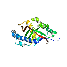
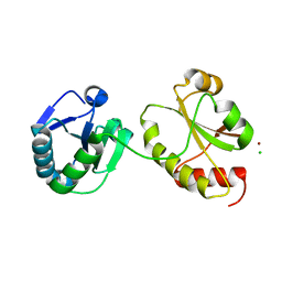
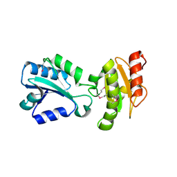
Crystal structure of the second RRM domain of human PABPC1 at pH 6.0
Descriptor: Polyadenylate-binding protein 1
Authors:Kozlov, G, Safaee, N, Gehring, K.
Deposit date:2012-05-07
Release date:2012-08-08
Last modified:2023-09-13
Method:X-RAY DIFFRACTION (1.9 Å)
Cite:Interdomain Allostery Promotes Assembly of the Poly(A) mRNA Complex with PABP and eIF4G.
Mol.Cell, 48, 2012
1FC8
DownloadVisualize
BU of 1fc8 by Molmil
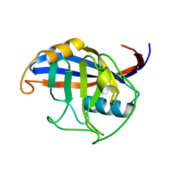
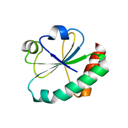
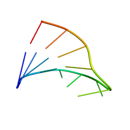
NMR SOLUTION STRUCTURE OF A CHIMERIC OLIGONUCLEOTIDE HAIRPIN R(GGAC)D(TTCG)2'F-A(GTCC)
Descriptor: 5'-R(*GP*GP*AP*CP)D(*TP*TP*CP*GP*(GFL)P*(TAF)P*(CFL)P*(CFL))-3'
Authors:Trempe, J.-F, Gehring, K.
Deposit date:2000-07-18
Release date:2001-05-30
Last modified:2024-05-22
Method:SOLUTION NMR
Cite:NMR solution structure of an oligonucleotide hairpin with a 2'F-ANA/RNA stem: implications for RNase H specificity toward DNA/RNA hybrid duplexes.
J.Am.Chem.Soc., 123, 2001
1GH8
DownloadVisualize
BU of 1gh8 by Molmil
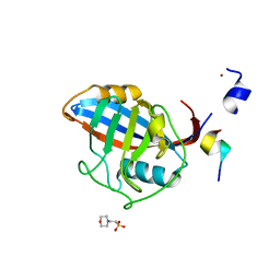
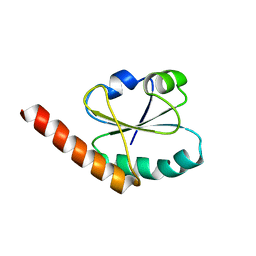
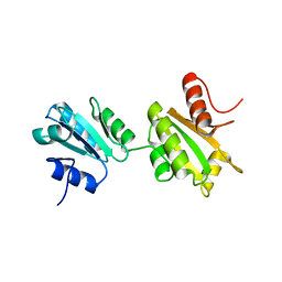
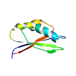
SOLUTION STRUCTURE OF THE ARCHAEAL TRANSLATION ELONGATION FACTOR 1BETA FROM METHANOBACTERIUM THERMOAUTOTROPHICUM
Descriptor: TRANSLATION ELONGATION FACTOR 1BETA
Authors:Kozlov, G, Ekiel, I, Gehring, K, Northeast Structural Genomics Consortium (NESG)
Deposit date:2000-11-30
Release date:2000-12-13
Last modified:2023-12-27
Method:SOLUTION NMR
Cite:Rapid fold and structure determination of the archaeal translation elongation factor 1beta from Methanobacterium thermoautotrophicum.
J.Biomol.NMR, 17, 2000
3PDZ
DownloadVisualize
BU of 3pdz by Molmil
SOLUTION STRUCTURE OF THE PDZ2 DOMAIN FROM HUMAN PHOSPHATASE HPTP1E
Descriptor: PROTEIN (TYROSINE PHOSPHATASE (PTP-BAS, TYPE 1))
Authors:Kozlov, G, Gehring, K, Ekiel, I.
Deposit date:1999-05-10
Release date:2000-03-17
Last modified:2023-12-27
Method:SOLUTION NMR
Cite:Solution structure of the PDZ2 domain from human phosphatase hPTP1E and its interactions with C-terminal peptides from the Fas receptor.
Biochemistry, 39, 2000
1DU6
DownloadVisualize
BU of 1du6 by Molmil
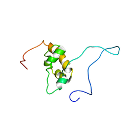
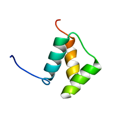
SOLUTION STRUCTURE OF THE TRUNCATED PBX HOMEODOMAIN
Descriptor: HOMEOBOX PROTEIN PBX1
Authors:Sprules, T, Green, N, Featherstone, M, Gehring, K.
Deposit date:2000-01-14
Release date:2000-08-16
Last modified:2024-05-22
Method:SOLUTION NMR
Cite:Conformational changes in the PBX homeodomain and C-terminal extension upon binding DNA and HOX-derived YPWM peptides.
Biochemistry, 39, 2000
2ILX
DownloadVisualize
BU of 2ilx by Molmil
Solution structure of catalytic domain of rat 2',3'-cyclic-nucleotide 3'-phosphodiesterase (CNP) protein
Descriptor: 2',3'-cyclic-nucleotide 3'-phosphodiesterase
Authors:Denisov, A.Y, Kozlov, G, Gehring, K.
Deposit date:2006-10-03
Release date:2007-03-06
Last modified:2024-05-29
Method:SOLUTION NMR
Cite:Solution structure of the catalytic domain of RICH protein from goldfish.
Febs J., 274, 2007
2K18
DownloadVisualize
BU of 2k18 by Molmil
Solution structure of bb' domains of human protein disulfide isomerase
Descriptor: Protein disulfide-isomerase
Authors:Denisov, A.Y, Maattanen, P, Dabrowski, C, Kozlov, G, Thomas, D.Y, Gehring, K.
Deposit date:2008-02-22
Release date:2008-04-29
Last modified:2024-05-29
Method:SOLUTION NMR
Cite:Solution structure of the bb' domains of human protein disulfide isomerase.
Febs J., 276, 2009
2KBW
DownloadVisualize
BU of 2kbw by Molmil
Solution Structure of human Mcl-1 complexed with human Bid_BH3 peptide
Descriptor: BH3-interacting domain death agonist, Induced myeloid leukemia cell differentiation protein Mcl-1
Authors:Liu, Q, Moldoveanu, T, Sprules, T, Matta-Camacho, E, Mansur-Azzam, N, Gehring, K.
Deposit date:2008-12-09
Release date:2009-12-15
Last modified:2024-05-22
Method:SOLUTION NMR
Cite:Apoptotic regulation by MCL-1 through heterodimerization.
J.Biol.Chem., 285, 2010
2I3E
DownloadVisualize
BU of 2i3e by Molmil
Solution structure of catalytic domain of goldfish RICH protein
Descriptor: G-RICH
Authors:Denisov, A.Y, Kozlov, G, Gehring, K.
Deposit date:2006-08-18
Release date:2007-03-06
Last modified:2024-05-29
Method:SOLUTION NMR
Cite:Solution structure of the catalytic domain of RICH protein from goldfish.
Febs J., 274, 2007
2H8L
DownloadVisualize
BU of 2h8l by Molmil
Crystal structure of the bb' fragment of ERp57
Descriptor: Protein disulfide-isomerase A3
Authors:Kozlov, G, Schrag, J.D, Cygler, M, Gehring, K.
Deposit date:2006-06-07
Release date:2006-08-29
Last modified:2011-07-13
Method:X-RAY DIFFRACTION (2 Å)
Cite:Crystal Structure of the bb' Domains of the Protein Disulfide Isomerase ERp57.
Structure, 14, 2006
3IQL
DownloadVisualize
BU of 3iql by Molmil
Crystal structure of the rat endophilin-A1 SH3 domain
Descriptor: CHLORIDE ION, Endophilin-A1
Authors:Trempe, J.F, Kozlov, G, Camacho, E.M, Gehring, K.
Deposit date:2009-08-20
Release date:2009-11-10
Last modified:2023-09-06
Method:X-RAY DIFFRACTION (1.4 Å)
Cite:SH3 domains from a subset of BAR proteins define a Ubl-binding domain and implicate parkin in synaptic ubiquitination.
Mol.Cell, 36, 2009
3NTW
DownloadVisualize
BU of 3ntw by Molmil
Structure of the MLLE domain of EDD in complex with a PAM2 peptide from Paip1
Descriptor: E3 ubiquitin-protein ligase UBR5, Polyadenylate-binding protein-interacting protein 1
Authors:Kozlov, G, Gehring, K.
Deposit date:2010-07-05
Release date:2011-07-06
Last modified:2023-09-06
Method:X-RAY DIFFRACTION (2.6 Å)
Cite:The MLLE domain of the ubiquitin ligase UBR5 binds to its catalytic domain to regulate substrate binding.
J. Biol. Chem., 290, 2015
3NY1
DownloadVisualize
BU of 3ny1 by Molmil
Structure of the ubr-box of the UBR1 ubiquitin ligase
Descriptor: E3 ubiquitin-protein ligase UBR1, ZINC ION
Authors:Matta-Camacho, E, Kozlov, G, Li, F, Gehring, K.
Deposit date:2010-07-14
Release date:2010-08-11
Last modified:2024-02-21
Method:X-RAY DIFFRACTION (2.085 Å)
Cite:Structural basis of substrate recognition and specificity in the N-end rule pathway.
Nat.Struct.Mol.Biol., 17, 2010
3O0X
DownloadVisualize
BU of 3o0x by Molmil
Structural basis of carbohydrate recognition by calreticulin
Descriptor: CALCIUM ION, Calreticulin, alpha-D-glucopyranose-(1-3)-alpha-D-mannopyranose-(1-2)-alpha-D-mannopyranose-(1-2)-alpha-D-mannopyranose
Authors:Kozlov, G, Gehring, K.
Deposit date:2010-07-20
Release date:2010-09-29
Last modified:2023-12-06
Method:X-RAY DIFFRACTION (2.01 Å)
Cite:Structural basis of carbohydrate recognition by calreticulin.
J.Biol.Chem., 285, 2010
3O0W
DownloadVisualize
BU of 3o0w by Molmil
Structural basis of carbohydrate recognition by calreticulin
Descriptor: CALCIUM ION, Calreticulin, alpha-D-glucopyranose-(1-3)-alpha-D-mannopyranose-(1-2)-alpha-D-mannopyranose-(1-2)-alpha-D-mannopyranose
Authors:Kozlov, G, Gehring, K.
Deposit date:2010-07-20
Release date:2010-09-29
Last modified:2020-07-29
Method:X-RAY DIFFRACTION (1.95 Å)
Cite:Structural basis of carbohydrate recognition by calreticulin.
J.Biol.Chem., 285, 2010
3NY3
DownloadVisualize
BU of 3ny3 by Molmil
Structure of the ubr-box of UBR2 in complex with N-degron
Descriptor: E3 ubiquitin-protein ligase UBR2, N-degron, ZINC ION
Authors:Matta-Camacho, E, Kozlov, G, Li, F, Gehring, K.
Deposit date:2010-07-14
Release date:2010-08-11
Last modified:2024-02-21
Method:X-RAY DIFFRACTION (1.6 Å)
Cite:Structural basis of substrate recognition and specificity in the N-end rule pathway.
Nat.Struct.Mol.Biol., 17, 2010
3O0V
DownloadVisualize
BU of 3o0v by Molmil
Crystal structure of the calreticulin lectin domain
Descriptor: CALCIUM ION, Calreticulin
Authors:Kozlov, G, Gehring, K.
Deposit date:2010-07-20
Release date:2010-09-29
Last modified:2023-09-06
Method:X-RAY DIFFRACTION (2.3 Å)
Cite:Structural basis of carbohydrate recognition by calreticulin.
J.Biol.Chem., 285, 2010
3KUR
DownloadVisualize
BU of 3kur by Molmil
Crystal structure of the MLLE domain of poly(A)-binding protein
Descriptor: CHLORIDE ION, Polyadenylate-binding protein 1
Authors:Kozlov, G, Gehring, K.
Deposit date:2009-11-27
Release date:2010-02-09
Last modified:2023-09-06
Method:X-RAY DIFFRACTION (2.5 Å)
Cite:Molecular Determinants of PAM2 Recognition by the MLLE Domain of Poly(A)-Binding Protein.
J.Mol.Biol., 397, 2010
3PT3
DownloadVisualize
BU of 3pt3 by Molmil
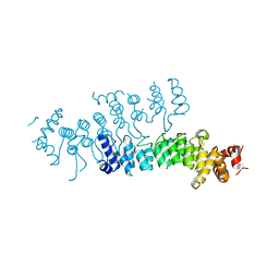
Crystal structure of the C-terminal lobe of the human UBR5 HECT domain
Descriptor: E3 ubiquitin-protein ligase UBR5
Authors:Matta-Camacho, E, Kozlov, G, Menade, M, Gehring, K.
Deposit date:2010-12-02
Release date:2012-01-25
Last modified:2023-09-06
Method:X-RAY DIFFRACTION (1.97 Å)
Cite:Structure of the HECT C-lobe of the UBR5 E3 ubiquitin ligase.
Acta Crystallogr.,Sect.F, 68, 2012
5TDA
DownloadVisualize
BU of 5tda by Molmil
Crystal structure of the UBR-box domain from UBR2 in complex with RLWS N-degron
Descriptor: ARG-LEU-TRP-SER peptide, E3 ubiquitin-protein ligase UBR2, ZINC ION
Authors:Munoz-Escobar, J, Kozlov, G, Gehring, K.
Deposit date:2016-09-19
Release date:2017-03-22
Last modified:2023-10-04
Method:X-RAY DIFFRACTION (0.79 Å)
Cite:Bound Waters Mediate Binding of Diverse Substrates to a Ubiquitin Ligase.
Structure, 25, 2017
1LFU
DownloadVisualize
BU of 1lfu by Molmil
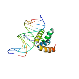
NMR Solution Structure of the Extended PBX Homeodomain Bound to DNA
Descriptor: 5'-D(*GP*CP*GP*CP*AP*TP*GP*AP*TP*TP*GP*CP*CP*C)-3', 5'-D(*GP*GP*GP*CP*AP*AP*TP*CP*AP*TP*GP*CP*GP*C)-3', homeobox protein PBX1
Authors:Sprules, T, Green, N, Featherstone, M, Gehring, K.
Deposit date:2002-04-12
Release date:2003-01-14
Last modified:2024-05-22
Method:SOLUTION NMR
Cite:Lock and Key Binding of the HOX YPWM Peptide to the PBX Homeodomain
J.Biol.Chem., 278, 2003
1ME1
DownloadVisualize
BU of 1me1 by Molmil
Chimeric hairpin with 2',5'-linked RNA loop and RNA stem
Descriptor: 5'-R(*GP*GP*AP*CP*(U25)P*(U25)P*(C25)P*(5GP)P*GP*UP*CP*C)-3'
Authors:Denisov, A.Y, Hannoush, R.N, Gehring, K, Damha, M.J.
Deposit date:2002-08-07
Release date:2003-08-19
Last modified:2024-05-01
Method:SOLUTION NMR
Cite:A Novel RNA Motif Based on the Structure of Unusually Stable 2',5'-Linked r(UUCG) Loops
J.AM.CHEM.SOC., 125, 2003
1IFW
DownloadVisualize
BU of 1ifw by Molmil
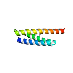
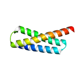
SOLUTION STRUCTURE OF C-TERMINAL DOMAIN OF POLY(A) BINDING PROTEIN FROM SACCHAROMYCES CEREVISIAE
Descriptor: POLYADENYLATE-BINDING PROTEIN, CYTOPLASMIC AND NUCLEAR
Authors:Kozlov, G, Siddiqui, N, Coillet-Matillon, S, Sprules, T, Ekiel, I, Gehring, K.
Deposit date:2001-04-13
Release date:2002-07-24
Last modified:2024-05-22
Method:SOLUTION NMR
Cite:Solution structure of the orphan PABC domain from Saccharomyces cerevisiae poly(A)-binding protein.
J.Biol.Chem., 277, 2002
1JDQ
DownloadVisualize
BU of 1jdq by Molmil
Solution Structure of TM006 Protein from Thermotoga maritima
Descriptor: HYPOTHETICAL PROTEIN TM0983
Authors:Denisov, A.Y, Finak, G, Yee, A, Kozlov, G, Gehring, K, Arrowsmith, C.H.
Deposit date:2001-06-14
Release date:2002-02-27
Last modified:2024-05-22
Method:SOLUTION NMR
Cite:An NMR approach to structural proteomics.
Proc.Natl.Acad.Sci.USA, 99, 2002

223790

數據於2024-08-14公開中

PDB statisticsPDBj update infoContact PDBjnumon