3M0R
 
 | |
4C6Y
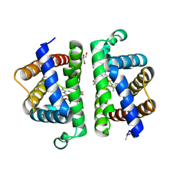
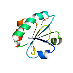
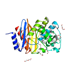 | | Ancestral PNCA (last common ancestors of Gram-positive and Gram- negative bacteria) beta-lactamase class A | | 分子名称: | ACETATE ION, BETA-LACTAMASE, CHLORIDE ION, ... | | 著者 | Gavira, J.A, Risso, V.A, Sanchez-Ruiz, J.M. | | 登録日 | 2013-09-19 | | 公開日 | 2014-04-16 | | 最終更新日 | 2023-12-20 | | 実験手法 | X-RAY DIFFRACTION (1.799 Å) | | 主引用文献 | Phenotypic Comparisons of Consensus Variants Versus Laboratory Resurrections of Precambrian Proteins.
Proteins, 82, 2014
|
|
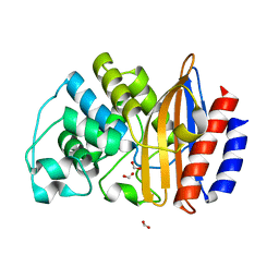
4C75
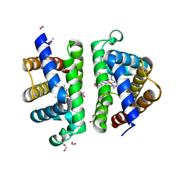
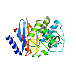 | | Consensus (ALL-CON) beta-lactamase class A | | 分子名称: | ACETATE ION, BETA-LACTAMASE, DI(HYDROXYETHYL)ETHER, ... | | 著者 | Gavira, J.A, Risso, V.A, Sanchez-Ruiz, J.M. | | 登録日 | 2013-09-19 | | 公開日 | 2014-04-16 | | 最終更新日 | 2023-12-20 | | 実験手法 | X-RAY DIFFRACTION (2.2 Å) | | 主引用文献 | Phenotypic Comparisons of Consensus Variants Versus Laboratory Resurrections of Precambrian Proteins.
Proteins, 82, 2014
|
|
2FCH
 | |
2FD3
 | |
2H73
 | |
2H71
 | |
2H6X
 | |
2H76
 | |
2H75
 | |
2H74
 | |
2H72
 | |
2H6Y
 | |
3PT7
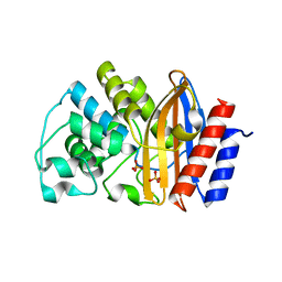
 | | Structure of HbII-III-Oxy from Lucina pectinata at pH 5.0 | | 分子名称: | GLYCEROL, Hemoglobin II, Hemoglobin III, ... | | 著者 | Gavira, J.A, Ruiz-Martinez, C.R, Nieves-Marrero, C.A, Estremera-Andujar, R.A, Lopez-Garriga, J, Garcia-Ruiz, J.M. | | 登録日 | 2010-12-02 | | 公開日 | 2011-12-07 | | 最終更新日 | 2023-11-01 | | 実験手法 | X-RAY DIFFRACTION (2.15 Å) | | 主引用文献 | New Crystallographic Structure of HbII-III-Oxy and CN forms from Lucina pectinata.
To be Published
|
|
3PT8
 | | Structure of HbII-III-CN from Lucina pectinata at pH 5.0 | | 分子名称: | CYANIDE ION, FORMIC ACID, GLYCEROL, ... | | 著者 | Gavira, J.A, Ruiz-Martinez, C.R, Nieves-Marrero, C.A, Estremera-Andujar, R.A, Lopez-Garriga, J, Garcia-Ruiz, J.M. | | 登録日 | 2010-12-02 | | 公開日 | 2011-12-07 | | 最終更新日 | 2023-11-01 | | 実験手法 | X-RAY DIFFRACTION (1.762 Å) | | 主引用文献 | New Crystallographic Structure of HbII-III-Oxy and CN forms from Lucina pectinata.
To be Published
|
|
3ZDJ
 | |
3ZIV
 | |
4BA7
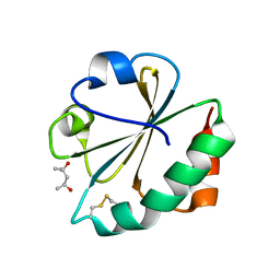
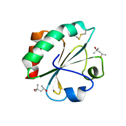
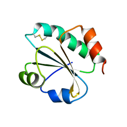
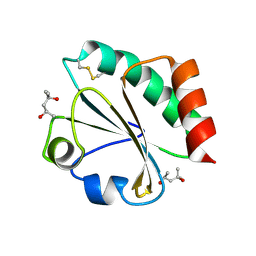
 | | Crystal Structure of Ancestral Thioredoxin Relative to Last Bacteria Common Ancestor (LBCA) from the Precambrian Period | | 分子名称: | 1,2-ETHANEDIOL, LBCA THIOREDOXIN | | 著者 | Gavira, J.A, Ingles-Prieto, A, Ibarra-Molero, B, Sanchez-Ruiz, J.M. | | 登録日 | 2012-09-12 | | 公開日 | 2013-08-21 | | 最終更新日 | 2023-12-20 | | 実験手法 | X-RAY DIFFRACTION (2.45 Å) | | 主引用文献 | Conservation of Protein Structure Over Four Billion Years
Structure, 21, 2013
|
|
4B88
 | |
2H6Z
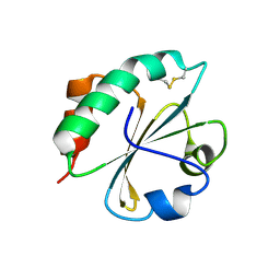
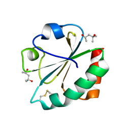
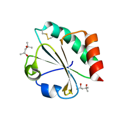
 | | Crystal Structure of Thioredoxin Mutant E44D in Hexagonal (p61) Space Group | | 分子名称: | (4S)-2-METHYL-2,4-PENTANEDIOL, Thioredoxin | | 著者 | Gavira, J.A, Godoy-Ruiz, R, Ibarra-Molero, B, Sanchez-Ruiz, J.M. | | 登録日 | 2006-06-01 | | 公開日 | 2007-05-15 | | 最終更新日 | 2023-08-30 | | 実験手法 | X-RAY DIFFRACTION (2.25 Å) | | 主引用文献 | A stability pattern of protein hydrophobic mutations that reflects evolutionary structural optimization.
Biophys.J., 89, 2005
|
|
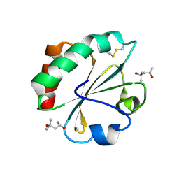
2H70
 | | Crystal Structure of Thioredoxin Mutant D9E in Hexagonal (p61) Space Group | | 分子名称: | (4S)-2-METHYL-2,4-PENTANEDIOL, Thioredoxin | | 著者 | Gavira, J.A, Godoy-Ruiz, R, Ibarra-Molero, B, Sanchez-Ruiz, J.M. | | 登録日 | 2006-06-01 | | 公開日 | 2007-05-15 | | 最終更新日 | 2023-08-30 | | 実験手法 | X-RAY DIFFRACTION (2.7 Å) | | 主引用文献 | A stability pattern of protein hydrophobic mutations that reflects evolutionary structural optimization.
Biophys.J., 89, 2005
|
|
7ZR2
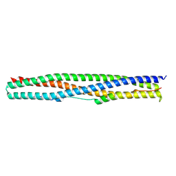 | | Crystal structure of a chimeric protein mimic of SARS-CoV-2 Spike HR1 in complex with HR2 | | 分子名称: | Spike protein S2', Spike protein S2',Chimeric protein mimic of SARS-CoV-2 Spike HR1 | | 著者 | Camara-Artigas, A, Gavira, J.A, Cano-Munoz, M, Polo-Megias, D, Conejero-Lara, F. | | 登録日 | 2022-05-03 | | 公開日 | 2022-11-09 | | 最終更新日 | 2024-01-31 | | 実験手法 | X-RAY DIFFRACTION (1.45 Å) | | 主引用文献 | Novel chimeric proteins mimicking SARS-CoV-2 spike epitopes with broad inhibitory activity.
Int.J.Biol.Macromol., 222, 2022
|
|
2YFA
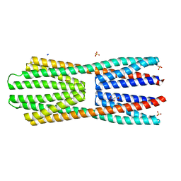 | | X-ray structure of McpS ligand binding domain in complex with malate | | 分子名称: | (2S)-2-hydroxybutanedioic acid, ACETATE ION, METHYL-ACCEPTING CHEMOTAXIS TRANSDUCER, ... | | 著者 | Pineda-Molina, E, Gavira, J.A, Krell, T. | | 登録日 | 2011-04-05 | | 公開日 | 2012-04-18 | | 最終更新日 | 2024-05-08 | | 実験手法 | X-RAY DIFFRACTION (1.8 Å) | | 主引用文献 | Evidence for Chemoreceptors with Bimodular Ligand-Binding Regions Harboring Two Signal-Binding Sites.
Proc.Natl.Acad.Sci.USA, 109, 2012
|
|
3N5F
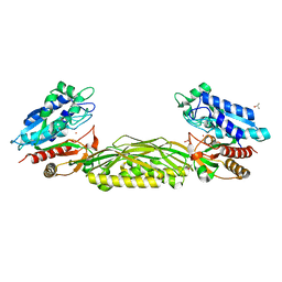 | | Crystal Structure of L-N-carbamoylase from Geobacillus stearothermophilus CECT43 | | 分子名称: | CACODYLATE ION, COBALT (II) ION, ISOPROPYL ALCOHOL, ... | | 著者 | Garcia-Pino, A, Martinez-Rodriguez, S, Gavira, J.A. | | 登録日 | 2010-05-25 | | 公開日 | 2011-05-25 | | 最終更新日 | 2023-09-06 | | 実験手法 | X-RAY DIFFRACTION (2.75 Å) | | 主引用文献 | Mutational and structural analysis of L-N-carbamoylase reveals new insights into a peptidase m20/m25/m40 family member.
J.Bacteriol., 194, 2012
|
|
4F17
 | |


