Loading
PDBj
メニューPDBj@FacebookPDBj@TwitterPDBj@YouTubewwPDB FoundationwwPDB
RCSB PDBPDBeBMRBAdv. SearchSearch help
PDB: 112 件

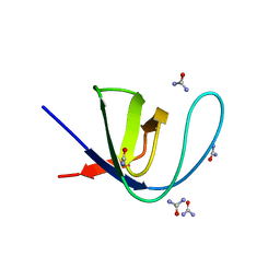
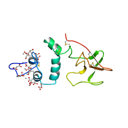
2PF2
DownloadVisualize
BU of 2pf2 by Molmil
THE CA+2 ION AND MEMBRANE BINDING STRUCTURE OF THE GLA DOMAIN OF CA-PROTHROMBIN FRAGMENT 1
分子名称: CALCIUM ION, PROTHROMBIN FRAGMENT 1
著者Soriano-Garcia, M, Padmanabhan, K, De vos, A.M, Tulinsky, A.
登録日1991-12-08
公開日1994-01-31
最終更新日2017-11-29
実験手法X-RAY DIFFRACTION (2.2 Å)
主引用文献The Ca2+ ion and membrane binding structure of the Gla domain of Ca-prothrombin fragment 1.
Biochemistry, 31, 1992
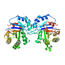
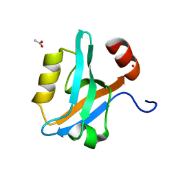
1TCD
DownloadVisualize
BU of 1tcd by Molmil
TRYPANOSOMA CRUZI TRIOSEPHOSPHATE ISOMERASE
分子名称: TRIOSEPHOSPHATE ISOMERASE
著者Maldonado, E, Soriano-Garcia, M, Cabrera, N, Garza-Ramos, G, Tuena De Gomez-Puyou, M, Gomez-Puyou, A, Perez-Montfort, R.
登録日1998-01-29
公開日1999-01-13
最終更新日2024-05-22
実験手法X-RAY DIFFRACTION (1.83 Å)
主引用文献Differences in the intersubunit contacts in triosephosphate isomerase from two closely related pathogenic trypanosomes.
J.Mol.Biol., 283, 1998
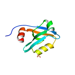
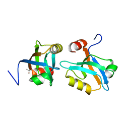
8AH7
DownloadVisualize
BU of 8ah7 by Molmil
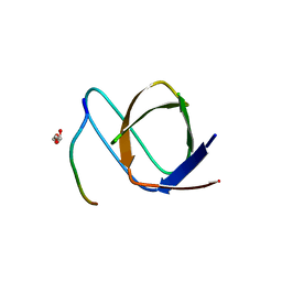
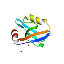
Crystal Structure of the third PDZ domain of PSD-95 protein in the space group P212121 at pH 4.0
分子名称: SULFATE ION, cDNA FLJ50577, highly similar to Discs large homolog 4
著者Camara-Artigas, A, Salinas-Garcia, M.C.
登録日2022-07-20
公開日2023-03-08
最終更新日2024-02-07
実験手法X-RAY DIFFRACTION (1.25 Å)
主引用文献pH-Driven Polymorphic Behaviour of the Third PDZ Domain of PSD95: The Role of Electrostatic Interactions
Crystals, 2023
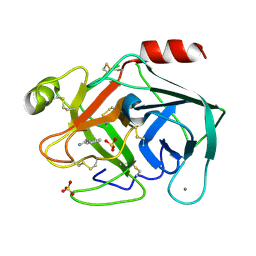
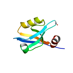
6SUX
DownloadVisualize
BU of 6sux by Molmil
Terahertz irradiated structure of bovine trypsin (even frames of crystal x37)
分子名称: BENZAMIDINE, CALCIUM ION, Cationic trypsin, ...
著者Ahlberg Gagner, V, Lundholm, I, Jose-Garcia, M.J, Rodilla, H, Friedman, R, Zhaunerchyk, V, Bourenkov, G, Schneider, T, Stake, J, Katona, G.
登録日2019-09-17
公開日2020-01-22
最終更新日2020-01-29
実験手法X-RAY DIFFRACTION (1.16 Å)
主引用文献Clustering of atomic displacement parameters in bovine trypsin reveals a distributed lattice of atoms with shared chemical properties.
Sci Rep, 9, 2019
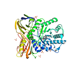
5NDX
DownloadVisualize
BU of 5ndx by Molmil
The bacterial orthologue of Human a-L-iduronidase does not need N-glycan post-translational modifications to be catalytically competent: Crystallography and QM/MM insights into Mucopolysaccharidosis I
分子名称: (2~{R},3~{S},4~{S},5~{R},6~{S})-6-(4-methyl-2-oxidanylidene-chromen-7-yl)oxy-3,4,5-tris(oxidanyl)oxane-2-carboxylic acid, Glycosyl hydrolase, SULFATE ION
著者Raich, L, Valero-Gonzalez, J, Castro-Lopez, J, Millan, C, Jimenez-Garcia, M.J, Nieto, P, Uson, I, Hurtado-Guerrero, R, Rovira, C.
登録日2017-03-09
公開日2018-07-11
最終更新日2024-05-01
実験手法X-RAY DIFFRACTION (2.2 Å)
主引用文献The bacterial orthologue of Human a-L-iduronidase does not need N-glycan post-translational modifications to be catalytically competent: Crystallography and QM/MM insights into Mucopolysaccharidosis I.
To Be Published
6FWW
DownloadVisualize
BU of 6fww by Molmil
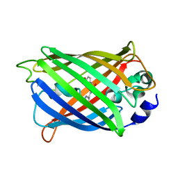
GFP/KKK. A redesigned GFP with improved solubility
分子名称: Green fluorescent protein
著者Varejao, N, Lascorz, J, Gil-Garcia, M, Diaz-Caballero, M, Navarro, S, Ventura, S, Reverter, D.
登録日2018-03-07
公開日2018-08-01
最終更新日2024-01-17
実験手法X-RAY DIFFRACTION (1.131 Å)
主引用文献Combining Structural Aggregation Propensity and Stability Predictions To Redesign Protein Solubility.
Mol. Pharm., 15, 2018
6SYC
DownloadVisualize
BU of 6syc by Molmil
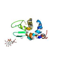
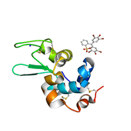
Crystal structure of the lysozyme in presence of bromophenol blue at pH 6.5
分子名称: CHLORIDE ION, IMIDAZOLE, Lysozyme, ...
著者Camara-Artigas, A, Plaza-Garrido, M, Salinas-Garcia, M.C.
登録日2019-09-27
公開日2020-09-09
最終更新日2024-01-24
実験手法X-RAY DIFFRACTION (1.38 Å)
主引用文献Lysozyme crystals dyed with bromophenol blue: where has the dye gone?
Acta Crystallogr D Struct Biol, 76, 2020
6SYE
DownloadVisualize
BU of 6sye by Molmil
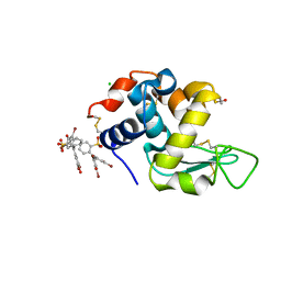
Crystal structure of orthorhombic lysozyme in presence of the dye bromophenol blue at pH 7.0
分子名称: CHLORIDE ION, Lysozyme C, bromophenol blue
著者Camara-Artigas, A, Plaza-Garrido, M, Salinas-Garcia, M.C.
登録日2019-09-27
公開日2020-09-09
最終更新日2024-01-24
実験手法X-RAY DIFFRACTION (0.97 Å)
主引用文献Lysozyme crystals dyed with bromophenol blue: where has the dye gone?
Acta Crystallogr D Struct Biol, 76, 2020
7PVT
DownloadVisualize
BU of 7pvt by Molmil
Crystal structure of the v-Src SH3 domain Q128R mutant in complex with the synthetic peptide VSL12
分子名称: DI(HYDROXYETHYL)ETHER, GLYCEROL, Tyrosine-protein kinase transforming protein Src, ...
著者Camara-Artigas, A, Salinas-Garcia, M.C.
登録日2021-10-05
公開日2022-09-14
最終更新日2024-01-31
実験手法X-RAY DIFFRACTION (1.6 Å)
主引用文献Crystal structure of the v-Src SH3 domain Q128R mutant in complex with the synthetic peptide VSL12
To be published
6SYD
DownloadVisualize
BU of 6syd by Molmil
Crystal structure of the lysozyme in presence of bromophenol blue at pH 5.5
分子名称: Lysozyme, SODIUM ION, bromophenol blue
著者Camara-Artigas, A, Plaza-Garrido, M, Salinas-Garcia, M.C.
登録日2019-09-27
公開日2020-09-09
最終更新日2024-01-24
実験手法X-RAY DIFFRACTION (1.1 Å)
主引用文献Lysozyme crystals dyed with bromophenol blue: where has the dye gone?
Acta Crystallogr D Struct Biol, 76, 2020
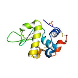
6S7N
DownloadVisualize
BU of 6s7n by Molmil
Crystal structure of orthorhombic lysozyme grown at pH 5.5 with a 26% of solvent content
分子名称: Lysozyme C, SULFATE ION
著者Camara-Artigas, A, Plaza-Garrido, M, Salinas-Garcia, M.C.
登録日2019-07-05
公開日2019-11-20
最終更新日2024-01-24
実験手法X-RAY DIFFRACTION (1.2 Å)
主引用文献Major conformational changes in the structure of lysozyme obtained from a crystal with a very low solvent content.
Acta Crystallogr.,Sect.F, 75, 2019
8AH4
DownloadVisualize
BU of 8ah4 by Molmil
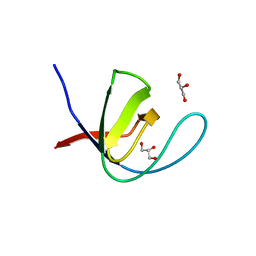
Crystal Structure of the third PDZ domain of PSD-95 protein in the space group P3112 at pH 4.0
分子名称: ACETATE ION, cDNA FLJ50577, highly similar to Discs large homolog 4
著者Camara-Artigas, A, Salinas Garcia, M.C.
登録日2022-07-20
公開日2023-03-08
最終更新日2024-02-07
実験手法X-RAY DIFFRACTION (1.48 Å)
主引用文献pH-Driven Polymorphic Behaviour of the Third PDZ Domain of PSD95: The Role of Electrostatic Interactions
Crystals, 2023
7A36
DownloadVisualize
BU of 7a36 by Molmil
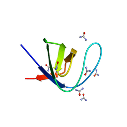
Crystal structure of the c-Src SH3 domain mutant V111L-N113S-T114S in 7 M urea
分子名称: Proto-oncogene tyrosine-protein kinase Src, SODIUM ION, UREA
著者Camara-Artigas, A, Plaza-Garrido, M, Salinas-Garcia, M.C.
登録日2020-08-18
公開日2021-08-25
最終更新日2024-01-31
実験手法X-RAY DIFFRACTION (1.5 Å)
主引用文献Crystal structure of the c-Src SH3 domain mutant V111L-N113S-T114S in 7 M urea
To be published
7A37
DownloadVisualize
BU of 7a37 by Molmil
Crystal structure of the c-Src SH3 domain mutant V111L-N113S-T114S in 3 M urea
分子名称: Proto-oncogene tyrosine-protein kinase Src, UREA
著者Camara-Artigas, A, Plaza-Garrido, M, Salinas-Garcia, M.C.
登録日2020-08-18
公開日2021-08-25
最終更新日2024-01-31
実験手法X-RAY DIFFRACTION (1.52 Å)
主引用文献Crystal structure of the c-Src SH3 domain mutant V111L-N113S-T114S in 3 M urea
To be published
8AH5
DownloadVisualize
BU of 8ah5 by Molmil
Crystal Structure of the third PDZ domain of PSD-95 protein in the space group P212121 at pH 4.6
分子名称: ACETATE ION, cDNA FLJ50577, highly similar to Discs large homolog 4
著者Camara-Artigas, A, Salinas Garcia, M.C.
登録日2022-07-20
公開日2023-03-08
最終更新日2024-02-07
実験手法X-RAY DIFFRACTION (1.25 Å)
主引用文献pH-Driven Polymorphic Behaviour of the Third PDZ Domain of PSD95: The Role of Electrostatic Interactions
Crystals, 2023
8AH6
DownloadVisualize
BU of 8ah6 by Molmil
Crystal Structure of the third PDZ domain of PSD-95 protein in the space group P21 at pH 4.0
分子名称: ACETATE ION, cDNA FLJ50577, highly similar to Discs large homolog 4
著者Camara-Artigas, A, Salinas Garcia, M.C.
登録日2022-07-20
公開日2023-03-08
最終更新日2024-02-07
実験手法X-RAY DIFFRACTION (1.63 Å)
主引用文献pH-Driven Polymorphic Behaviour of the Third PDZ Domain of PSD95: The Role of Electrostatic Interactions
Crystals, 2023
8AH8
DownloadVisualize
BU of 8ah8 by Molmil
Crystal Structure of the third PDZ domain of PSD-95 protein in the space group P3121 at pH 3.7
分子名称: cDNA FLJ50577, highly similar to Discs large homolog 4
著者Camara-Artigas, A, Salinas Garcia, M.C.
登録日2022-07-20
公開日2023-03-08
最終更新日2024-02-07
実験手法X-RAY DIFFRACTION (1.5 Å)
主引用文献pH-Driven Polymorphic Behaviour of the Third PDZ Domain of PSD95: The Role of Electrostatic Interactions
Crystals, 2023
7A3C
DownloadVisualize
BU of 7a3c by Molmil
Crystal structure of the c-Src SH3 domain mutant L100I at pH 3.0
分子名称: GLYCEROL, Proto-oncogene tyrosine-protein kinase Src, SULFATE ION
著者Camara-Artigas, A, Plaza-Garrido, M, Salinas-Garcia, M.C.
登録日2020-08-18
公開日2021-08-25
最終更新日2024-01-31
実験手法X-RAY DIFFRACTION (1.8 Å)
主引用文献Crystal structure of the c-Src SH3 domain mutant L100I at pH 3.0
To be published
7A2N
DownloadVisualize
BU of 7a2n by Molmil
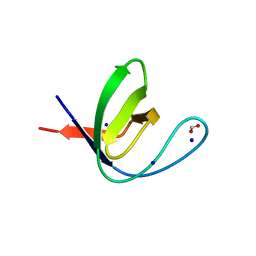
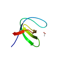
Crystal structure of the Fyn SH3 domain A95S-D99T mutant in space group P21
分子名称: FORMIC ACID, SODIUM ION, Tyrosine-protein kinase Fyn
著者Camara-Artigas, A, Plaza-Garrido, M, Salinas-Garcia, M.C.
登録日2020-08-18
公開日2021-08-25
最終更新日2024-01-31
実験手法X-RAY DIFFRACTION (1.4 Å)
主引用文献Crystal structure of the Fyn SH3 domain A95S-D99T mutant in space group P21
To be published
7A39
DownloadVisualize
BU of 7a39 by Molmil
Crystal structure of the c-Src SH3 domain mutant V111L-N113S-T114S-Q128E at pH 7.0
分子名称: Proto-oncogene tyrosine-protein kinase Src, SULFATE ION
著者Camara-Artigas, A, Plaza-Garrido, M, Salinas-Garcia, M.C.
登録日2020-08-18
公開日2021-08-25
最終更新日2024-01-31
実験手法X-RAY DIFFRACTION (1.65 Å)
主引用文献Crystal structure of the c-Src SH3 domain mutant V111L-N113S-T114S-Q128E at pH 7.0
To be published
7A3E
DownloadVisualize
BU of 7a3e by Molmil
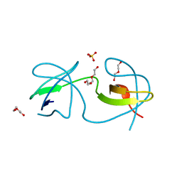
Intertwined dimer of the c-Src SH3 domain mutant T126S
分子名称: DI(HYDROXYETHYL)ETHER, Proto-oncogene tyrosine-protein kinase Src, SULFATE ION, ...
著者Camara-Artigas, A, Plaza-Garrido, M, Salinas-Garcia, M.C.
登録日2020-08-18
公開日2021-08-25
最終更新日2024-01-31
実験手法X-RAY DIFFRACTION (1.52 Å)
主引用文献Intertwined dimer of the c-Src SH3 domain mutant T126S
To be published
7A2T
DownloadVisualize
BU of 7a2t by Molmil
Crystal structure of the Fyn SH3 domain L112V-S114N-S115T-E121L-R123H mutant at pH 4.0
分子名称: ACETATE ION, Tyrosine-protein kinase Fyn
著者Camara-Artigas, A, Plaza-Garrido, M, Salinas-Garcia, M.C.
登録日2020-08-18
公開日2021-08-25
最終更新日2024-01-31
実験手法X-RAY DIFFRACTION (1.22 Å)
主引用文献Crystal structure of the Fyn SH3 domain L112V-S114N-S115T-E121L-R123H mutant at pH 4.0
To be published
7A2Y
DownloadVisualize
BU of 7a2y by Molmil
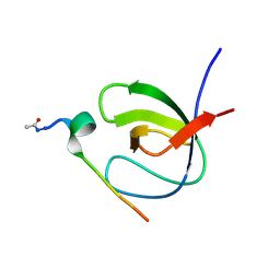
Crystal structure of the Fyn SH3 domain L112V-S114N-S115T-E121L-R123H mutant in complex with VSL12 at pH 4.0
分子名称: Tyrosine-protein kinase Fyn, VSL12
著者Camara-Artigas, A, Plaza-Garrido, M, Salinas-Garcia, M.C.
登録日2020-08-18
公開日2021-08-25
最終更新日2024-01-31
実験手法X-RAY DIFFRACTION (0.97 Å)
主引用文献Crystal structure of the Fyn SH3 domain L112V-S114N-S115T-E121L-R123H mutant in complex with VSL12 at pH 4.0
To be published
7A2Z
DownloadVisualize
BU of 7a2z by Molmil
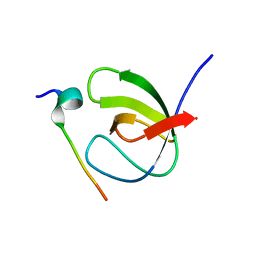
Crystal structure of the Fyn SH3 domain L112V-S114N-S115T-E121L-R123H mutant in complex with VSL12 at pH 6.0
分子名称: Tyrosine-protein kinase Fyn, VSL12
著者Camara-Artigas, A, Plaza-Garrido, M, Salinas-Garcia, M.C.
登録日2020-08-18
公開日2021-08-25
最終更新日2024-01-31
実験手法X-RAY DIFFRACTION (1.14 Å)
主引用文献Crystal structure of the Fyn SH3 domain L112V-S114N-S115T-E121L-R123H mutant in complex with VSL12 at pH 6.0
To be published
7A31
DownloadVisualize
BU of 7a31 by Molmil
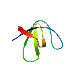
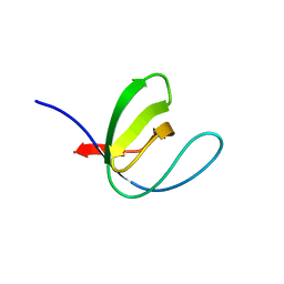
Crystal structure of the c-Src SH3 domain mutant S94A-T98D-V111L-N113S-T114S at pH 4.5
分子名称: Proto-oncogene tyrosine-protein kinase Src
著者Camara-Artigas, A, Plaza-Garrido, M, Salinas-Garcia, M.C.
登録日2020-08-18
公開日2021-08-25
最終更新日2024-01-31
実験手法X-RAY DIFFRACTION (0.94 Å)
主引用文献Crystal structure of the c-Src SH3 domain mutant S94A-T98D-V111L-N113S-T114S at pH 4.5
To be published

222624

件を2024-07-17に公開中

PDB statisticsPDBj update infoContact PDBjnumon