Loading
PDBj
MenuPDBj@FacebookPDBj@X(formerly Twitter)PDBj@BlueSkyPDBj@YouTubewwPDB FoundationwwPDB
RCSB PDBPDBeBMRBAdv. SearchSearch help
PDB: 23 results

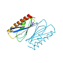
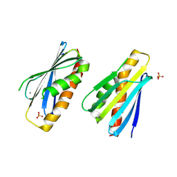
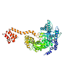
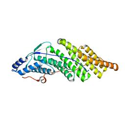
3IIR
DownloadVisualize
BU of 3iir by Molmil
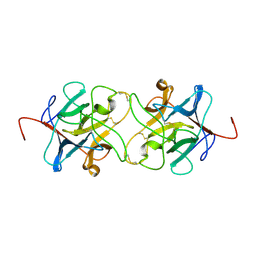
Crystal Structure of Miraculin like protein from seeds of Murraya koenigii
Descriptor: Trypsin inhibitor
Authors:Gahloth, D, Selvakumar, P, Shee, C, Kumar, P, Sharma, A.K.
Deposit date:2009-08-03
Release date:2009-12-08
Last modified:2024-10-30
Method:X-RAY DIFFRACTION (2.9 Å)
Cite:Cloning, sequence analysis and crystal structure determination of a miraculin-like protein from Murraya koenigii
Arch.Biochem.Biophys., 494, 2010
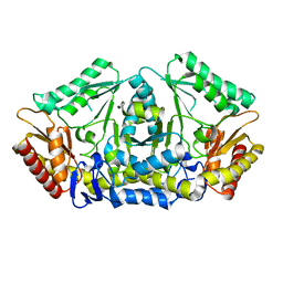
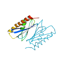
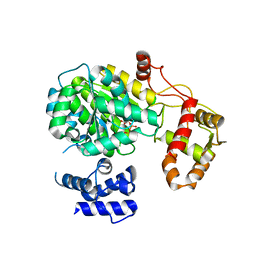
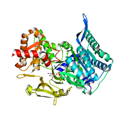
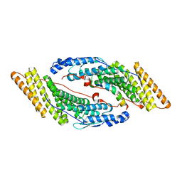
6HX9
DownloadVisualize
BU of 6hx9 by Molmil
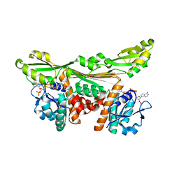
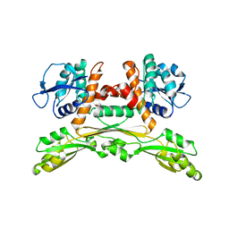
Putrescine transaminase from Pseudomonas putida
Descriptor: Aspartate aminotransferase family protein
Authors:Gahloth, D.
Deposit date:2018-10-16
Release date:2019-06-12
Last modified:2024-01-24
Method:X-RAY DIFFRACTION (2.05 Å)
Cite:Characterization of a Putrescine Transaminase FromPseudomonas putidaand its Application to the Synthesis of Benzylamine Derivatives.
Front Bioeng Biotechnol, 6, 2018
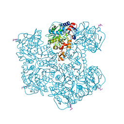
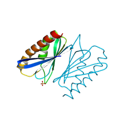
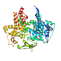
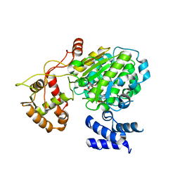
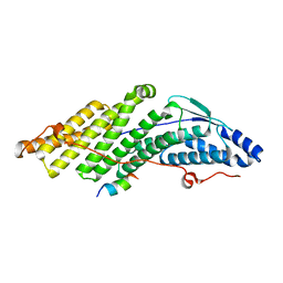
7PDA
DownloadVisualize
BU of 7pda by Molmil
Crystal structure of Phenazine 1-carboxylic acid decarboxylase from Mycobacterium fortuitum
Descriptor: 1-deoxy-5-O-phosphono-1-(3,3,4,5-tetramethyl-9,11-dioxo-2,3,8,9,10,11-hexahydro-7H-quinolino[1,8-fg]pteridin-12-ium-7-y l)-D-ribitol, MANGANESE (II) ION, SODIUM ION, ...
Authors:Gahloth, D, Leys, D.
Deposit date:2021-08-05
Release date:2022-08-24
Last modified:2024-01-31
Method:X-RAY DIFFRACTION (2.65 Å)
Cite:Crystal structure of Phenazine 1-carboxylic acid decarboxylase from Mycobacterium fortuitum
To Be Published
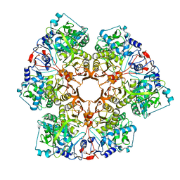
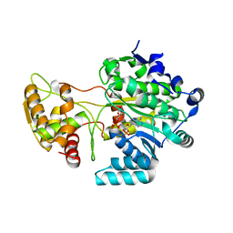
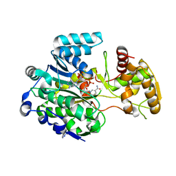
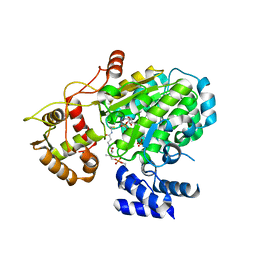
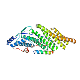
7P9Q
DownloadVisualize
BU of 7p9q by Molmil
Crystal structure of Indole 3-Carboxylic acid decarboxylase from Arthrobacter nicotianae FI1612 in complex with co-factor prFMN.
Descriptor: 1-deoxy-5-O-phosphono-1-(3,3,4,5-tetramethyl-9,11-dioxo-2,3,8,9,10,11-hexahydro-7H-quinolino[1,8-fg]pteridin-12-ium-7-y l)-D-ribitol, AnInD, MANGANESE (II) ION, ...
Authors:Gahloth, D, Leys, D.
Deposit date:2021-07-27
Release date:2022-03-02
Last modified:2024-01-31
Method:X-RAY DIFFRACTION (2.53 Å)
Cite:Structural and biochemical characterization of the prenylated flavin mononucleotide-dependent indole-3-carboxylic acid decarboxylase.
J.Biol.Chem., 298, 2022
8PZO
DownloadVisualize
BU of 8pzo by Molmil
LpdD
Descriptor: Protein LpdD, SODIUM ION
Authors:Gahloth, D, Leys, D.
Deposit date:2023-07-27
Release date:2024-01-17
Method:X-RAY DIFFRACTION (2 Å)
Cite:Structure of LpdD from Lactobacillus plantarum.
To Be Published
8PZH
DownloadVisualize
BU of 8pzh by Molmil
LpdD (H61A) mutant
Descriptor: MANGANESE (II) ION, PHOSPHATE ION, Protein LpdD
Authors:Gahloth, D, Leys, D.
Deposit date:2023-07-27
Release date:2024-01-17
Method:X-RAY DIFFRACTION (2.02 Å)
Cite:Structure of LpdD (H61A) mutant from Lactobacillus plantarum.
To Be Published
8PO5
DownloadVisualize
BU of 8po5 by Molmil
Lactobacillus plantarum LpdD
Descriptor: MANGANESE (II) ION, Protein LpdD
Authors:Gahloth, D, Leys, D.
Deposit date:2023-07-03
Release date:2024-01-17
Method:X-RAY DIFFRACTION (2.1 Å)
Cite:Gallate decarboxylase subunit D, LpdD
To Be Published
8P4W
DownloadVisualize
BU of 8p4w by Molmil
Lactobacillus plantarum LpdD mutant - H35A
Descriptor: PHOSPHATE ION, Protein LpdD
Authors:Gahloth, D, Leys, D.
Deposit date:2023-05-23
Release date:2024-06-12
Method:X-RAY DIFFRACTION (1.78 Å)
Cite:Gallate decarboxylase subunit D, LpdD
To Be Published
5MSO
DownloadVisualize
BU of 5mso by Molmil
Structure of the R domain of carboxylic acid reductase (CAR) from Mycobacterium marinum in complex with NADP
Descriptor: Carboxylic acid reductase, NADP NICOTINAMIDE-ADENINE-DINUCLEOTIDE PHOSPHATE
Authors:Gahloth, D, Leys, D.
Deposit date:2017-01-05
Release date:2017-07-05
Last modified:2024-01-17
Method:X-RAY DIFFRACTION (1.2 Å)
Cite:Structures of carboxylic acid reductase reveal domain dynamics underlying catalysis.
Nat. Chem. Biol., 13, 2017
5MSW
DownloadVisualize
BU of 5msw by Molmil
Structure of the A-PCP didomain of carboxylic acid reductase (CAR) from Segniliparus rugosus in complex with AMP
Descriptor: ADENOSINE MONOPHOSPHATE, Thioester reductase domain-containing protein
Authors:Gahloth, D, Leys, D.
Deposit date:2017-01-05
Release date:2017-07-05
Last modified:2024-01-17
Method:X-RAY DIFFRACTION (2.33 Å)
Cite:Structures of carboxylic acid reductase reveal domain dynamics underlying catalysis.
Nat. Chem. Biol., 13, 2017
5MSP
DownloadVisualize
BU of 5msp by Molmil
Structure of the unmodified PCP-R didomain of carboxylic acid reductase (CAR) from Segniliparus rugosus in complex with NADP, F2221 form
Descriptor: NADP NICOTINAMIDE-ADENINE-DINUCLEOTIDE PHOSPHATE, Thioester reductase domain-containing protein
Authors:Gahloth, D, Leys, D.
Deposit date:2017-01-05
Release date:2017-07-05
Last modified:2024-01-17
Method:X-RAY DIFFRACTION (2.41 Å)
Cite:Structures of carboxylic acid reductase reveal domain dynamics underlying catalysis.
Nat. Chem. Biol., 13, 2017
5MSS
DownloadVisualize
BU of 5mss by Molmil
Structure of the A-PCP didomain of carboxylic acid reductase (CAR) from Segniliparus rugosus in complex with AMP
Descriptor: ADENOSINE MONOPHOSPHATE, SODIUM ION, Thioester reductase domain-containing protein
Authors:Gahloth, D, Leys, D.
Deposit date:2017-01-05
Release date:2017-07-05
Last modified:2024-01-17
Method:X-RAY DIFFRACTION (1.96 Å)
Cite:Structures of carboxylic acid reductase reveal domain dynamics underlying catalysis.
Nat. Chem. Biol., 13, 2017
5MSU
DownloadVisualize
BU of 5msu by Molmil
Structure of the R domain of carboxylic acid reductase (CAR) from Mycobacterium marinum in complex with NADP, P21 form
Descriptor: Carboxylic acid reductase, NADP NICOTINAMIDE-ADENINE-DINUCLEOTIDE PHOSPHATE
Authors:Gahloth, D, Leys, D.
Deposit date:2017-01-05
Release date:2017-07-05
Last modified:2024-01-17
Method:X-RAY DIFFRACTION (1.74 Å)
Cite:Structures of carboxylic acid reductase reveal domain dynamics underlying catalysis.
Nat. Chem. Biol., 13, 2017
5MST
DownloadVisualize
BU of 5mst by Molmil
Structure of the A domain of carboxylic acid reductase (CAR) from Segniliparus rugosus in complex with AMP and a co-purified carboxylic acid
Descriptor: ADENOSINE MONOPHOSPHATE, CALCIUM ION, FUMARIC ACID, ...
Authors:Gahloth, D, Leys, D.
Deposit date:2017-01-05
Release date:2017-07-05
Last modified:2024-01-17
Method:X-RAY DIFFRACTION (1.72 Å)
Cite:Structures of carboxylic acid reductase reveal domain dynamics underlying catalysis.
Nat. Chem. Biol., 13, 2017
5MSR
DownloadVisualize
BU of 5msr by Molmil
Structure of the unmodified PCP-R domain of carboxylic acid reductase (CAR) from Segniliparus rugosus in complex with NADPH, P43 form
Descriptor: NADP NICOTINAMIDE-ADENINE-DINUCLEOTIDE PHOSPHATE, Thioester reductase domain-containing protein
Authors:Gahloth, D, Leys, D.
Deposit date:2017-01-05
Release date:2017-07-05
Last modified:2024-01-17
Method:X-RAY DIFFRACTION (2.37 Å)
Cite:Structures of carboxylic acid reductase reveal domain dynamics underlying catalysis.
Nat. Chem. Biol., 13, 2017
5MSV
DownloadVisualize
BU of 5msv by Molmil
Structure of the phosphopantetheine modified PCP-R didomain of carboxylic acid reductase (CAR) in complex with NADP
Descriptor: 4'-PHOSPHOPANTETHEINE, NADP NICOTINAMIDE-ADENINE-DINUCLEOTIDE PHOSPHATE, Thioester reductase domain-containing protein
Authors:Gahloth, D, Leys, D.
Deposit date:2017-01-05
Release date:2017-07-05
Last modified:2024-01-17
Method:X-RAY DIFFRACTION (2.34 Å)
Cite:Structures of carboxylic acid reductase reveal domain dynamics underlying catalysis.
Nat. Chem. Biol., 13, 2017
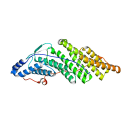
5MJZ
DownloadVisualize
BU of 5mjz by Molmil
Crystal structure of the His Domain Protein Tyrosine Phosphatase (HD-PTP/PTPN23) Bro1 domain (Apo structure)
Descriptor: Tyrosine-protein phosphatase non-receptor type 23
Authors:Levy, C, Gahloth, D.
Deposit date:2016-12-02
Release date:2017-08-16
Last modified:2024-01-17
Method:X-RAY DIFFRACTION (1.867 Å)
Cite:Structural Basis for Specific Interaction of TGF beta Signaling Regulators SARA/Endofin with HD-PTP.
Structure, 25, 2017
5MK2
DownloadVisualize
BU of 5mk2 by Molmil
Crystal structure of the His Domain Protein Tyrosine Phosphatase (HD-PTP/PTPN23) Bro1 domain (CHMP4B peptide complex structure)
Descriptor: Charged multivesicular body protein 4b, Tyrosine-protein phosphatase non-receptor type 23
Authors:Levy, C, Gahloth, D.
Deposit date:2016-12-02
Release date:2017-08-16
Last modified:2024-01-17
Method:X-RAY DIFFRACTION (1.7 Å)
Cite:Structural Basis for Specific Interaction of TGF beta Signaling Regulators SARA/Endofin with HD-PTP.
Structure, 25, 2017
5MJY
DownloadVisualize
BU of 5mjy by Molmil
Crystal structure of the His Domain Protein Tyrosine Phosphatase (HD-PTP/PTPN23) Bro1 domain (SARA complex structure)
Descriptor: Tyrosine-protein phosphatase non-receptor type 23, Zinc finger FYVE domain-containing protein 9
Authors:Levy, C, Gahloth, D.
Deposit date:2016-12-02
Release date:2017-08-16
Last modified:2024-01-17
Method:X-RAY DIFFRACTION (2.25 Å)
Cite:Structural Basis for Specific Interaction of TGF beta Signaling Regulators SARA/Endofin with HD-PTP.
Structure, 25, 2017
5MK1
DownloadVisualize
BU of 5mk1 by Molmil
Crystal structure of the His Domain Protein Tyrosine Phosphatase (HD-PTP/PTPN23) Bro1 domain (CHMP4A peptide complex structure)
Descriptor: Charged multivesicular body protein 4a, Tyrosine-protein phosphatase non-receptor type 23
Authors:Levy, C, Gahloth, D.
Deposit date:2016-12-02
Release date:2017-08-16
Last modified:2024-01-17
Method:X-RAY DIFFRACTION (2.5 Å)
Cite:Structural Basis for Specific Interaction of TGF beta Signaling Regulators SARA/Endofin with HD-PTP.
Structure, 25, 2017
5MK0
DownloadVisualize
BU of 5mk0 by Molmil
Crystal structure of the His Domain Protein Tyrosine Phosphatase (HD-PTP/PTPN23) Bro1 domain (Endofin peptide complex)
Descriptor: Tyrosine-protein phosphatase non-receptor type 23, Zinc finger FYVE domain-containing protein 16
Authors:Levy, C, Gahloth, D.
Deposit date:2016-12-02
Release date:2017-08-16
Last modified:2024-01-17
Method:X-RAY DIFFRACTION (1.765 Å)
Cite:Structural Basis for Specific Interaction of TGF beta Signaling Regulators SARA/Endofin with HD-PTP.
Structure, 25, 2017
5LOA
DownloadVisualize
BU of 5loa by Molmil
Crystal structure of the engineered D-Amino Acid Dehydrogenase (DAADH) bound to NADP+
Descriptor: Meso-diaminopimelate D-dehydrogenase, NADP NICOTINAMIDE-ADENINE-DINUCLEOTIDE PHOSPHATE
Authors:Dunstan, M.S, Gahloth, D.
Deposit date:2016-08-09
Release date:2017-08-16
Last modified:2024-05-08
Method:X-RAY DIFFRACTION (1.84 Å)
Cite:Single-biocatalyst synthesis of enantiopure D-arylalanines exploiting an engineered D-amino acid dehydrogenase
To Be Published
5LOC
DownloadVisualize
BU of 5loc by Molmil
Crystal structure of the engineered D-Amino Acid Dehydrogenase (DAADH)
Descriptor: Meso-diaminopimelate D-dehydrogenase
Authors:Dunstan, M.S, Gahloth, D.
Deposit date:2016-08-09
Release date:2017-08-16
Last modified:2024-05-08
Method:X-RAY DIFFRACTION (2.04 Å)
Cite:Single-biocatalyst synthesis of enantiopure D-arylalanines exploiting an engineered D-amino acid dehydrogenase
To Be Published

227344

건을2024-11-13부터공개중

PDB statisticsPDBj update infoContact PDBjnumon