8H5V
 
 | |
7YRT
 | |
8X1A
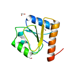
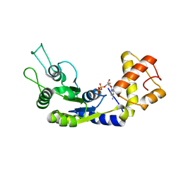
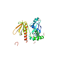
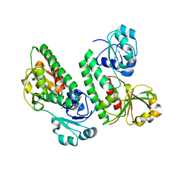
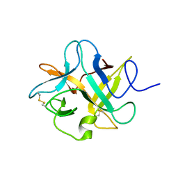
 | | Crystal structure of periplasmic G6P binding protein VcA0625 | | 分子名称: | 6-O-phosphono-alpha-D-glucopyranose, Iron(III) ABC transporter, periplasmic iron-compound-binding protein | | 著者 | Dasgupta, J, Saha, I. | | 登録日 | 2023-11-06 | | 公開日 | 2024-04-17 | | 最終更新日 | 2024-05-22 | | 実験手法 | X-RAY DIFFRACTION (1.604 Å) | | 主引用文献 | Structural insights in to the atypical type-I ABC Glucose-6-phosphate importer VCA0625-27 of Vibrio cholerae.
Biochem.Biophys.Res.Commun., 716, 2024
|
|
6LUA
 | |
6LUF
 | |
5W1O
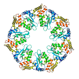
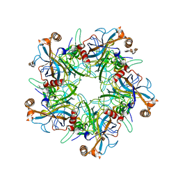
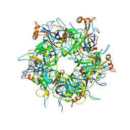 | | Crystal Structure of HPV16 L1 Pentamer Bound to Heparin Oligosaccharides | | 分子名称: | 2-O-sulfo-alpha-L-idopyranuronic acid-(1-4)-2-deoxy-6-O-sulfo-alpha-D-glucopyranose, 2-O-sulfo-alpha-L-idopyranuronic acid-(1-4)-2-deoxy-6-O-sulfo-alpha-D-glucopyranose-(1-4)-2-O-sulfo-alpha-L-idopyranuronic acid-(1-4)-2-deoxy-6-O-sulfo-alpha-D-glucopyranose-(1-4)-2-O-sulfo-alpha-L-idopyranuronic acid-(1-4)-2-deoxy-6-O-sulfo-alpha-D-glucopyranose, Major capsid protein L1 | | 著者 | Dasgupta, J, Chen, X.S. | | 登録日 | 2017-06-04 | | 公開日 | 2017-10-18 | | 最終更新日 | 2023-10-04 | | 実験手法 | X-RAY DIFFRACTION (2.8 Å) | | 主引用文献 | Structural basis of oligosaccharide receptor recognition by human papillomavirus.
J. Biol. Chem., 286, 2011
|
|
4LX8
 | |
3TO5
 | |
1NQP
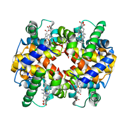
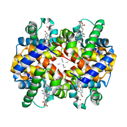
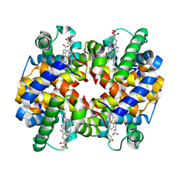 | | Crystal structure of Human hemoglobin E at 1.73 A resolution | | 分子名称: | CYANIDE ION, Hemoglobin alpha chain, Hemoglobin beta chain, ... | | 著者 | Dasgupta, J, Sen, U, Choudhury, D, Dutta, P, Basu, S, Chakrabarti, A, Chakrabarty, A, Dattagupta, J.K. | | 登録日 | 2003-01-22 | | 公開日 | 2004-03-02 | | 最終更新日 | 2023-08-16 | | 実験手法 | X-RAY DIFFRACTION (1.73 Å) | | 主引用文献 | Crystallization and preliminary X-ray structural Studies of Hemoglobin A2 and Hemoglobin E, isolated from the blood samples of Beta-thalassemic patients
Biochem.Biophys.Res.Commun., 303, 2004
|
|
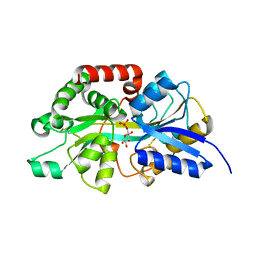
7W61
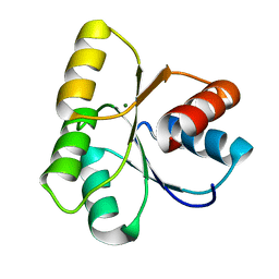
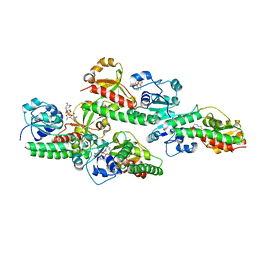
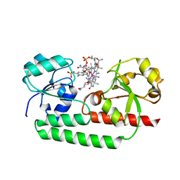
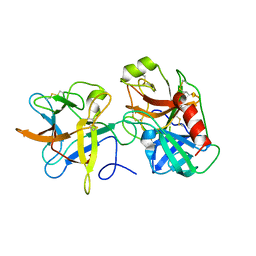
 | | Crystal structure of farnesol dehydrogenase from Helicoverpa armigera | | 分子名称: | 1,2-ETHANEDIOL, ACETATE ION, Farnesol dehydrogenase, ... | | 著者 | Kumar, R, Das, J, Mahto, J.K, Sharma, M, Kumar, P, Sharma, A.K. | | 登録日 | 2021-12-01 | | 公開日 | 2022-07-27 | | 最終更新日 | 2023-11-29 | | 実験手法 | X-RAY DIFFRACTION (1.6 Å) | | 主引用文献 | Crystal structure and molecular characterization of NADP + -farnesol dehydrogenase from cotton bollworm, Helicoverpaarmigera.
Insect Biochem.Mol.Biol., 147, 2022
|
|
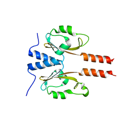
2LMA
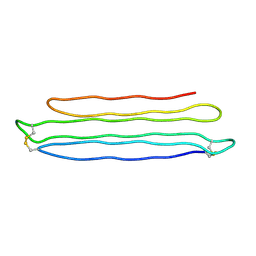
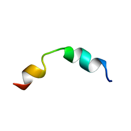 | | Solution structure of CD4+ T cell derived peptide Thp5 | | 分子名称: | Thp5 peptide | | 著者 | Pandey, N.K, Khan, M.M, Chatterjee, S, Dwivedi, V.P, Tousif, S, Das, J, Van Kaer, L. | | 登録日 | 2011-11-29 | | 公開日 | 2011-12-28 | | 最終更新日 | 2024-05-15 | | 実験手法 | SOLUTION NMR | | 主引用文献 | CD4+ T cell derived novel peptide Thp5 induces IL-4 production in CD4+ T cells to direct T helper 2 cell differentiation
J.Biol.Chem., 2011
|
|
4FKD
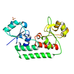
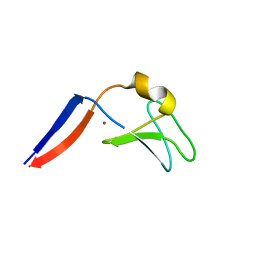 | | Identification of the Activator Binding Residues in the Second Cysteine-Rich Regulatory Domain of Protein Kinase C Theta | | 分子名称: | Protein kinase C theta type, ZINC ION | | 著者 | Rahman, G.M, Shanker, S, Lewin, N.E, Prasad, B.V.V, Blumberg, P.M, Das, J. | | 登録日 | 2012-06-13 | | 公開日 | 2013-01-23 | | 最終更新日 | 2024-02-28 | | 実験手法 | X-RAY DIFFRACTION (1.633 Å) | | 主引用文献 | Identification of the Activator Binding Residues in the Second Cysteine-Rich Regulatory Domain of Protein Kinase C Theta.
Biochem.J., 451, 2013
|
|
7CT1
 | |
5GGX
 | |
5GGY
 | |
5W1X
 | | Crystal Structure of Humanpapillomavirus18 (HPV18) Capsid L1 Pentamers Bound to Heparin Oligosaccharides | | 分子名称: | 2-O-sulfo-alpha-L-idopyranuronic acid-(1-4)-2-deoxy-6-O-sulfo-alpha-D-glucopyranose, 2-O-sulfo-alpha-L-idopyranuronic acid-(1-4)-2-deoxy-6-O-sulfo-alpha-D-glucopyranose-(1-4)-2-O-sulfo-alpha-L-idopyranuronic acid-(1-4)-2-deoxy-6-O-sulfo-alpha-D-glucopyranose, 2-O-sulfo-alpha-L-idopyranuronic acid-(1-4)-2-deoxy-6-O-sulfo-alpha-D-glucopyranose-(1-4)-2-O-sulfo-alpha-L-idopyranuronic acid-(1-4)-2-deoxy-6-O-sulfo-alpha-D-glucopyranose-(1-4)-2-O-sulfo-alpha-L-idopyranuronic acid-(1-4)-2-deoxy-6-O-sulfo-alpha-D-glucopyranose, ... | | 著者 | Chen, X.S, Dasgupta, J. | | 登録日 | 2017-06-05 | | 公開日 | 2018-12-12 | | 最終更新日 | 2023-10-04 | | 実験手法 | X-RAY DIFFRACTION (3.374 Å) | | 主引用文献 | Structural basis of oligosaccharide receptor recognition by human papillomavirus.
J. Biol. Chem., 286, 2011
|
|
5YSC
 | |
5KHL
 | |
3I2X
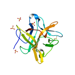
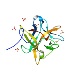
 | | Crystal structure of a chimeric trypsin inhibitor having reactive site loop of ETI on the scaffold of WCI | | 分子名称: | Chymotrypsin inhibitor 3 | | 著者 | Sen, U, Khamrui, S, Dasgupta, J, Dattagupta, J.K, Majumder, S. | | 登録日 | 2009-06-30 | | 公開日 | 2010-02-02 | | 最終更新日 | 2023-11-01 | | 実験手法 | X-RAY DIFFRACTION (2.85 Å) | | 主引用文献 | Identification of a novel set of scaffolding residues that are instrumental for the inhibitory property of Kunitz (STI) inhibitors.
Protein Sci., 19, 2010
|
|
3I29
 | | Crystal structure of a binary complex between an mutant trypsin inhibitor with bovine trypsin | | 分子名称: | CALCIUM ION, Cationic trypsin, Chymotrypsin inhibitor 3 | | 著者 | Khamrui, S, Majumder, S, Dasgupta, J, Dattagupta, J.K, Sen, U. | | 登録日 | 2009-06-29 | | 公開日 | 2010-06-16 | | 最終更新日 | 2023-11-01 | | 実験手法 | X-RAY DIFFRACTION (2.4 Å) | | 主引用文献 | Role of remote scaffolding residues in the inhibitory loop pre-organization, flexibility, rigidification and enzyme inhibition of serine protease inhibitors
Biochim.Biophys.Acta, 1824, 2012
|
|
1SI4
 | | Crystal structure of Human hemoglobin A2 (in R2 state) at 2.2 A resolution | | 分子名称: | CYANIDE ION, Hemoglobin alpha chain, Hemoglobin delta chain, ... | | 著者 | Sen, U, Dasgupta, J, Choudhury, D, Datta, P, Chakrabarti, A, Chakrabarty, S.B, Chakrabarty, A, Dattagupta, J.K. | | 登録日 | 2004-02-27 | | 公開日 | 2004-10-26 | | 最終更新日 | 2023-10-25 | | 実験手法 | X-RAY DIFFRACTION (2.2 Å) | | 主引用文献 | Crystal structures of HbA2 and HbE and modeling of hemoglobin delta4: interpretation of the thermal stability and the antisickling effect of HbA2 and identification of the ferrocyanide binding site in Hb.
Biochemistry, 43, 2004
|
|
1FN0
 | | STRUCTURE OF A MUTANT WINGED BEAN CHYMOTRYPSIN INHIBITOR PROTEIN, N14D. | | 分子名称: | CHYMOTRYPSIN INHIBITOR 3, SULFATE ION | | 著者 | Dattagupta, J.K, Chakrabarti, C, Ravichandran, S, Dasgupta, J, Ghosh, S. | | 登録日 | 2000-08-19 | | 公開日 | 2001-02-19 | | 最終更新日 | 2021-11-03 | | 実験手法 | X-RAY DIFFRACTION (2 Å) | | 主引用文献 | The role of Asn14 in the stability and conformation of the reactive-site loop of winged bean chymotrypsin inhibitor: crystal structures of two point mutants Asn14-->Lys and Asn14-->Asp.
PROTEIN ENG., 14, 2001
|
|
1SHR
 | | Crystal structure of ferrocyanide bound human hemoglobin A2 at 1.88A resolution | | 分子名称: | CYANIDE ION, FE (III) ION, Hemoglobin alpha chain, ... | | 著者 | Sen, U, Dasgupta, J, Choudhury, D, Datta, P, Chakrabarti, A, Chakrabarty, S.B, Chakrabarty, A, Dattagupta, J.K. | | 登録日 | 2004-02-26 | | 公開日 | 2004-10-26 | | 最終更新日 | 2023-10-25 | | 実験手法 | X-RAY DIFFRACTION (1.88 Å) | | 主引用文献 | Crystal structures of HbA2 and HbE and modeling of hemoglobin delta4: interpretation of the thermal stability and the antisickling effect of HbA2 and identification of the ferrocyanide binding site in Hb
Biochemistry, 43, 2004
|
|
1FMZ
 | | CRYSTAL STRUCTURE OF A MUTANT WINGED BEAN CHYMOTRYPSIN INHIBITOR PROTEIN, N14K. | | 分子名称: | CHYMOTRYPSIN INHIBITOR 3, SULFATE ION | | 著者 | Dattagupta, J.K, Chakrabarti, C, Ravichandran, S, Dasgupta, J, Ghosh, S. | | 登録日 | 2000-08-19 | | 公開日 | 2001-02-19 | | 最終更新日 | 2021-11-03 | | 実験手法 | X-RAY DIFFRACTION (2.05 Å) | | 主引用文献 | The role of Asn14 in the stability and conformation of the reactive-site loop of winged bean chymotrypsin inhibitor: crystal structures of two point mutants Asn14-->Lys and Asn14-->Asp.
PROTEIN ENG., 14, 2001
|
|
2PNE
 | | Crystal Structure of the Snow Flea Antifreeze Protein | | 分子名称: | 6.5 kDa glycine-rich antifreeze protein | | 著者 | Pentelute, B.L, Kent, S.B.H, Gates, Z.P, Tereshko, V, Kossiakoff, A.A, Kurutz, J, Dashnau, J, Vaderkooi, J.M. | | 登録日 | 2007-04-24 | | 公開日 | 2008-04-29 | | 最終更新日 | 2023-08-30 | | 実験手法 | X-RAY DIFFRACTION (0.98 Å) | | 主引用文献 | X-ray structure of snow flea antifreeze protein determined by racemic crystallization of synthetic protein enantiomers
J.Am.Chem.Soc., 130, 2008
|
|


