Loading
PDBj
MenuPDBj@FacebookPDBj@TwitterPDBj@YouTubewwPDB FoundationwwPDB
RCSB PDBPDBeBMRBAdv. SearchSearch help
PDB: 60 results

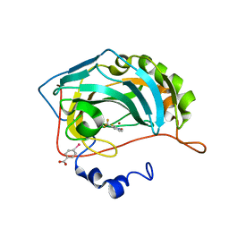
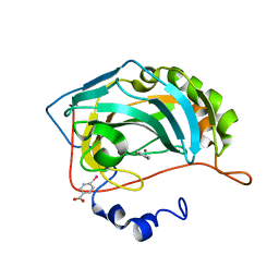
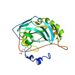
4Q8Y
DownloadVisualize
BU of 4q8y by Molmil
Crystal structure of 2-hydroxyisoquinoline-1(2H)-thione bound to human carbonic anhydrase II
Descriptor: 2-hydroxyisoquinoline-1(2H)-thione, Carbonic anhydrase 2, MERCURIBENZOIC ACID, ...
Authors:Martin, D.P, Cohen, S.M.
Deposit date:2014-04-28
Release date:2015-03-11
Last modified:2023-09-20
Method:X-RAY DIFFRACTION (1.45 Å)
Cite:Exploring the influence of the protein environment on metal-binding pharmacophores.
J.Med.Chem., 57, 2014
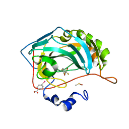
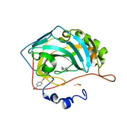
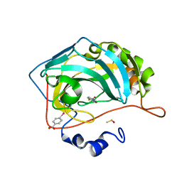
4Q7V
DownloadVisualize
BU of 4q7v by Molmil
Crystal structure of 1-hydroxy-5-methylpyridine-2(1H)-thione bound to human carbonic anhydrase II
Descriptor: 1-hydroxy-5-methylpyridine-2(1H)-thione, Carbonic anhydrase 2, DIMETHYL SULFOXIDE, ...
Authors:Martin, D.P, Cohen, S.M.
Deposit date:2014-04-25
Release date:2015-03-11
Last modified:2023-09-20
Method:X-RAY DIFFRACTION (1.6 Å)
Cite:Exploring the influence of the protein environment on metal-binding pharmacophores.
J.Med.Chem., 57, 2014
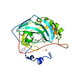
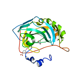
4Q83
DownloadVisualize
BU of 4q83 by Molmil
Crystal structure of 1-hydroxy-3-(trifluoromethyl)pyridine-2(1H)-thione bound to human carbonic anhydrase II
Descriptor: 1-hydroxy-3-(trifluoromethyl)pyridine-2(1H)-thione, Carbonic anhydrase 2, MERCURIBENZOIC ACID, ...
Authors:Martin, D.P, Cohen, S.M.
Deposit date:2014-04-25
Release date:2015-03-11
Last modified:2023-09-20
Method:X-RAY DIFFRACTION (1.55 Å)
Cite:Exploring the influence of the protein environment on metal-binding pharmacophores.
J.Med.Chem., 57, 2014
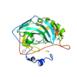
4Q8X
DownloadVisualize
BU of 4q8x by Molmil
Crystal structure of 1-hydroxy-5-(trifluoromethyl)pyridine-2(1H)-thione bound to human carbonic anhydrase II
Descriptor: 1-hydroxy-5-(trifluoromethyl)pyridine-2(1H)-thione, Carbonic anhydrase 2, DIMETHYL SULFOXIDE, ...
Authors:Martin, D.P, Cohen, S.M.
Deposit date:2014-04-28
Release date:2015-03-11
Last modified:2023-09-20
Method:X-RAY DIFFRACTION (1.55 Å)
Cite:Exploring the influence of the protein environment on metal-binding pharmacophores.
J.Med.Chem., 57, 2014
4Q99
DownloadVisualize
BU of 4q99 by Molmil
Crystal structure of 2-mercapto-4-methylphenol bound to human carbonic anhydrase II
Descriptor: 4-methyl-2-sulfanylphenol, Carbonic anhydrase 2, DIMETHYL SULFOXIDE, ...
Authors:Martin, D.P, Cohen, S.M.
Deposit date:2014-04-29
Release date:2015-03-11
Last modified:2023-09-20
Method:X-RAY DIFFRACTION (1.5 Å)
Cite:Exploring the influence of the protein environment on metal-binding pharmacophores.
J.Med.Chem., 57, 2014
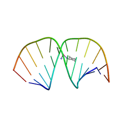
1LU5
DownloadVisualize
BU of 1lu5 by Molmil
2.4 Angstrom Crystal Structure of the Asymmetric Platinum Complex {Pt(ammine)(cyclohexylamine)}2+ Bound to a Dodecamer DNA Duplex
Descriptor: 5'-D(*CP*CP*TP*CP*TP*GP*GP*TP*CP*TP*CP*C)-3', 5'-D(*GP*GP*AP*GP*AP*CP*CP*AP*GP*AP*GP*G)-3', CIS-(AMMINE)(CYCLOHEXYLAMINE)PLATINUM(II) COMPLEX
Authors:Silverman, A.P, Bu, W, Cohen, S.M, Lippard, S.J.
Deposit date:2002-05-21
Release date:2002-12-20
Last modified:2024-02-14
Method:X-RAY DIFFRACTION (2.4 Å)
Cite:2.4-A Crystal Structure of the Asymmetric Platinum Complex {Pt(ammine)(cyclohexylamine)}2+ Bound to a Dodecamer DNA Duplex
J.Biol.Chem., 277, 2002
4Q7S
DownloadVisualize
BU of 4q7s by Molmil
Crystal Structure of 1-hydroxy-4-methylpyridine-2(1H)-thione bound to human carbonic anhydrase II
Descriptor: 1-hydroxy-4-methylpyridine-2(1H)-thione, Carbonic anhydrase 2, MERCURIBENZOIC ACID, ...
Authors:Martin, D.P, Cohen, S.M.
Deposit date:2014-04-25
Release date:2015-03-11
Last modified:2023-09-20
Method:X-RAY DIFFRACTION (1.8 Å)
Cite:Exploring the influence of the protein environment on metal-binding pharmacophores.
J.Med.Chem., 57, 2014
4Q81
DownloadVisualize
BU of 4q81 by Molmil
Crystal structure of 1-hydroxy-4,6-dimethylpyridine-2(1H)-thione bound to human carbonic anhydrase II
Descriptor: 1-hydroxy-4,6-dimethylpyridine-2(1H)-thione, Carbonic anhydrase 2, DIMETHYL SULFOXIDE, ...
Authors:Martin, D.P, Cohen, S.M.
Deposit date:2014-04-25
Release date:2015-03-11
Last modified:2023-09-20
Method:X-RAY DIFFRACTION (1.55 Å)
Cite:Exploring the influence of the protein environment on metal-binding pharmacophores.
J.Med.Chem., 57, 2014
4Q8Z
DownloadVisualize
BU of 4q8z by Molmil
Crystal structure of 1-hydroxy-4-methylpyridin-2(1H)-one bound to human carbonic anhydrase II
Descriptor: 1-hydroxy-4-methylpyridin-2(1H)-one, Carbonic anhydrase 2, MERCURIBENZOIC ACID, ...
Authors:Martin, D.P, Cohen, S.M.
Deposit date:2014-04-28
Release date:2015-03-11
Last modified:2023-09-20
Method:X-RAY DIFFRACTION (1.5 Å)
Cite:Exploring the influence of the protein environment on metal-binding pharmacophores.
J.Med.Chem., 57, 2014
4Q9Y
DownloadVisualize
BU of 4q9y by Molmil
Crystal structure of 3-methylthiophenol bound to human carbonic anhydrase II
Descriptor: 3-methylbenzenethiol, Carbonic anhydrase 2, DIMETHYL SULFOXIDE, ...
Authors:Martin, D.P, Cohen, S.M.
Deposit date:2014-05-02
Release date:2015-03-11
Last modified:2023-09-20
Method:X-RAY DIFFRACTION (1.55 Å)
Cite:Exploring the influence of the protein environment on metal-binding pharmacophores.
J.Med.Chem., 57, 2014
<123


226707

건을2024-10-30부터공개중

PDB statisticsPDBj update infoContact PDBjnumon