4B62
 
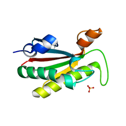 | | The structure of the cell wall anchor of the T6SS from Pseudomonas aeruginosa | | 分子名称: | 1,2-ETHANEDIOL, PHOSPHATE ION, TSSL1 | | 著者 | Robb, C.S, Carlson, M, Nano, F.E, Boraston, A.B. | | 登録日 | 2012-08-08 | | 公開日 | 2013-10-02 | | 最終更新日 | 2023-12-20 | | 実験手法 | X-RAY DIFFRACTION (1.45 Å) | | 主引用文献 | Crystal Structure of the Periplasmic Peptidoglycan Binding Anchor of a T6Ss from Pseudomonas Aeruginosa.
To be Published
|
|
2NYE
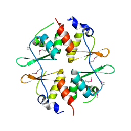 | | Crystal structure of the Bateman2 domain of yeast Snf4 | | 分子名称: | Nuclear protein SNF4 | | 著者 | Rudolph, M.J, Amodeo, G.A, Iram, S, Hong, S, Pirino, G, Carlson, M, Tong, L. | | 登録日 | 2006-11-20 | | 公開日 | 2006-12-12 | | 最終更新日 | 2023-11-15 | | 実験手法 | X-RAY DIFFRACTION (2.5 Å) | | 主引用文献 | Structure of the Bateman2 domain of yeast Snf4: dimeric association and relevance for AMP binding.
Structure, 15, 2007
|
|
2NYC
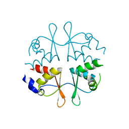 | | Crystal structure of the Bateman2 domain of yeast Snf4 | | 分子名称: | Nuclear protein SNF4 | | 著者 | Rudolph, M.J, Amodeo, G.A, Iram, S, Hong, S, Pirino, G, Carlson, M, Tong, L. | | 登録日 | 2006-11-20 | | 公開日 | 2006-12-12 | | 最終更新日 | 2023-12-27 | | 実験手法 | X-RAY DIFFRACTION (1.9 Å) | | 主引用文献 | Structure of the Bateman2 domain of yeast Snf4: dimeric association and relevance for AMP binding.
Structure, 15, 2007
|
|


