Loading
PDBj
MenuPDBj@FacebookPDBj@TwitterPDBj@YouTubewwPDB FoundationwwPDB
RCSB PDBPDBeBMRBAdv. SearchSearch help
PDB: 185 results

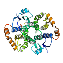
1U88
DownloadVisualize
BU of 1u88 by Molmil
Crystal Structure Of The 26 Kda Glutathione S-Transferase Y7F mutant From Schistosoma Japonicum Complexed With S-Octyl Glutathione
Descriptor: Glutathione S-transferase 26 kDa, L-GAMMA-GLUTAMYL-S-OCTYL-D-CYSTEINYLGLYCINE
Authors:Smith, A.W, Camara-Artigas, A.
Deposit date:2004-08-05
Release date:2005-02-22
Last modified:2024-04-03
Method:X-RAY DIFFRACTION (3.5 Å)
Cite:Crystallographic and Thermodynamic Analysis of the Binding of S-Octylglutathione to the Tyr 7 to Phe Mutant of Glutathione S-Transferase from Schistosoma japonicum(,)
Biochemistry, 44, 2005
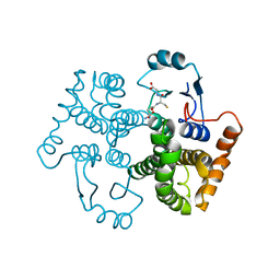
1U87
DownloadVisualize
BU of 1u87 by Molmil
Crystal Structure Of The 26 Kda Glutathione S-Transferase Y7F mutant From Schistosoma Japonicum Complexed With Glutathione
Descriptor: GLUTATHIONE, Glutathione S-Transferase 26 kDa
Authors:Smith, A.W, Camara-Artigas, A.
Deposit date:2004-08-05
Release date:2005-02-22
Last modified:2023-10-25
Method:X-RAY DIFFRACTION (3.5 Å)
Cite:Crystallographic and Thermodynamic Analysis of the Binding of S-Octylglutathione to the Tyr 7 to Phe Mutant of Glutathione S-Transferase from Schistosoma japonicum(,)
Biochemistry, 44, 2005
1Z9J
DownloadVisualize
BU of 1z9j by Molmil
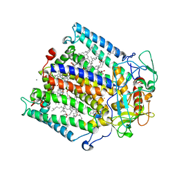
Photosynthetic Reaction Center from Rhodobacter sphaeroides
Descriptor: BACTERIOCHLOROPHYLL A, BACTERIOPHEOPHYTIN A, FE (III) ION, ...
Authors:Thielges, M, Uyeda, G, Camara-Artigas, A, Kalman, L, Williams, J.C, Allen, J.P.
Deposit date:2005-04-02
Release date:2005-06-07
Last modified:2023-08-23
Method:X-RAY DIFFRACTION (4.5 Å)
Cite:Design of a Redox-Linked Active Metal Site: Manganese Bound to Bacterial Reaction Centers at a Site Resembling That of Photosystem II
Biochemistry, 44, 2005
3UA7
DownloadVisualize
BU of 3ua7 by Molmil
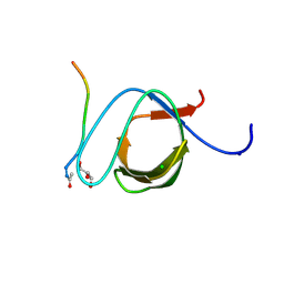
Crystal Structure of the Human Fyn SH3 domain in complex with a peptide from the Hepatitis C virus NS5A-protein
Descriptor: CHLORIDE ION, FORMIC ACID, GLYCEROL, ...
Authors:Martin-Garcia, J.M, Ruiz-Sanz, J, Luque, I, Camara-Artigas, A.
Deposit date:2011-10-21
Release date:2012-07-25
Last modified:2024-10-30
Method:X-RAY DIFFRACTION (1.5 Å)
Cite:The promiscuous binding of the Fyn SH3 domain to a peptide from the NS5A protein.
Acta Crystallogr.,Sect.D, 68, 2012
3M0U
DownloadVisualize
BU of 3m0u by Molmil
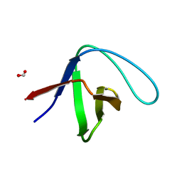
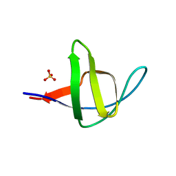
Crystal Structure of the R21D mutant of alpha-spectrin SH3 domain. Hexagonal crystal obtained in sodium formate at pH 6.5.
Descriptor: FORMIC ACID, Spectrin alpha chain, brain
Authors:Gavira, J.A, Camara-Artigas, A.
Deposit date:2010-03-03
Release date:2011-01-19
Last modified:2023-09-06
Method:X-RAY DIFFRACTION (1.1 Å)
Cite:Understanding the polymorphic behaviour of a mutant of the alpha-spectrin SH3 domain by means of two 1.1 A structures
Acta Crystallogr.,Sect.D, 2011
3GYR
DownloadVisualize
BU of 3gyr by Molmil
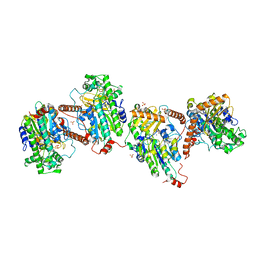
Structure of Phenoxazinone synthase from Streptomyces antibioticus reveals a new type 2 copper center.
Descriptor: COPPER (II) ION, CU-O-CU LINKAGE, GLYCEROL, ...
Authors:Smith, A.W, Camara-Artigas, A, Wang, M, Francisco, W.A, Allen, J.P.
Deposit date:2009-04-05
Release date:2009-04-28
Last modified:2023-09-06
Method:X-RAY DIFFRACTION (2.3 Å)
Cite:Structure of Phenoxazinone Synthase from Streptomyces Antibioticus Reveals a New Type 2 Copper Center.
Biochemistry, 45, 2006
3M0R
DownloadVisualize
BU of 3m0r by Molmil
Crystal Structure of the R21D mutant of alpha-spectrin SH3 domain. Crystal obtained in ammonium sulphate at pH 6.
Descriptor: SULFATE ION, Spectrin alpha chain, brain
Authors:Gavira, J.A, Camara-Artigas, A.
Deposit date:2010-03-03
Release date:2011-01-19
Last modified:2023-09-06
Method:X-RAY DIFFRACTION (1.1 Å)
Cite:Understanding the polymorphic behaviour of a mutant of the alpha-spectrin SH3 domain by means of two 1.1 A structures
Acta Crystallogr.,Sect.D, 2011
3ENI
DownloadVisualize
BU of 3eni by Molmil
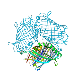
Crystal structure of the Fenna-Matthews-Olson Protein from Chlorobaculum Tepidum
Descriptor: BACTERIOCHLOROPHYLL A, Bacteriochlorophyll a protein
Authors:Tronrud, D, Camara-Artigas, A, Blankenship, R, Allen, J.P.
Deposit date:2008-09-25
Release date:2009-05-12
Last modified:2024-02-21
Method:X-RAY DIFFRACTION (2.2 Å)
Cite:The structural basis for the difference in absorbance spectra for the FMO antenna protein from various green sulfur bacteria.
Photosynth.Res., 100, 2009
6G60
DownloadVisualize
BU of 6g60 by Molmil
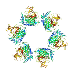
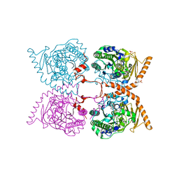
Choline sulfatase from Ensifer (Sinorhizobium) meliloti cocrystalized with choline
Descriptor: CHOLINE ION, Choline-sulfatase, MAGNESIUM ION, ...
Authors:Martinez-Rodriguez, S, Camara-Artigas, A.
Deposit date:2018-03-30
Release date:2019-04-10
Last modified:2023-03-08
Method:X-RAY DIFFRACTION (1.84 Å)
Cite:Structural insights into choline-O-sulfatase reveal the molecular determinants for ligand binding.
Acta Crystallogr D Struct Biol, 78, 2022
6G5Z
DownloadVisualize
BU of 6g5z by Molmil
Choline sulfatase from Ensifer (Sinorhizobium) meliloti
Descriptor: Choline-sulfatase, MAGNESIUM ION, SULFATE ION
Authors:Martinez-Rodriguez, S, Camara-Artigas, A.
Deposit date:2018-03-30
Release date:2019-04-10
Last modified:2024-10-23
Method:X-RAY DIFFRACTION (1.98 Å)
Cite:Structural insights into choline-O-sulfatase reveal the molecular determinants for ligand binding.
Acta Crystallogr D Struct Biol, 78, 2022

226707

PDB entries from 2024-10-30

PDB statisticsPDBj update infoContact PDBjnumon