1B95
 
 | |
1BD9
 | |
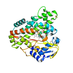
1BEH
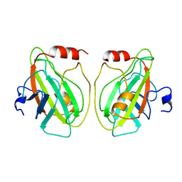
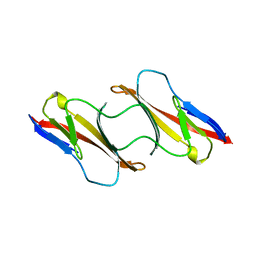
 | | HUMAN PHOSPHATIDYLETHANOLAMINE BINDING PROTEIN IN COMPLEX WITH CACODYLATE | | 分子名称: | CACODYLATE ION, PHOSPHATIDYLETHANOLAMINE BINDING PROTEIN | | 著者 | Banfield, M.J, Barker, J.J, Perry, A, Brady, R.L. | | 登録日 | 1998-05-14 | | 公開日 | 1998-09-16 | | 最終更新日 | 2023-08-02 | | 実験手法 | X-RAY DIFFRACTION (1.75 Å) | | 主引用文献 | Function from structure? The crystal structure of human phosphatidylethanolamine-binding protein suggests a role in membrane signal transduction.
Structure, 6, 1998
|
|
3PR7
 | |
1CDC
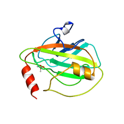
 | | CD2, N-TERMINAL DOMAIN (1-99), TRUNCATED FORM | | 分子名称: | CD2 | | 著者 | Murray, A.J, Barclay, A.N, Brady, R.L. | | 登録日 | 1995-05-23 | | 公開日 | 1995-09-15 | | 最終更新日 | 2024-02-07 | | 実験手法 | X-RAY DIFFRACTION (2 Å) | | 主引用文献 | One sequence, two folds: a metastable structure of CD2.
Proc.Natl.Acad.Sci.USA, 92, 1995
|
|
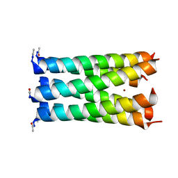
3R3K
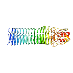
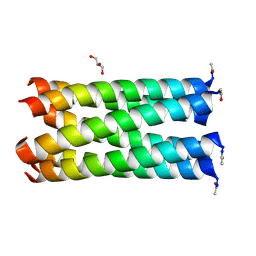
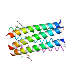 | | Crystal structure of a parallel 6-helix coiled coil | | 分子名称: | 1,2-ETHANEDIOL, CChex-Phi22 helix, CHLORIDE ION, ... | | 著者 | Zaccai, N.R, Chi, B.H.C, Woolfson, D.N, Brady, R.L. | | 登録日 | 2011-03-16 | | 公開日 | 2011-11-16 | | 最終更新日 | 2011-11-30 | | 実験手法 | X-RAY DIFFRACTION (2.2009 Å) | | 主引用文献 | A de novo peptide hexamer with a mutable channel.
Nat.Chem.Biol., 7, 2011
|
|
3R46
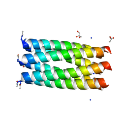 | | Crystal structure of a parallel 6-helix coiled coil CC-hex-D24 | | 分子名称: | CHLORIDE ION, GLYCEROL, SODIUM ION, ... | | 著者 | Zaccai, N.R, Chi, B.H.C, Woolfson, D.N, Brady, R.L. | | 登録日 | 2011-03-17 | | 公開日 | 2011-11-16 | | 最終更新日 | 2023-09-13 | | 実験手法 | X-RAY DIFFRACTION (1.751 Å) | | 主引用文献 | A de novo peptide hexamer with a mutable channel.
Nat.Chem.Biol., 7, 2011
|
|
3R4A
 | |
3R48
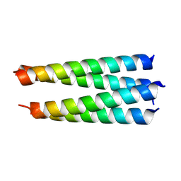
 | | Crystal structure of a hetero-hexamer coiled coil | | 分子名称: | GLYCEROL, coiled coil helix W22-L24H, coiled coil helix Y15-L24D | | 著者 | Zaccai, N.R, Chi, B.H.C, Woolfson, D.N, Brady, R.L. | | 登録日 | 2011-03-17 | | 公開日 | 2011-11-16 | | 最終更新日 | 2023-09-13 | | 実験手法 | X-RAY DIFFRACTION (2.0011 Å) | | 主引用文献 | A de novo peptide hexamer with a mutable channel.
Nat.Chem.Biol., 7, 2011
|
|
3R4H
 | |
3RA3
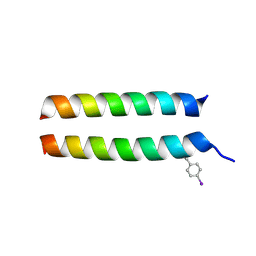 | | Crystal structure of a section of a de novo design gigaDalton protein fibre | | 分子名称: | SODIUM ION, p1c, p2f | | 著者 | Zaccai, N.R, Sharp, T.H, Bruning, M, Woolfson, D.N, Brady, R.L. | | 登録日 | 2011-03-27 | | 公開日 | 2012-08-08 | | 最終更新日 | 2013-06-19 | | 実験手法 | X-RAY DIFFRACTION (2.31 Å) | | 主引用文献 | Cryo-transmission electron microscopy structure of a gigadalton peptide fiber of de novo design
Proc.Natl.Acad.Sci.USA, 109, 2012
|
|
3R8Q
 | |
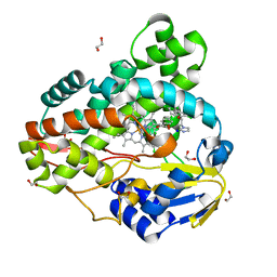
3R9B
 | | Crystal structure of Mycobacterium smegmatis CYP164A2 in ligand free state | | 分子名称: | 1,2-ETHANEDIOL, CYTOCHROME P450 164A2, DODECANE, ... | | 著者 | Agnew, C.R.J, Warrilow, A.G.S, Kelly, S.L, Brady, R.L. | | 登録日 | 2011-03-25 | | 公開日 | 2012-04-04 | | 最終更新日 | 2024-03-20 | | 実験手法 | X-RAY DIFFRACTION (1.89 Å) | | 主引用文献 | An enlarged, adaptable active site in CYP164 family P450 enzymes, the sole P450 in Mycobacterium leprae.
Antimicrob.Agents Chemother., 56, 2012
|
|
3R47
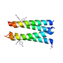
 | | Crystal structure of a 6-helix coiled coil CC-hex-H24 | | 分子名称: | BROMIDE ION, coiled coil helix L24H | | 著者 | Zaccai, N.R, Chi, B.H.C, Woolfson, D.N, Brady, R.L. | | 登録日 | 2011-03-17 | | 公開日 | 2011-11-16 | | 最終更新日 | 2023-09-13 | | 実験手法 | X-RAY DIFFRACTION (2.5002 Å) | | 主引用文献 | A de novo peptide hexamer with a mutable channel.
Nat.Chem.Biol., 7, 2011
|
|
3R9C
 | | Crystal structure of Mycobacterium smegmatis CYP164A2 with Econazole bound | | 分子名称: | 1,2-ETHANEDIOL, 1-[(2R)-2-[(4-chlorobenzyl)oxy]-2-(2,4-dichlorophenyl)ethyl]-1H-imidazole, Cytochrome P450 164A2, ... | | 著者 | Agnew, C.R.J, Kelly, S.L, Brady, R.L. | | 登録日 | 2011-03-25 | | 公開日 | 2012-03-28 | | 最終更新日 | 2023-11-01 | | 実験手法 | X-RAY DIFFRACTION (2.14 Å) | | 主引用文献 | An enlarged, adaptable active site in CYP164 family P450 enzymes, the sole P450 in Mycobacterium leprae.
Antimicrob.Agents Chemother., 56, 2012
|
|
1R8H
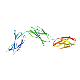
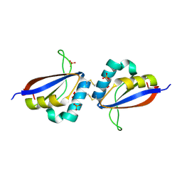 | | Comparison of the structure and DNA binding properties of the E2 proteins from an oncogenic and a non-oncogenic human papillomavirus | | 分子名称: | PHOSPHATE ION, Regulatory protein E2 | | 著者 | Dell, G, Wilkinson, K.W, Tranter, R, Parish, J, Brady, R.L, Gaston, K. | | 登録日 | 2003-10-24 | | 公開日 | 2003-12-23 | | 最終更新日 | 2011-07-13 | | 実験手法 | X-RAY DIFFRACTION (1.9 Å) | | 主引用文献 | Comparison of the structure and DNA-binding properties of the E2 proteins from an oncogenic and a non-oncogenic human papillomavirus.
J.Mol.Biol., 334, 2003
|
|


