3NX5
 
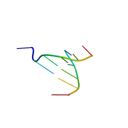 | | The crystal structure of Sanguinarine bound to DNA d(CGTACG) | | 分子名称: | 13-methyl[1,3]benzodioxolo[5,6-c][1,3]dioxolo[4,5-i]phenanthridin-13-ium, 5'-D(*CP*GP*TP*AP*CP*G)-3', CALCIUM ION | | 著者 | Ferraroni, M, Bazzicalupi, C, Gratteri, P, Bilia, A.R. | | 登録日 | 2010-07-13 | | 公開日 | 2011-05-04 | | 最終更新日 | 2023-11-01 | | 実験手法 | X-RAY DIFFRACTION (2.31 Å) | | 主引用文献 | X-Ray diffraction analyses of the natural isoquinoline alkaloids Berberine and Sanguinarine complexed with double helix DNA d(CGTACG)
Chem.Commun.(Camb.), 47, 2011
|
|
3NP6
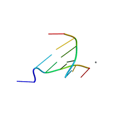 | | The crystal structure of Berberine bound to DNA d(CGTACG) | | 分子名称: | 5'-D(*CP*GP*TP*AP*CP*G)-3', BERBERINE, CALCIUM ION | | 著者 | Ferraroni, M, Bazzicalupi, C, Gratteri, P, Bilia, A.R. | | 登録日 | 2010-06-28 | | 公開日 | 2011-05-04 | | 最終更新日 | 2023-11-01 | | 実験手法 | X-RAY DIFFRACTION (2.3 Å) | | 主引用文献 | X-Ray diffraction analyses of the natural isoquinoline alkaloids Berberine and Sanguinarine complexed with double helix DNA d(CGTACG)
Chem.Commun.(Camb.), 47, 2011
|
|
3R6R
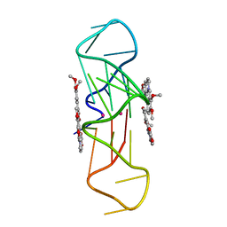 | | Structure of the complex of an intramolecular human telomeric DNA with Berberine formed in K+ solution | | 分子名称: | BERBERINE, DNA (22-mer), POTASSIUM ION | | 著者 | Ferraroni, M, Bazzicalupi, C, Gratteri, P, Bilia, A.R. | | 登録日 | 2011-03-22 | | 公開日 | 2012-02-29 | | 最終更新日 | 2023-09-13 | | 実験手法 | X-RAY DIFFRACTION (2.4 Å) | | 主引用文献 | The crystal structure of human telomeric DNA complexed with berberine: an interesting case of stacked ligand to G-tetrad ratio higher than 1:1.
Nucleic Acids Res., 41, 2013
|
|
4D9Y
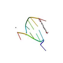 | | The crystal structure of Chelerythrine bound to DNA d(CGTACG) | | 分子名称: | 1,2-dimethoxy-12-methyl[1,3]benzodioxolo[5,6-c]phenanthridin-12-ium, CALCIUM ION, DNA (5'-D(*CP*GP*TP*AP*CP*G)-3') | | 著者 | Ferraroni, M, Bazzicalupi, C, Gratteri, P, Bilia, A.R. | | 登録日 | 2012-01-12 | | 公開日 | 2013-01-23 | | 最終更新日 | 2023-09-13 | | 実験手法 | X-RAY DIFFRACTION (2.1 Å) | | 主引用文献 | The crystal structure of Chelerythrine bound to DNA d(CGTACG)
To be Published
|
|
4D9X
 | | The crystal structure of Coptisine bound to DNA d(CGTACG) | | 分子名称: | 6,7-dihydro[1,3]dioxolo[4,5-g][1,3]dioxolo[7,8]isoquino[3,2-a]isoquinolin-5-ium, CALCIUM ION, DNA (5'-D(*CP*GP*TP*AP*CP*G)-3') | | 著者 | Ferraroni, M, Bazzicalupi, C, Gratteri, P, Bilia, A.R. | | 登録日 | 2012-01-12 | | 公開日 | 2013-01-23 | | 最終更新日 | 2023-09-13 | | 実験手法 | X-RAY DIFFRACTION (2.44 Å) | | 主引用文献 | The crystal structure of Coptisine bound to DNA d(CGTACG)
to be published
|
|


