Loading
PDBj
MenuPDBj@FacebookPDBj@X(formerly Twitter)PDBj@BlueSkyPDBj@YouTubewwPDB FoundationwwPDB
RCSB PDBPDBeBMRBAdv. SearchSearch help
PDB: 15 results

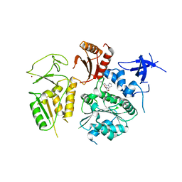
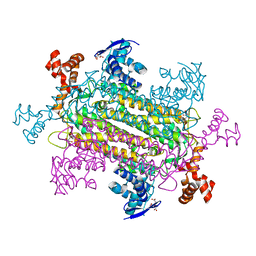
4NL4
DownloadVisualize
BU of 4nl4 by Molmil
PriA Helicase Bound to ADP
Descriptor: ADENOSINE-5'-DIPHOSPHATE, Primosome assembly protein PriA, ZINC ION
Authors:Bhattacharyya, B, George, N.P, Keck, J.L.
Deposit date:2013-11-13
Release date:2014-01-08
Last modified:2024-02-28
Method:X-RAY DIFFRACTION (2.65 Å)
Cite:Structural mechanisms of PriA-mediated DNA replication restart.
Proc.Natl.Acad.Sci.USA, 111, 2014
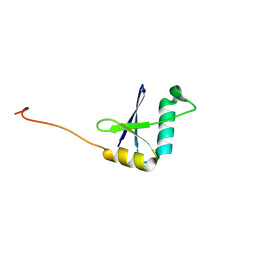
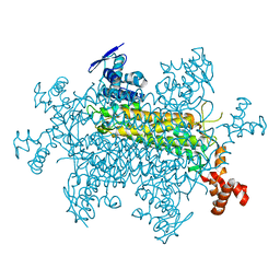
4NL8
DownloadVisualize
BU of 4nl8 by Molmil
PriA Helicase Bound to SSB C-terminal Tail Peptide
Descriptor: Primosome assembly protein PriA, Single-stranded DNA-binding protein, ZINC ION
Authors:Bhattacharyya, B, George, N.P, Thurmes, T.M, Keck, J.L.
Deposit date:2013-11-13
Release date:2014-01-08
Last modified:2024-02-28
Method:X-RAY DIFFRACTION (4.08 Å)
Cite:Structural mechanisms of PriA-mediated DNA replication restart.
Proc.Natl.Acad.Sci.USA, 111, 2014
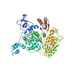
9E7C
DownloadVisualize
BU of 9e7c by Molmil
TRK-fused Gene (TFG) PB1 Domain D60A/D62R Variant
Descriptor: Protein TFG
Authors:Schuh, A.L, Bhattacharyya, B, Keck, J.L, Audhya, A.
Deposit date:2024-11-01
Release date:2024-11-20
Method:X-RAY DIFFRACTION (1.906 Å)
Cite:Multiple roles for TFG ring complexes in neuronal cargo trafficking
Biorxiv, 2024
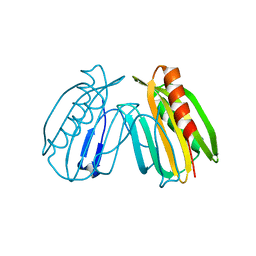
6E8A
DownloadVisualize
BU of 6e8a by Molmil
Crystal structure of DcrB from Salmonella enterica at 1.92 Angstroms resolution
Descriptor: DUF1795 domain-containing protein
Authors:Rasmussen, D.M, Soens, R.W, Bhattacharyya, B, May, J.F.
Deposit date:2018-07-27
Release date:2018-10-31
Last modified:2024-03-13
Method:X-RAY DIFFRACTION (1.92 Å)
Cite:The structure of DcrB, a lipoprotein from Salmonella enterica, reveals flexibility in the N-terminal segment of the Mog1p/PsbP-like fold.
J. Struct. Biol., 204, 2018
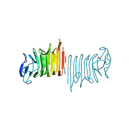
6PZL
DownloadVisualize
BU of 6pzl by Molmil
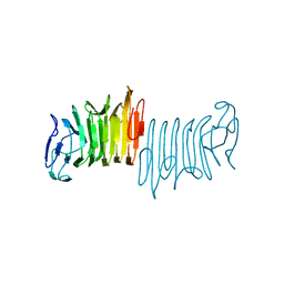
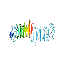
P. mirabilis hemolysin A mutant - Q125A
Descriptor: GLYCEROL, Hemolysin
Authors:Weaver, T.M, Novak, W.R.P, Bhattacharyya, B.
Deposit date:2019-08-01
Release date:2020-08-05
Last modified:2024-10-23
Method:X-RAY DIFFRACTION (1.17 Å)
Cite:Structure of the HpmA265 Q125A variant
To Be Published
6PYK
DownloadVisualize
BU of 6pyk by Molmil
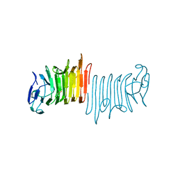
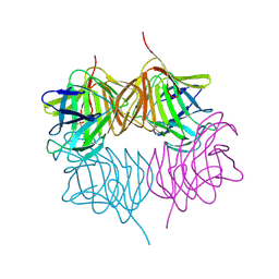
P. mirabilis hemolysin A mutant - F80L
Descriptor: Hemolysin
Authors:Weaver, T.M, Novak, W.R.P, Bhattacharyya, B.
Deposit date:2019-07-30
Release date:2020-08-05
Last modified:2024-11-20
Method:X-RAY DIFFRACTION (1.35 Å)
Cite:Structure of truncated hemolysin A variant F80L
To Be Published
5SZ8
DownloadVisualize
BU of 5sz8 by Molmil
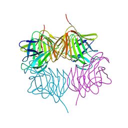
Truncated hemolysin A Q125A/Y134A from P. mirabilis at 1.8 Angstroms resolution crystallized in a high salt condition
Descriptor: Hemolysin, SULFATE ION
Authors:Novak, W.R.P, Bhattacharyya, B, Weaver, T.M.
Deposit date:2016-08-12
Release date:2017-03-22
Last modified:2024-11-20
Method:X-RAY DIFFRACTION (1.83 Å)
Cite:Proteolysis of truncated hemolysin A yields a stable dimerization interface.
Acta Crystallogr F Struct Biol Commun, 73, 2017
5KDK
DownloadVisualize
BU of 5kdk by Molmil
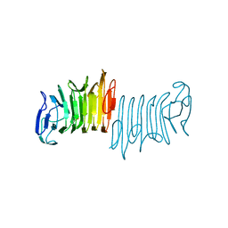
Truncated hemolysin A from P. mirabilis at 2.0 Angstroms resolution crystallized in a high salt condition
Descriptor: Hemolysin
Authors:Novak, W.R.P, Bhattacharyya, B, Weaver, T.M.
Deposit date:2016-06-08
Release date:2017-06-14
Last modified:2024-10-16
Method:X-RAY DIFFRACTION (2.002 Å)
Cite:Crystal structure of truncated hemolysin A from P. mirabilis at 2.0 Angstroms in high salt
To Be Published
5KEH
DownloadVisualize
BU of 5keh by Molmil
Truncated hemolysin A from P. mirabilis at 2.0 Angstroms resolution crystallized in a high salt condition
Descriptor: Hemolysin
Authors:Novak, W.R.P, Bhattacharyya, B, Weaver, T.M.
Deposit date:2016-06-09
Release date:2017-03-22
Last modified:2024-10-23
Method:X-RAY DIFFRACTION (1.551 Å)
Cite:Proteolysis of truncated hemolysin A yields a stable dimerization interface.
Acta Crystallogr F Struct Biol Commun, 73, 2017
5KF3
DownloadVisualize
BU of 5kf3 by Molmil
Truncated hemolysin A from P. mirabilis Y134A at 2.2 Angstroms resolution
Descriptor: Hemolysin
Authors:Novak, W.R.P, Bhattacharyya, B, Weaver, T.M.
Deposit date:2016-06-11
Release date:2017-03-22
Last modified:2024-10-16
Method:X-RAY DIFFRACTION (2.2 Å)
Cite:Proteolysis of truncated hemolysin A yields a stable dimerization interface.
Acta Crystallogr F Struct Biol Commun, 73, 2017
5KKD
DownloadVisualize
BU of 5kkd by Molmil
Truncated hemolysin A Y134A from P. mirabilis at 2.1 Angstroms resolution crystallized in a high salt condition
Descriptor: Hemolysin, SULFATE ION
Authors:Novak, W.R.P, Bhattacharyya, B, Weaver, T.M.
Deposit date:2016-06-21
Release date:2017-03-22
Last modified:2024-11-06
Method:X-RAY DIFFRACTION (2.13 Å)
Cite:Proteolysis of truncated hemolysin A yields a stable dimerization interface.
Acta Crystallogr F Struct Biol Commun, 73, 2017
6NZC
DownloadVisualize
BU of 6nzc by Molmil
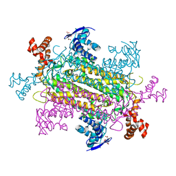
Crystal structure of E. coli fumarase C N326A variant with closed SS Loop at 1.40 angstrom resolution
Descriptor: CITRIC ACID, Fumarate hydratase class II
Authors:Weaver, T.M, May, J.F, Bhattacharyya, B.
Deposit date:2019-02-13
Release date:2019-09-25
Last modified:2023-10-11
Method:X-RAY DIFFRACTION (1.403 Å)
Cite:Closed fumarase C active-site structures reveal SS Loop residue contribution in catalysis.
Febs Lett., 594, 2020
6NZA
DownloadVisualize
BU of 6nza by Molmil
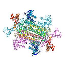
Crystal structure of E. coli fumarase C K324A variant with closed SS Loop at 1.41 angstrom resolution
Descriptor: CITRIC ACID, Fumarate hydratase class II
Authors:Weaver, T.M, May, J.F, Bhattacharyya, B.
Deposit date:2019-02-13
Release date:2019-09-25
Last modified:2023-10-11
Method:X-RAY DIFFRACTION (1.406 Å)
Cite:Closed fumarase C active-site structures reveal SS Loop residue contribution in catalysis.
Febs Lett., 594, 2020
6NZB
DownloadVisualize
BU of 6nzb by Molmil
Crystal structure of E. coli fumarase C S318A variant with closed SS Loop at 1.37 angstrom resolution
Descriptor: CITRIC ACID, Fumarate hydratase class II
Authors:Weaver, T.M, May, J.F, Bhattacharyya, B.
Deposit date:2019-02-13
Release date:2019-09-25
Last modified:2023-10-11
Method:X-RAY DIFFRACTION (1.37 Å)
Cite:Closed fumarase C active-site structures reveal SS Loop residue contribution in catalysis.
Febs Lett., 594, 2020
8SBS
DownloadVisualize
BU of 8sbs by Molmil
Fumarate C - R126A in (3-(N-morpholino)propanesulfonic acid) at pH 7.5
Descriptor: Fumarate hydratase class II
Authors:Weaver, T.M, May, J, Bhattacharyya, B.
Deposit date:2023-04-04
Release date:2023-11-15
Method:X-RAY DIFFRACTION (1.91 Å)
Cite:Fumarate C - R126A in (3-(N-morpholino)propanesulfonic acid) at pH 7.5
To Be Published

227561

PDB entries from 2024-11-20

PDB statisticsPDBj update infoContact PDBjnumon