7FJ7
 
 | |
7FJ8
 | |
7FJB
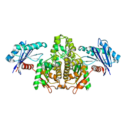
 | | KpAckA (PduW) with AMPPNP, sodium acetate complex structure | | 分子名称: | ACETATE ION, PHOSPHOAMINOPHOSPHONIC ACID-ADENYLATE ESTER, Probable propionate kinase, ... | | 著者 | Wenyue, W, Zhang, Q, Bartlam, M. | | 登録日 | 2021-08-03 | | 公開日 | 2022-08-10 | | 最終更新日 | 2023-11-29 | | 実験手法 | X-RAY DIFFRACTION (2.44 Å) | | 主引用文献 | KpAckA (PduW) with AMPPNP, sodium acetate complex structure
To Be Published
|
|
7EPO
 | |
7EPN
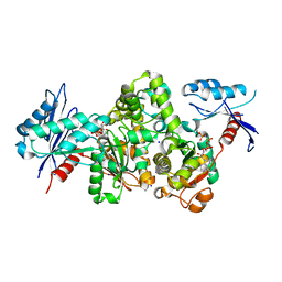
 | | Ketosteroid Isomerase KSI native | | 分子名称: | SnoaL-like domain-containing protein | | 著者 | Liang, Y, Zhang, Q, Bartlam, M. | | 登録日 | 2021-04-27 | | 公開日 | 2021-12-22 | | 最終更新日 | 2023-11-29 | | 実験手法 | X-RAY DIFFRACTION (1.9 Å) | | 主引用文献 | Structural characterization and Kemp eliminase activity of the Mycobacterium smegmatis Ketosteroid Isomerase.
Biochem.Biophys.Res.Commun., 560, 2021
|
|
3HF2
 | | Crystal structure of the I401P mutant of cytochrome P450 BM3 | | 分子名称: | Bifunctional P-450/NADPH-P450 reductase, PROTOPORPHYRIN IX CONTAINING FE | | 著者 | Yang, W, Whitehouse, C.J.C, Bell, S.G, Bartlam, M, Wong, L.L, Rao, Z. | | 登録日 | 2009-05-10 | | 公開日 | 2009-06-30 | | 最終更新日 | 2023-11-01 | | 実験手法 | X-RAY DIFFRACTION (2.2 Å) | | 主引用文献 | A Highly Active Single-Mutation Variant of P450(BM3) (CYP102A1)
Chembiochem, 10, 2009
|
|
3JU5
 | | Crystal Structure of Dimeric Arginine Kinase at 1.75-A Resolution | | 分子名称: | Arginine kinase, MAGNESIUM ION | | 著者 | Wu, X, Ye, S, Guo, S, Yan, W, Bartlam, M, Rao, Z. | | 登録日 | 2009-09-14 | | 公開日 | 2009-09-29 | | 最終更新日 | 2011-07-13 | | 実験手法 | X-RAY DIFFRACTION (1.75 Å) | | 主引用文献 | Structural basis for a reciprocating mechanism of negative cooperativity in dimeric phosphagen kinase activity
Faseb J., 24, 2010
|
|
3JU6
 | | Crystal Structure of Dimeric Arginine Kinase in Complex with AMPPNP and Arginine | | 分子名称: | ARGININE, Arginine kinase, PHOSPHOAMINOPHOSPHONIC ACID-ADENYLATE ESTER | | 著者 | Wu, X, Ye, S, Guo, S, Yan, W, Bartlam, M, Rao, Z. | | 登録日 | 2009-09-14 | | 公開日 | 2009-09-29 | | 最終更新日 | 2023-11-22 | | 実験手法 | X-RAY DIFFRACTION (2.45 Å) | | 主引用文献 | Structural basis for a reciprocating mechanism of negative cooperativity in dimeric phosphagen kinase activity
Faseb J., 24, 2010
|
|
3L1N
 | | Crystal structure of Mp1p ligand binding domain 2 complexd with palmitic acid | | 分子名称: | Cell wall antigen, PALMITIC ACID | | 著者 | Liao, S, Tung, E.T, Zheng, W, Chong, K, Xu, Y, Bartlam, M, Rao, Z, Yuen, K.Y. | | 登録日 | 2009-12-14 | | 公開日 | 2010-01-05 | | 最終更新日 | 2021-11-10 | | 実験手法 | X-RAY DIFFRACTION (1.3 Å) | | 主引用文献 | Crystal structure of the Mp1p ligand binding domain 2 reveals its function as a fatty acid-binding protein.
J.Biol.Chem., 285, 2010
|
|
3LD1
 | | Crystal Structure of IBV Nsp2a | | 分子名称: | Replicase polyprotein 1a | | 著者 | Xu, Y, Cong, L, Wei, L, Fu, J, Chen, C, Yang, A, Tang, H, Bartlam, M, Rao, Z. | | 登録日 | 2010-01-12 | | 公開日 | 2011-05-25 | | 最終更新日 | 2024-02-21 | | 実験手法 | X-RAY DIFFRACTION (2.498 Å) | | 主引用文献 | IBV nsp2 is an endosome-associated protein and viral pathogenicity factor
To be Published
|
|
3LXF
 | | Crystal Structure of [2Fe-2S] Ferredoxin Arx from Novosphingobium aromaticivorans | | 分子名称: | FE2/S2 (INORGANIC) CLUSTER, Ferredoxin | | 著者 | Yang, W, Bell, S.G, Wang, H, Bartlam, M, Wong, L.L, Rao, Z. | | 登録日 | 2010-02-25 | | 公開日 | 2010-06-23 | | 最終更新日 | 2014-02-12 | | 実験手法 | X-RAY DIFFRACTION (2.3 Å) | | 主引用文献 | Molecular characterization of a class I P450 electron transfer system from Novosphingobium aromaticivorans DSM12444
J.Biol.Chem., 285, 2010
|
|
3LXI
 | | Crystal Structure of Camphor-Bound CYP101D1 | | 分子名称: | CAMPHOR, Cytochrome P450, PHOSPHATE ION, ... | | 著者 | Yang, W, Bell, S.G, Wang, H, Bartlam, M, Wong, L.L, Rao, Z. | | 登録日 | 2010-02-25 | | 公開日 | 2010-06-23 | | 最終更新日 | 2023-11-01 | | 実験手法 | X-RAY DIFFRACTION (2.2 Å) | | 主引用文献 | Molecular characterization of a class I P450 electron transfer system from Novosphingobium aromaticivorans DSM12444
J.Biol.Chem., 285, 2010
|
|
3LXH
 | | Crystal Structure of Cytochrome P450 CYP101D1 | | 分子名称: | 1,4-DIETHYLENE DIOXIDE, Cytochrome P450, PHOSPHATE ION, ... | | 著者 | Yang, W, Bell, S.G, Wang, H, Bartlam, M, Wong, L.L, Rao, Z. | | 登録日 | 2010-02-25 | | 公開日 | 2010-06-23 | | 最終更新日 | 2023-11-01 | | 実験手法 | X-RAY DIFFRACTION (2.2 Å) | | 主引用文献 | Molecular characterization of a class I P450 electron transfer system from Novosphingobium aromaticivorans DSM12444
J.Biol.Chem., 285, 2010
|
|
3LXD
 | | Crystal Structure of Ferredoxin Reductase ArR from Novosphingobium aromaticivorans | | 分子名称: | FAD-dependent pyridine nucleotide-disulphide oxidoreductase, FLAVIN-ADENINE DINUCLEOTIDE | | 著者 | Yang, W, Bell, S.G, Wang, H, Bartlam, M, Wong, L.L, Rao, Z. | | 登録日 | 2010-02-25 | | 公開日 | 2010-06-23 | | 最終更新日 | 2023-11-01 | | 実験手法 | X-RAY DIFFRACTION (2.5 Å) | | 主引用文献 | Molecular characterization of a class I P450 electron transfer system from Novosphingobium aromaticivorans DSM12444
J.Biol.Chem., 285, 2010
|
|
3M4V
 | | Crystal structure of the A330P mutant of cytochrome P450 BM3 | | 分子名称: | Bifunctional P-450/NADPH-P450 reductase, PROTOPORPHYRIN IX CONTAINING FE | | 著者 | Yang, W, Whitehouse, C.J.C, Bell, S.G, Bartlam, M, Wong, L.L, Rao, Z. | | 登録日 | 2010-03-12 | | 公開日 | 2011-03-23 | | 最終更新日 | 2023-11-01 | | 実験手法 | X-RAY DIFFRACTION (1.9 Å) | | 主引用文献 | Structural basis for the properties of two single-site proline mutants of CYP102A1 (P450BM3)
Chembiochem, 11, 2010
|
|
6IN8
 | | Crystal structure of MucB | | 分子名称: | Sigma factor AlgU regulatory protein MucB | | 著者 | Li, S, Zhang, Q, Bartlam, M. | | 登録日 | 2018-10-24 | | 公開日 | 2019-07-24 | | 最終更新日 | 2023-11-22 | | 実験手法 | X-RAY DIFFRACTION (2.2 Å) | | 主引用文献 | Structural basis for the recognition of MucA by MucB and AlgU in Pseudomonas aeruginosa.
Febs J., 286, 2019
|
|
6IKJ
 | | Crystal structure of YfiB(F48S) | | 分子名称: | GLYCEROL, SULFATE ION, YfiB | | 著者 | Li, S, Zhang, Q, Bartlam, M. | | 登録日 | 2018-10-16 | | 公開日 | 2019-03-13 | | 最終更新日 | 2023-11-22 | | 実験手法 | X-RAY DIFFRACTION (1.76 Å) | | 主引用文献 | Structural analysis of activating mutants of YfiB from Pseudomonas aeruginosa PAO1.
Biochem. Biophys. Res. Commun., 506, 2018
|
|
6IN9
 | |
6INB
 | |
6IKK
 | |
6IKI
 | | Crystal structure of YfiB(W55L) | | 分子名称: | GLYCEROL, SULFATE ION, YfiB | | 著者 | Li, S, Zhang, Q, Bartlam, M. | | 登録日 | 2018-10-16 | | 公開日 | 2019-03-13 | | 最終更新日 | 2023-11-22 | | 実験手法 | X-RAY DIFFRACTION (2.204 Å) | | 主引用文献 | Structural analysis of activating mutants of YfiB from Pseudomonas aeruginosa PAO1.
Biochem. Biophys. Res. Commun., 506, 2018
|
|
6IN7
 | |
6INC
 | | Crystal structure of an acetolactate decarboxylase from Klebsiella pneumoniae | | 分子名称: | 1,2-ETHANEDIOL, Alpha-acetolactate decarboxylase, CHLORIDE ION, ... | | 著者 | Wu, W, Zhang, Q, Bartlam, M. | | 登録日 | 2018-10-24 | | 公開日 | 2019-01-16 | | 最終更新日 | 2023-11-22 | | 実験手法 | X-RAY DIFFRACTION (1.604 Å) | | 主引用文献 | Structural characterization of an acetolactate decarboxylase from Klebsiella pneumoniae
Biochem. Biophys. Res. Commun., 509, 2019
|
|
6JC4
 | |
6JYV
 | | Structure of an isopenicillin N synthase from Pseudomonas aeruginosa PAO1 | | 分子名称: | Probable iron/ascorbate oxidoreductase, SODIUM ION | | 著者 | Hao, Z, Che, S, Wang, R, Liu, R, Zhang, Q, Bartlam, M. | | 登録日 | 2019-04-28 | | 公開日 | 2019-05-22 | | 最終更新日 | 2023-11-22 | | 実験手法 | X-RAY DIFFRACTION (1.651 Å) | | 主引用文献 | Structural characterization of an isopenicillin N synthase family oxygenase from Pseudomonas aeruginosa PAO1.
Biochem.Biophys.Res.Commun., 514, 2019
|
|


