2K8G
 
 | |
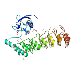
5G4X
 | |
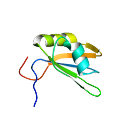
1RE6
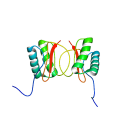 | | Localisation of Dynein Light Chains 1 and 2 and their Pro-apoptotic Ligands | | 分子名称: | dynein light chain 2 | | 著者 | Day, C.L, Puthalakath, H, Skea, G, Strasser, A, Barsukov, I, Lian, L.Y, Huang, D.C, Hinds, M.G. | | 登録日 | 2003-11-06 | | 公開日 | 2004-03-23 | | 最終更新日 | 2024-05-22 | | 実験手法 | SOLUTION NMR | | 主引用文献 | Localization of dynein light chains 1 and 2 and their pro-apoptotic ligands.
Biochem.J., 377, 2004
|
|


