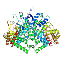
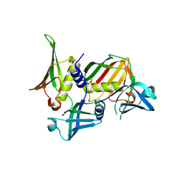
4XOM
 
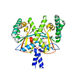 | | Coenzyme F420:L-glutamate ligase (FbiB) from Mycobacterium tuberculosis (C-terminal domain). | | 分子名称: | Coenzyme F420:L-glutamate ligase, SULFATE ION | | 著者 | Rehan, A.M, Bashiri, G, Baker, H.M, Baker, E.N, Squire, C.J. | | 登録日 | 2015-01-16 | | 公開日 | 2016-02-17 | | 最終更新日 | 2024-02-28 | | 実験手法 | X-RAY DIFFRACTION (1.9 Å) | | 主引用文献 | Elongation of the Poly-gamma-glutamate Tail of F420 Requires Both Domains of the F420: gamma-Glutamyl Ligase (FbiB) of Mycobacterium tuberculosis.
J.Biol.Chem., 291, 2016
|
|
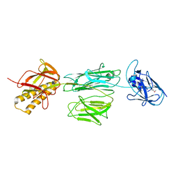
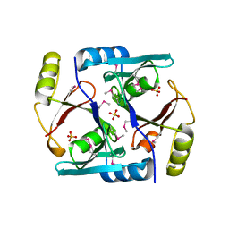
4DXF
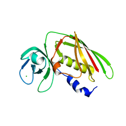 | | Crystal structure of Staphylococcal Superantigen-Like protein 4 | | 分子名称: | CHLORIDE ION, GLYCEROL, Staphylococcal enterotoxin-like toxin | | 著者 | Hermans, S.J, Baker, H.M, Langley, R.J, Baker, E.N, Fraser, J.F. | | 登録日 | 2012-02-27 | | 公開日 | 2012-09-19 | | 最終更新日 | 2023-11-08 | | 実験手法 | X-RAY DIFFRACTION (1.7 Å) | | 主引用文献 | Structural and functional properties of staphylococcal superantigen-like protein 4
Infect.Immun., 80, 2012
|
|
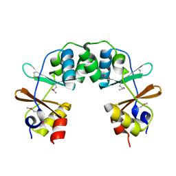
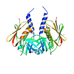
1LFH
 | | MOLECULAR REPLACEMENT SOLUTION OF THE STRUCTURE OF APOLACTOFERRIN, A PROTEIN DISPLAYING LARGE-SCALE CONFORMATIONAL CHANGE | | 分子名称: | CHLORIDE ION, LACTOFERRIN | | 著者 | Anderson, B.F, Baker, E.N, Norris, G.E. | | 登録日 | 1991-09-04 | | 公開日 | 1993-10-31 | | 最終更新日 | 2017-11-29 | | 実験手法 | X-RAY DIFFRACTION (2.8 Å) | | 主引用文献 | Molecular replacement solution of the structure of apolactoferrin, a protein displaying large-scale conformational change.
Acta Crystallogr.,Sect.B, 47, 1991
|
|
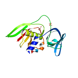
4DXG
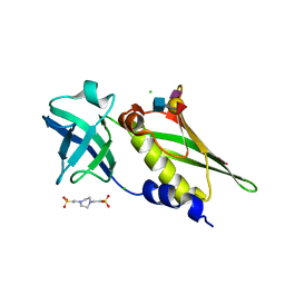 | | Crystal structure of Staphylococcal Superantigen-Like protein 4 complexed with sialyl Lewis X | | 分子名称: | CHLORIDE ION, N-acetyl-alpha-neuraminic acid-(2-3)-beta-D-galactopyranose-(1-4)-[alpha-L-fucopyranose-(1-3)]2-acetamido-2-deoxy-alpha-D-glucopyranose, PIPERAZINE-N,N'-BIS(2-ETHANESULFONIC ACID), ... | | 著者 | Hermans, S.J, Baker, H.M, Langley, R.J, Baker, E.N, Fraser, J.F. | | 登録日 | 2012-02-27 | | 公開日 | 2012-09-19 | | 最終更新日 | 2023-11-08 | | 実験手法 | X-RAY DIFFRACTION (2.5 Å) | | 主引用文献 | Structural and functional properties of staphylococcal superantigen-like protein 4
Infect.Immun., 80, 2012
|
|
1LCF
 | | CRYSTAL STRUCTURE OF COPPER-AND OXALATE-SUBSTITUTED HUMAN LACTOFERRIN AT 2.0 ANGSTROMS RESOLUTION | | 分子名称: | 2-acetamido-2-deoxy-beta-D-glucopyranose, CARBONATE ION, COPPER (II) ION, ... | | 著者 | Smith, C.A, Anderson, B.F, Baker, H.M, Baker, E.N. | | 登録日 | 1994-01-11 | | 公開日 | 1994-08-31 | | 最終更新日 | 2020-07-29 | | 実験手法 | X-RAY DIFFRACTION (2 Å) | | 主引用文献 | Structure of copper- and oxalate-substituted human lactoferrin at 2.0 A resolution.
Acta Crystallogr.,Sect.D, 50, 1994
|
|
1LFI
 | | METAL SUBSTITUTION IN TRANSFERRINS: THE CRYSTAL STRUCTURE OF HUMAN COPPER-LACTOFERRIN AT 2.1 ANGSTROMS RESOLUTION | | 分子名称: | 2-acetamido-2-deoxy-beta-D-glucopyranose-(1-3)-[beta-L-fucopyranose-(1-6)]2-acetamido-2-deoxy-beta-D-glucopyranose, CARBONATE ION, COPPER (II) ION, ... | | 著者 | Smith, C.A, Anderson, B.F, Baker, H.M, Baker, E.N. | | 登録日 | 1992-02-10 | | 公開日 | 1993-10-31 | | 最終更新日 | 2020-07-29 | | 実験手法 | X-RAY DIFFRACTION (2.1 Å) | | 主引用文献 | Metal substitution in transferrins: the crystal structure of human copper-lactoferrin at 2.1-A resolution.
Biochemistry, 31, 1992
|
|
1Y5H
 | |
2B7O
 | | The Structure of 3-Deoxy-D-Arabino-Heptulosonate 7-Phosphate Synthase from Mycobacterium tuberculosis | | 分子名称: | 3-deoxy-D-arabino-heptulosonate 7-phosphate synthase AroG, MANGANESE (II) ION, O-DODECANYL OCTAETHYLENE GLYCOL, ... | | 著者 | Webby, C.J, Baker, H.M, Lott, J.S, Baker, E.N, Parker, E.J, Mycobacterium Tuberculosis Structural Proteomics Project (XMTB) | | 登録日 | 2005-10-05 | | 公開日 | 2005-10-18 | | 最終更新日 | 2011-07-13 | | 実験手法 | X-RAY DIFFRACTION (2.3 Å) | | 主引用文献 | The structure of 3-deoxy-d-arabino-heptulosonate 7-phosphate synthase from Mycobacterium tuberculosis reveals a common catalytic scaffold and ancestry for type I and type II enzymes
J.Mol.Biol., 354, 2005
|
|
3RPK
 | |
1CB6
 | | STRUCTURE OF HUMAN APOLACTOFERRIN AT 2.0 A RESOLUTION. | | 分子名称: | CHLORIDE ION, Lactotransferrin | | 著者 | Jameson, G.B, Anderson, B.F, Norris, G.E, Thomas, D.H, Baker, E.N. | | 登録日 | 1999-03-01 | | 公開日 | 1999-03-12 | | 最終更新日 | 2023-08-09 | | 実験手法 | X-RAY DIFFRACTION (2 Å) | | 主引用文献 | Structure of human apolactoferrin at 2.0 A resolution. Refinement and analysis of ligand-induced conformational change.
Acta Crystallogr.,Sect.D, 54, 1998
|
|
1BKA
 | | OXALATE-SUBSTITUTED DIFERRIC LACTOFERRIN | | 分子名称: | FE (III) ION, LACTOFERRIN, OXALATE ION | | 著者 | Baker, H.M, Smith, C.A, Baker, E.N. | | 登録日 | 1996-04-15 | | 公開日 | 1996-11-08 | | 最終更新日 | 2024-06-05 | | 実験手法 | X-RAY DIFFRACTION (2.4 Å) | | 主引用文献 | Anion binding by transferrins: importance of second-shell effects revealed by the crystal structure of oxalate-substituted diferric lactoferrin.
Biochemistry, 35, 1996
|
|
1EVJ
 | | CRYSTAL STRUCTURE OF GLUCOSE-FRUCTOSE OXIDOREDUCTASE (GFOR) DELTA1-22 S64D | | 分子名称: | GLUCOSE-FRUCTOSE OXIDOREDUCTASE, NICOTINAMIDE-ADENINE-DINUCLEOTIDE | | 著者 | Lott, J.S, Halbig, D, Baker, H.M, Hardman, M.J, Sprenger, G.A, Baker, E.N. | | 登録日 | 2000-04-20 | | 公開日 | 2000-12-04 | | 最終更新日 | 2024-02-07 | | 実験手法 | X-RAY DIFFRACTION (2.7 Å) | | 主引用文献 | Crystal structure of a truncated mutant of glucose-fructose oxidoreductase shows that an N-terminal arm controls tetramer formation.
J.Mol.Biol., 304, 2000
|
|
1FQE
 | | CRYSTAL STRUCTURES OF MUTANT (K206A) THAT ABOLISH THE DILYSINE INTERACTION IN THE N-LOBE OF HUMAN TRANSFERRIN | | 分子名称: | CARBONATE ION, FE (III) ION, POTASSIUM ION, ... | | 著者 | Nurizzo, D, Baker, H.M, Baker, E.N. | | 登録日 | 2000-09-04 | | 公開日 | 2001-05-16 | | 最終更新日 | 2021-11-03 | | 実験手法 | X-RAY DIFFRACTION (1.8 Å) | | 主引用文献 | Crystal structures and iron release properties of mutants (K206A and K296A) that abolish the dilysine interaction in the N-lobe of human transferrin.
Biochemistry, 40, 2001
|
|
2O7U
 | | Crystal structure of K206E/K296E mutant of the N-terminal half molecule of human transferrin | | 分子名称: | CARBONATE ION, FE (III) ION, Serotransferrin | | 著者 | Baker, H.M, Nurizzo, D, Mason, A.B, Baker, E.N. | | 登録日 | 2006-12-11 | | 公開日 | 2007-01-23 | | 最終更新日 | 2023-08-30 | | 実験手法 | X-RAY DIFFRACTION (2.8 Å) | | 主引用文献 | Structures of two mutants that probe the role in iron release of the dilysine pair in the N-lobe of human transferrin.
Acta Crystallogr.,Sect.D, 63, 2007
|
|
1JC5
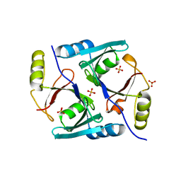 | | Crystal Structure of Native Methylmalonyl-CoA Epimerase | | 分子名称: | Methylmalonyl-CoA Epimerase, SULFATE ION | | 著者 | Mc Carthy, A.A, Baker, H.M, Shewry, S.C, Patchett, M.L, Baker, E.N. | | 登録日 | 2001-06-07 | | 公開日 | 2001-07-11 | | 最終更新日 | 2024-02-07 | | 実験手法 | X-RAY DIFFRACTION (2.2 Å) | | 主引用文献 | Crystal structure of methylmalonyl-coenzyme A epimerase from P. shermanii: a novel enzymatic function on an ancient metal binding scaffold.
Structure, 9, 2001
|
|
2RDG
 | | Crystal structure of Staphylococcal Superantigen-Like protein 11 in complex with Sialyl Lewis X | | 分子名称: | CITRIC ACID, N-acetyl-alpha-neuraminic acid-(2-3)-beta-D-galactopyranose-(1-4)-[alpha-L-fucopyranose-(1-3)]2-acetamido-2-deoxy-alpha-D-glucopyranose, POTASSIUM ION, ... | | 著者 | Chung, M.C, Wines, B.D, Baker, H, Langley, R.J, Baker, E.N, Fraser, J.D. | | 登録日 | 2007-09-24 | | 公開日 | 2007-12-18 | | 最終更新日 | 2023-10-25 | | 実験手法 | X-RAY DIFFRACTION (1.6 Å) | | 主引用文献 | The crystal structure of staphylococcal superantigen-like protein 11 in complex with sialyl Lewis X reveals the mechanism for cell binding and immune inhibition
Mol.Microbiol., 66, 2007
|
|
3VOM
 | | Structure of a putative phosphoserine aminotransferase from mycobacterium tuberculosis | | 分子名称: | GLYCEROL, PYRIDOXAL-5'-PHOSPHATE, Putative phosphoserine aminotransferase, ... | | 著者 | Coulibaly, F, Lassalle, E, Baker, H.M, Baker, E.N. | | 登録日 | 2012-01-30 | | 公開日 | 2012-02-22 | | 最終更新日 | 2023-11-08 | | 実験手法 | X-RAY DIFFRACTION (2.1 Å) | | 主引用文献 | Structure of phosphoserine aminotransferase from Mycobacterium tuberculosis.
Acta Crystallogr.,Sect.D, 68, 2012
|
|
2RDH
 | | Crystal structure of Staphylococcal Superantigen-Like protein 11 | | 分子名称: | PHOSPHATE ION, SODIUM ION, Superantigen-like protein 11 | | 著者 | Chung, M.C, Wines, B.D, Baker, H, Langley, R.J, Baker, E.N, Fraser, J.D. | | 登録日 | 2007-09-24 | | 公開日 | 2007-12-18 | | 最終更新日 | 2023-10-25 | | 実験手法 | X-RAY DIFFRACTION (1.7 Å) | | 主引用文献 | The crystal structure of staphylococcal superantigen-like protein 11 in complex with sialyl Lewis X reveals the mechanism for cell binding and immune inhibition
Mol.Microbiol., 66, 2007
|
|
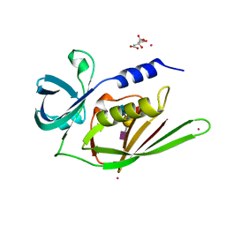
1JC4
 | | Crystal Structure of Se-Met Methylmalonyl-CoA Epimerase | | 分子名称: | Methylmalonyl-CoA epimerase, SULFATE ION | | 著者 | Mc Carthy, A.A, Baker, H.M, Shewry, S.C, Patchett, M.L, Baker, E.N. | | 登録日 | 2001-06-07 | | 公開日 | 2001-07-11 | | 最終更新日 | 2017-10-04 | | 実験手法 | X-RAY DIFFRACTION (2 Å) | | 主引用文献 | Crystal structure of methylmalonyl-coenzyme A epimerase from P. shermanii: a novel enzymatic function on an ancient metal binding scaffold.
Structure, 9, 2001
|
|
2Z8L
 | | Crystal Structure of the Staphylococcal superantigen-like protein SSL5 at pH 4.6 complexed with sialyl Lewis X | | 分子名称: | Exotoxin 3, GLYCEROL, N-acetyl-alpha-neuraminic acid-(2-3)-beta-D-galactopyranose-(1-4)-[alpha-L-fucopyranose-(1-3)]2-acetamido-2-deoxy-beta-D-glucopyranose, ... | | 著者 | Baker, H.M, Basu, I, Chung, M.C, Caradoc Davies, T, Fraser, J.D, Baker, E.N. | | 登録日 | 2007-09-06 | | 公開日 | 2007-11-27 | | 最終更新日 | 2023-11-01 | | 実験手法 | X-RAY DIFFRACTION (1.65 Å) | | 主引用文献 | Crystal structures of the staphylococcal toxin SSL5 in complex with sialyl Lewis X reveal a conserved binding site that shares common features with viral and bacterial sialic acid binding proteins
J.Mol.Biol., 374, 2007
|
|
2FYF
 | | Structure of a putative phosphoserine aminotransferase from Mycobacterium Tuberculosis | | 分子名称: | GLYCEROL, PYRIDOXAL-5'-PHOSPHATE, SULFATE ION, ... | | 著者 | Coulibaly, F, Lassalle, E, Baker, E.N, Mycobacterium Tuberculosis Structural Proteomics Project (XMTB) | | 登録日 | 2006-02-07 | | 公開日 | 2007-01-16 | | 最終更新日 | 2024-02-14 | | 実験手法 | X-RAY DIFFRACTION (1.5 Å) | | 主引用文献 | Structure of phosphoserine aminotransferase from Mycobacterium tuberculosis.
Acta Crystallogr.,Sect.D, 68, 2012
|
|
1TY2
 | | Crystal structure of the streptococcal pyrogenic exotoxin J (SPE-J) | | 分子名称: | ZINC ION, putative exotoxin (superantigen) | | 著者 | Baker, H.M, Proft, T, Webb, P.D, Arcus, V.L, Fraser, J.D, Baker, E.N. | | 登録日 | 2004-07-07 | | 公開日 | 2004-08-17 | | 最終更新日 | 2024-04-03 | | 実験手法 | X-RAY DIFFRACTION (2 Å) | | 主引用文献 | Crystallographic and mutational data show that the streptococcal pyrogenic exotoxin j can use a common binding surface for T-cell receptor binding and dimerization
J.Biol.Chem., 279, 2004
|
|
1TY0
 | | Crystal structure of the streptococcal pyrogenic exotoxin J (SPE-J) | | 分子名称: | putative exotoxin (superantigen) | | 著者 | Baker, H.M, Proft, T, Webb, P.D, Arcus, V.L, Fraser, J.D, Baker, E.N. | | 登録日 | 2004-07-06 | | 公開日 | 2004-08-03 | | 最終更新日 | 2024-04-03 | | 実験手法 | X-RAY DIFFRACTION (1.75 Å) | | 主引用文献 | Crystallographic and mutational data show that the streptococcal pyrogenic exotoxin j can use a common binding surface for T-cell receptor binding and dimerization
J.Biol.Chem., 279, 2004
|
|
1LCT
 | |
1HXN
 | |


