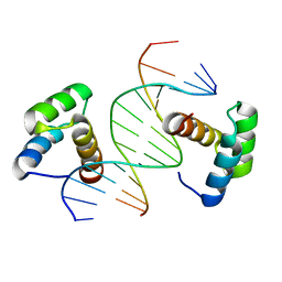
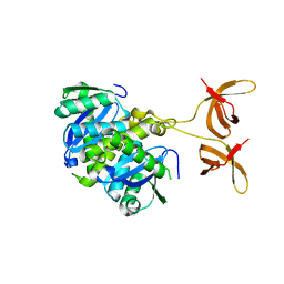
1P7D
 
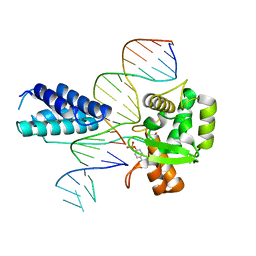 | | Crystal structure of the Lambda Integrase (residues 75-356) bound to DNA | | 分子名称: | 26-MER, 5'-D(*CP*AP*AP*TP*GP*CP*CP*AP*AP*CP*TP*TP*T)-3', Integrase | | 著者 | Aihara, H, Kwon, H.J, Nunes-Duby, S.E, Landy, A, Ellenberger, T. | | 登録日 | 2003-05-01 | | 公開日 | 2003-08-12 | | 最終更新日 | 2011-07-13 | | 実験手法 | X-RAY DIFFRACTION (2.95 Å) | | 主引用文献 | A Conformational Switch Controls the DNA Cleavage Activity of Lambda Integrase
Mol.Cell, 12, 2003
|
|
2V6E
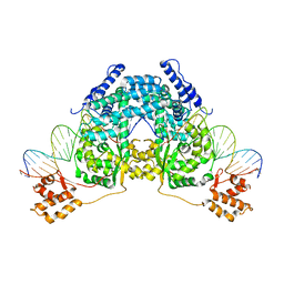 | |
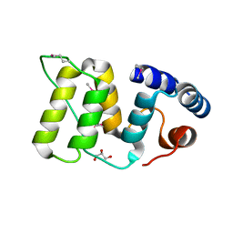
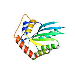
1AA3
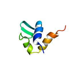 | | C-TERMINAL DOMAIN OF THE E. COLI RECA, NMR, MINIMIZED AVERAGE STRUCTURE | | 分子名称: | RECA | | 著者 | Aihara, H, Ito, Y, Kurumizaka, H, Terada, T, Yokoyama, S, Shibata, T, RIKEN Structural Genomics/Proteomics Initiative (RSGI) | | 登録日 | 1997-01-22 | | 公開日 | 1997-07-23 | | 最終更新日 | 2024-04-10 | | 実験手法 | SOLUTION NMR | | 主引用文献 | An interaction between a specified surface of the C-terminal domain of RecA protein and double-stranded DNA for homologous pairing.
J.Mol.Biol., 274, 1997
|
|
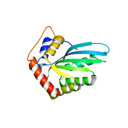
1B22
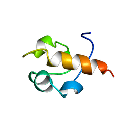 | | RAD51 (N-TERMINAL DOMAIN) | | 分子名称: | DNA REPAIR PROTEIN RAD51 | | 著者 | Aihara, H, Ito, Y, Kurumizaka, H, Yokoyama, S, Shibata, T, RIKEN Structural Genomics/Proteomics Initiative (RSGI) | | 登録日 | 1998-12-04 | | 公開日 | 1999-12-03 | | 最終更新日 | 2023-12-27 | | 実験手法 | SOLUTION NMR | | 主引用文献 | The N-terminal domain of the human Rad51 protein binds DNA: structure and a DNA binding surface as revealed by NMR.
J.Mol.Biol., 290, 1999
|
|
6DFY
 | | Remodeled crystal structure of DNA-bound DUX4-HD2 | | 分子名称: | DNA (5'-D(*AP*AP*GP*AP*TP*TP*AP*GP*AP*TP*TP*AP*GP*T)-3'), DNA (5'-D(*TP*TP*CP*TP*AP*AP*TP*CP*TP*AP*AP*TP*CP*A)-3'), Double homeobox protein 4 | | 著者 | Aihara, H, Shi, K. | | 登録日 | 2018-05-15 | | 公開日 | 2018-09-05 | | 最終更新日 | 2023-10-11 | | 実験手法 | X-RAY DIFFRACTION (2.623 Å) | | 主引用文献 | Comment on structural basis of DUX4/IGH-driven transactivation.
Leukemia, 32, 2018
|
|
6DT1
 | | Crystal structure of the ligase from bacteriophage T4 complexed with DNA intermediate | | 分子名称: | 2,3-DIHYDROXY-1,4-DITHIOBUTANE, ADENOSINE MONOPHOSPHATE, CHLORIDE ION, ... | | 著者 | Shi, K, Aihara, H. | | 登録日 | 2018-06-14 | | 公開日 | 2018-09-19 | | 最終更新日 | 2023-10-11 | | 実験手法 | X-RAY DIFFRACTION (2.75 Å) | | 主引用文献 | T4 DNA ligase structure reveals a prototypical ATP-dependent ligase with a unique mode of sliding clamp interaction.
Nucleic Acids Res., 46, 2018
|
|
6XC1
 | | Crystal structure of bacteriophage T4 spackle and lysozyme in orthorhombic form | | 分子名称: | 1,2-ETHANEDIOL, ISOPROPYL ALCOHOL, Lysozyme, ... | | 著者 | Shi, K, Oakland, J.T, Kurniawan, F, Moeller, N.H, Aihara, H. | | 登録日 | 2020-06-07 | | 公開日 | 2020-12-02 | | 最終更新日 | 2023-10-18 | | 実験手法 | X-RAY DIFFRACTION (1.92 Å) | | 主引用文献 | Structural basis of superinfection exclusion by bacteriophage T4 Spackle.
Commun Biol, 3, 2020
|
|
6X6O
 | | Crystal structure of T4 protein Spackle as determined by native SAD phasing | | 分子名称: | CHLORIDE ION, Protein spackle | | 著者 | Shi, K, Kurniawan, F, Banerjee, S, Moeller, N.H, Aihara, H. | | 登録日 | 2020-05-28 | | 公開日 | 2020-09-16 | | 実験手法 | X-RAY DIFFRACTION (1.52 Å) | | 主引用文献 | Crystal structure of bacteriophage T4 Spackle as determined by native SAD phasing.
Acta Crystallogr D Struct Biol, 76, 2020
|
|
6XC0
 | | Crystal structure of bacteriophage T4 spackle and lysozyme in monoclinic form | | 分子名称: | 1,2-ETHANEDIOL, 4-(2-HYDROXYETHYL)-1-PIPERAZINE ETHANESULFONIC ACID, CHLORIDE ION, ... | | 著者 | Shi, K, Oakland, J.T, Kurniawan, F, Moeller, N.H, Aihara, H. | | 登録日 | 2020-06-07 | | 公開日 | 2020-12-02 | | 最終更新日 | 2023-10-18 | | 実験手法 | X-RAY DIFFRACTION (1.78 Å) | | 主引用文献 | Structural basis of superinfection exclusion by bacteriophage T4 Spackle.
Commun Biol, 3, 2020
|
|
5WFY
 | |
5W8N
 | |
4FVA
 | | Crystal structure of truncated Caenorhabditis elegans TDP2 | | 分子名称: | 1,2-ETHANEDIOL, 5'-tyrosyl-DNA phosphodiesterase, MAGNESIUM ION, ... | | 著者 | Shi, K, Kurahashi, K, Aihara, H. | | 登録日 | 2012-06-29 | | 公開日 | 2012-10-31 | | 最終更新日 | 2024-02-28 | | 実験手法 | X-RAY DIFFRACTION (2.07 Å) | | 主引用文献 | Structural basis for recognition of 5'-phosphotyrosine adducts by Tdp2.
Nat.Struct.Mol.Biol., 19, 2012
|
|
4GEW
 | |
5HX5
 | | APOBEC3F Catalytic Domain Crystal Structure | | 分子名称: | DNA dC->dU-editing enzyme APOBEC-3F, ZINC ION | | 著者 | Shaban, N.M, Shi, K, Aihara, H, Harris, R.S. | | 登録日 | 2016-01-29 | | 公開日 | 2016-05-18 | | 最終更新日 | 2023-09-27 | | 実験手法 | X-RAY DIFFRACTION (2.33 Å) | | 主引用文献 | 1.92 Angstrom Zinc-Free APOBEC3F Catalytic Domain Crystal Structure.
J.Mol.Biol., 428, 2016
|
|
5HX4
 | |
5EJK
 | | Crystal structure of the Rous sarcoma virus intasome | | 分子名称: | DNA (5'-D(*AP*AP*TP*GP*TP*TP*GP*TP*CP*TP*TP*AP*TP*GP*CP*AP*AP*TP*AP*CP*TP*C)-3'), DNA (5'-D(*AP*GP*TP*GP*TP*CP*TP*T)-3'), DNA (5'-D(*CP*TP*TP*CP*TP*CP*TP*C)-3'), ... | | 著者 | Yin, Z, Shi, K, Banerjee, S, Aihara, H. | | 登録日 | 2015-11-02 | | 公開日 | 2016-02-17 | | 最終更新日 | 2023-11-15 | | 実験手法 | X-RAY DIFFRACTION (3.8 Å) | | 主引用文献 | Crystal structure of the Rous sarcoma virus intasome.
Nature, 530, 2016
|
|
8VQR
 | | Crystal structure of chimeric SARS-CoV-2 RBD complexed with chimeric raccoon dog ACE2 | | 分子名称: | 1,2-ETHANEDIOL, 2-acetamido-2-deoxy-beta-D-glucopyranose, 2-acetamido-2-deoxy-beta-D-glucopyranose-(1-4)-2-acetamido-2-deoxy-beta-D-glucopyranose, ... | | 著者 | Hsueh, F.-C, Shi, K, Aihara, H, Li, F. | | 登録日 | 2024-01-19 | | 公開日 | 2024-05-01 | | 最終更新日 | 2024-05-08 | | 実験手法 | X-RAY DIFFRACTION (2.565 Å) | | 主引用文献 | Structural basis for raccoon dog receptor recognition by SARS-CoV-2
To Be Published
|
|
4FW1
 | |
4YJ0
 | | Crystal structure of the DM domain of human DMRT1 bound to 25mer target DNA | | 分子名称: | DNA (25-MER), Doublesex- and mab-3-related transcription factor 1, ZINC ION | | 著者 | Murphy, M.W, Lee, J.K, Rojo, S, Gearhart, M.D, Kurahashi, K, Banerjee, S, Loeuille, G, Bashamboo, A, McElreavey, K, Zarkower, D, Aihara, H, Bardwell, V.J. | | 登録日 | 2015-03-02 | | 公開日 | 2015-05-27 | | 最終更新日 | 2024-06-19 | | 実験手法 | X-RAY DIFFRACTION (3.814 Å) | | 主引用文献 | An ancient protein-DNA interaction underlying metazoan sex determination.
Nat.Struct.Mol.Biol., 22, 2015
|
|
5IRS
 | | crystal structure of the proteasomal Rpn13 PRU-domain | | 分子名称: | 2,3-DIHYDROXY-1,4-DITHIOBUTANE, Proteasomal ubiquitin receptor ADRM1 | | 著者 | Chen, X, Shi, K, Walters, K, Aihara, H. | | 登録日 | 2016-03-14 | | 公開日 | 2016-07-20 | | 最終更新日 | 2023-09-27 | | 実験手法 | X-RAY DIFFRACTION (1.796 Å) | | 主引用文献 | Structures of Rpn1 T1:Rad23 and hRpn13:hPLIC2 Reveal Distinct Binding Mechanisms between Substrate Receptors and Shuttle Factors of the Proteasome.
Structure, 24, 2016
|
|
6E8C
 | | Crystal structure of the double homeodomain of DUX4 in complex with DNA | | 分子名称: | DNA (5'-D(*GP*CP*GP*TP*AP*AP*TP*CP*TP*AP*AP*TP*CP*AP*AP*CP*A)-3'), DNA (5'-D(*TP*GP*TP*TP*GP*AP*TP*TP*AP*GP*AP*TP*TP*AP*CP*GP*C)-3'), Double homeobox protein 4 | | 著者 | Lee, J.K, Bosnakovski, D, Toso, E.A, Dinh, T, Banerjee, S, Bohl, T.E, Shi, K, Kurahashi, K, Kyba, M, Aihara, H. | | 登録日 | 2018-07-27 | | 公開日 | 2018-12-26 | | 最終更新日 | 2023-10-11 | | 実験手法 | X-RAY DIFFRACTION (2.12 Å) | | 主引用文献 | Crystal Structure of the Double Homeodomain of DUX4 in Complex with DNA.
Cell Rep, 25, 2018
|
|
4NQ3
 | | Crystal structure of cyanuic acid hydrolase from A. caulinodans | | 分子名称: | BARBITURIC ACID, Cyanuric acid amidohydrolase, MAGNESIUM ION, ... | | 著者 | Cho, S, Shi, K, Aihara, H. | | 登録日 | 2013-11-23 | | 公開日 | 2014-09-10 | | 実験手法 | X-RAY DIFFRACTION (2.702 Å) | | 主引用文献 | Cyanuric acid hydrolase from Azorhizobium caulinodans ORS 571: crystal structure and insights into a new class of Ser-Lys dyad proteins.
Plos One, 9, 2014
|
|
7S6T
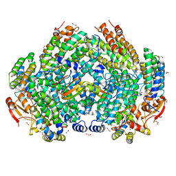 | | Complex structure of Methane monooxygenase hydroxylase and regulatory subunit H33A | | 分子名称: | 1,2-ETHANEDIOL, BENZOIC ACID, FE (III) ION, ... | | 著者 | Johns, J.C, Banerjee, R, Semonis, M.M, Shi, K, Aihara, H, Lipscomb, J.D. | | 登録日 | 2021-09-14 | | 公開日 | 2021-12-29 | | 最終更新日 | 2023-10-18 | | 実験手法 | X-RAY DIFFRACTION (1.82 Å) | | 主引用文献 | X-ray Crystal Structures of Methane Monooxygenase Hydroxylase Complexes with Variants of Its Regulatory Component: Correlations with Altered Reaction Cycle Dynamics.
Biochemistry, 61, 2022
|
|
7S6S
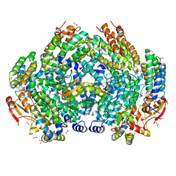 | | Complex structure of Methane monooxygenase hydroxylase and regulatory subunit DBL1 | | 分子名称: | 1,2-ETHANEDIOL, BENZOIC ACID, FE (III) ION, ... | | 著者 | Johns, J.C, Banerjee, R, Semonis, M.M, Shi, K, Aihara, H, Lipscomb, J.D. | | 登録日 | 2021-09-14 | | 公開日 | 2021-12-29 | | 最終更新日 | 2023-10-18 | | 実験手法 | X-RAY DIFFRACTION (1.98 Å) | | 主引用文献 | X-ray Crystal Structures of Methane Monooxygenase Hydroxylase Complexes with Variants of Its Regulatory Component: Correlations with Altered Reaction Cycle Dynamics.
Biochemistry, 61, 2022
|
|
7S7H
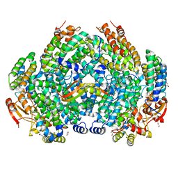 | | Complex structure of Methane monooxygenase hydroxylase and regulatory subunit DBL2 | | 分子名称: | 1,2-ETHANEDIOL, FE (III) ION, Methane monooxygenase beta chain, ... | | 著者 | Johns, J.C, Banerjee, R, Semonis, M.M, Shi, K, Aihara, H, Lipscomb, J.D. | | 登録日 | 2021-09-16 | | 公開日 | 2021-12-29 | | 最終更新日 | 2023-10-18 | | 実験手法 | X-RAY DIFFRACTION (2.4 Å) | | 主引用文献 | X-ray Crystal Structures of Methane Monooxygenase Hydroxylase Complexes with Variants of Its Regulatory Component: Correlations with Altered Reaction Cycle Dynamics.
Biochemistry, 61, 2022
|
|


