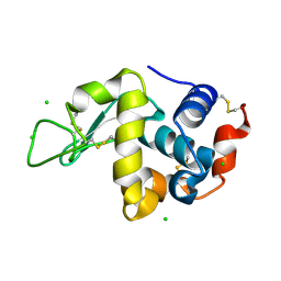
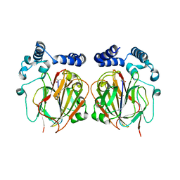
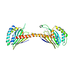
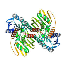
2RU8
 
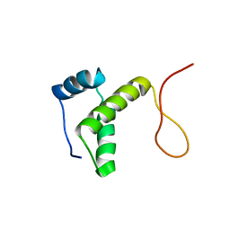 | | DnaT C-terminal domain | | 分子名称: | Primosomal protein 1 | | 著者 | Abe, Y, Tani, J, Fujiyama, S, Urabe, M, Sato, K, Aramaki, T, Katayama, T, Ueda, T. | | 登録日 | 2014-01-29 | | 公開日 | 2014-10-08 | | 最終更新日 | 2024-05-15 | | 実験手法 | SOLUTION NMR | | 主引用文献 | Structure and mechanism of the primosome protein DnaT-functional structures for homotrimerization, dissociation of ssDNA from the PriB·ssDNA complex, and formation of the DnaT·ssDNA complex.
Febs J., 281, 2014
|
|
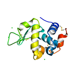
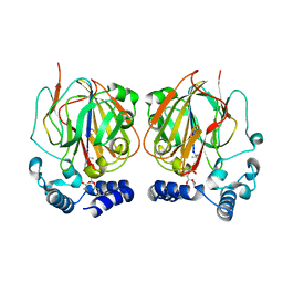
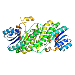
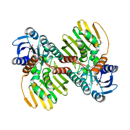
2RUP
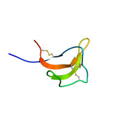 | | Solution structure of rat P2X4 receptor head domain | | 分子名称: | P2X purinoceptor 4 | | 著者 | Abe, Y, Igawa, T, Tsuda, M, Inoue, K, Ueda, T. | | 登録日 | 2014-11-12 | | 公開日 | 2015-02-04 | | 最終更新日 | 2023-06-14 | | 実験手法 | SOLUTION NMR | | 主引用文献 | Solution structure of the rat P2X4 receptor head domain involved in inhibitory metal binding
FEBS Lett., 589, 2015
|
|
1OM2
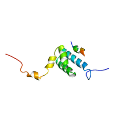 | | SOLUTION NMR STRUCTURE OF THE MITOCHONDRIAL PROTEIN IMPORT RECEPTOR TOM20 FROM RAT IN A COMPLEX WITH A PRESEQUENCE PEPTIDE DERIVED FROM RAT ALDEHYDE DEHYDROGENASE (ALDH) | | 分子名称: | PROTEIN (MITOCHONDRIAL ALDEHYDE DEHYDROGENASE), PROTEIN (MITOCHONDRIAL IMPORT RECEPTOR SUBUNIT TOM20) | | 著者 | Abe, Y, Shodai, T, Muto, T, Mihara, K, Torii, H, Nishikawa, S, Endo, T, Kohda, D. | | 登録日 | 1999-04-23 | | 公開日 | 2000-02-02 | | 最終更新日 | 2023-12-27 | | 実験手法 | SOLUTION NMR | | 主引用文献 | Structural basis of presequence recognition by the mitochondrial protein import receptor Tom20.
Cell(Cambridge,Mass.), 100, 2000
|
|
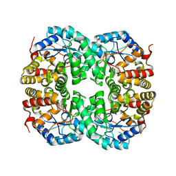
5YCQ
 | | Unique Specificity-Enhancing Factor for the AAA+ Lon Protease | | 分子名称: | Heat shock protein HspQ | | 著者 | Abe, Y, Shioi, S, Kita, S, Nakata, H, Maenaka, K, Kohda, D, Katayama, T, Ueda, T. | | 登録日 | 2017-09-08 | | 公開日 | 2018-04-11 | | 実験手法 | X-RAY DIFFRACTION (2.503 Å) | | 主引用文献 | X-ray crystal structure of Escherichia coli HspQ, a protein involved in the retardation of replication initiation
FEBS Lett., 591, 2017
|
|
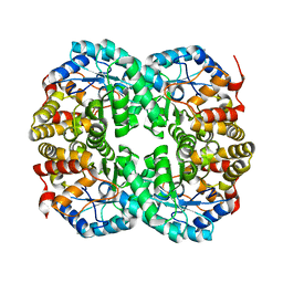
3WW6
 | | Crystal Structure of hen egg white lysozyme mutant N46D/D52S | | 分子名称: | CHLORIDE ION, Lysozyme C | | 著者 | Abe, Y, Kubota, M, Ito, Y, Imoto, T, Ueda, T. | | 登録日 | 2014-06-17 | | 公開日 | 2015-06-17 | | 最終更新日 | 2023-11-08 | | 実験手法 | X-RAY DIFFRACTION (1.53 Å) | | 主引用文献 | Effect on catalysis by replacement of catalytic residue from hen egg white lysozyme to Venerupis philippinarum lysozyme.
Protein Sci., 25, 2016
|
|
3WW5
 | | Crystal Structure of hen egg white lysozyme mutant N46E/D52S | | 分子名称: | CHLORIDE ION, Lysozyme C | | 著者 | Abe, Y, Kubota, M, Ito, Y, Imoto, T, Ueda, T. | | 登録日 | 2014-06-17 | | 公開日 | 2015-06-17 | | 最終更新日 | 2023-11-08 | | 実験手法 | X-RAY DIFFRACTION (1.53 Å) | | 主引用文献 | Effect on catalysis by replacement of catalytic residue from hen egg white lysozyme to Venerupis philippinarum lysozyme.
Protein Sci., 25, 2016
|
|
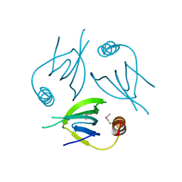
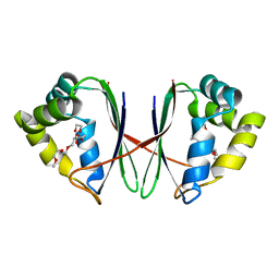
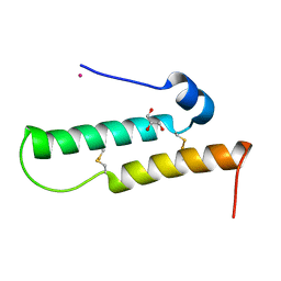
2D35
 | | Solution structure of Cell Division Reactivation Factor, CedA | | 分子名称: | Cell division activator cedA | | 著者 | Abe, Y, Watanabe, N, Matsuda, Y, Yoshida, Y, Katayama, T, Ueda, T. | | 登録日 | 2005-09-26 | | 公開日 | 2006-12-12 | | 最終更新日 | 2024-05-29 | | 実験手法 | SOLUTION NMR | | 主引用文献 | Structural Analysis and Molecular Interaction of Cell Division Reactivation Factor, CedA from Escherichia coli
To be Published
|
|
2E0G
 | |
6L06
 | |
6L07
 | |
7BYU
 | | Crystal structure of Acidovorax avenae L-fucose mutarotase (apo form) | | 分子名称: | 1,2-ETHANEDIOL, 2-(2-{2-[2-(2-METHOXY-ETHOXY)-ETHOXY]-ETHOXY}-ETHOXY)-ETHANOL, L-fucose mutarotase | | 著者 | Watanabe, Y, Fukui, Y, Watanabe, S. | | 登録日 | 2020-04-24 | | 公開日 | 2020-05-27 | | 最終更新日 | 2023-11-29 | | 実験手法 | X-RAY DIFFRACTION (2.206 Å) | | 主引用文献 | Functional and structural characterization of a novel L-fucose mutarotase involved in non-phosphorylative pathway of L-fucose metabolism.
Biochem.Biophys.Res.Commun., 528, 2020
|
|
7BYW
 | |
6J7C
 | | Crystal structure of proline racemase-like protein from Thermococcus litoralis in complex with proline | | 分子名称: | PROLINE, Proline racemase | | 著者 | Watanabe, Y, Watanabe, S, Itoh, Y, Watanabe, Y. | | 登録日 | 2019-01-17 | | 公開日 | 2019-02-27 | | 最終更新日 | 2023-11-22 | | 実験手法 | X-RAY DIFFRACTION (2.7 Å) | | 主引用文献 | Crystal structure of substrate-bound bifunctional proline racemase/hydroxyproline epimerase from a hyperthermophilic archaeon.
Biochem. Biophys. Res. Commun., 511, 2019
|
|
7C0D
 | |
7C0E
 | |
7C0C
 | |
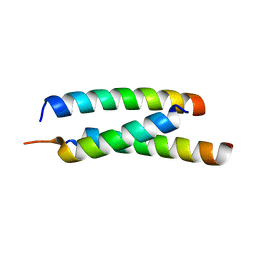
4YTV
 | | Crystal structure of Mdm35 | | 分子名称: | COBALT (II) ION, GLYCEROL, Mitochondrial distribution and morphology protein 35 | | 著者 | Watanabe, Y, Tamura, Y, Kawano, S, Endo, T. | | 登録日 | 2015-03-18 | | 公開日 | 2015-08-12 | | 最終更新日 | 2020-02-05 | | 実験手法 | X-RAY DIFFRACTION (1.45 Å) | | 主引用文献 | Structural and mechanistic insights into phospholipid transfer by Ups1-Mdm35 in mitochondria.
Nat Commun, 6, 2015
|
|
4YTW
 | | Crystal structure of Ups1-Mdm35 complex | | 分子名称: | Mitochondrial distribution and morphology protein 35, Protein UPS1, mitochondrial | | 著者 | Watanabe, Y, Tamura, Y, Kawano, S, Endo, T. | | 登録日 | 2015-03-18 | | 公開日 | 2015-08-12 | | 最終更新日 | 2020-02-05 | | 実験手法 | X-RAY DIFFRACTION (1.4 Å) | | 主引用文献 | Structural and mechanistic insights into phospholipid transfer by Ups1-Mdm35 in mitochondria.
Nat Commun, 6, 2015
|
|
4YTX
 | | Crystal structure of Ups1-Mdm35 complex with PA | | 分子名称: | 1,2-DILAUROYL-SN-GLYCERO-3-PHOSPHATE, Mitochondrial distribution and morphology protein 35, Protein UPS1, ... | | 著者 | Watanabe, Y, Tamura, Y, Kawano, S, Endo, T. | | 登録日 | 2015-03-18 | | 公開日 | 2015-08-12 | | 最終更新日 | 2023-11-08 | | 実験手法 | X-RAY DIFFRACTION (3.2 Å) | | 主引用文献 | Structural and mechanistic insights into phospholipid transfer by Ups1-Mdm35 in mitochondria.
Nat Commun, 6, 2015
|
|
7YPD
 | |
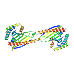
2KZB
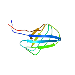 | | Solution structure of alpha-mannosidase binding domain of Atg19 | | 分子名称: | Autophagy-related protein 19 | | 著者 | Watanabe, Y, Noda, N, Kumeta, H, Suzuki, K, Ohsumi, Y, Inagaki, F. | | 登録日 | 2010-06-15 | | 公開日 | 2010-07-21 | | 最終更新日 | 2024-05-15 | | 実験手法 | SOLUTION NMR | | 主引用文献 | Selective transport of alpha-mannosidase by autophagic pathways: structural basis for cargo recognition by Atg19 and Atg34.
J.Biol.Chem., 285, 2010
|
|
2KZK
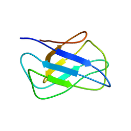 | | Solution structure of alpha-mannosidase binding domain of Atg34 | | 分子名称: | Uncharacterized protein YOL083W | | 著者 | Watanabe, Y, Noda, N, Kumeta, H, Suzuki, K, Ohsumi, Y, Inagaki, F. | | 登録日 | 2010-06-18 | | 公開日 | 2010-07-21 | | 最終更新日 | 2024-05-15 | | 実験手法 | SOLUTION NMR | | 主引用文献 | Selective transport of alpha-mannosidase by autophagic pathways: structural basis for cargo recognition by Atg19 and Atg34.
J.Biol.Chem., 285, 2010
|
|
6JNJ
 | |
5JGE
 | |
6JNK
 | |


