Loading
PDBj
MenuPDBj@FacebookPDBj@TwitterPDBj@YouTubewwPDB FoundationwwPDB
RCSB PDBPDBeBMRBAdv. SearchSearch help
PDB: 356 results

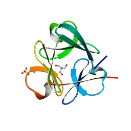
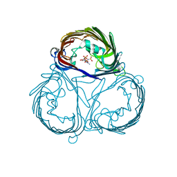
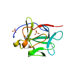
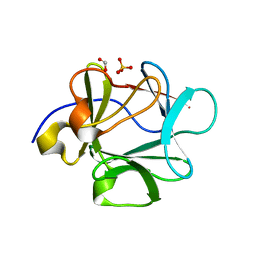
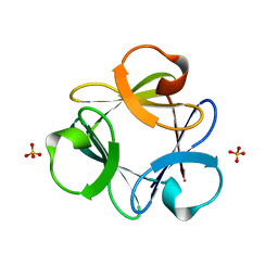
3Q7Y
DownloadVisualize
BU of 3q7y by Molmil
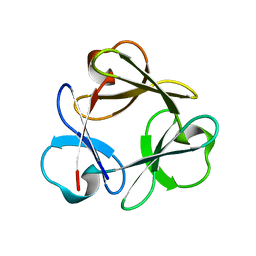
Crystal structure of K15R/E18D/Y22W/H41G/F44W/E51D/E53P/K57R/E60D/Y64W/H82G/F85W/E90D/E94P/K98R/E101D/Y108W/H129G/F132W/E137D Symfoil-4P: de novo designed beta-trefoil architecture with symmetric primary structure
Descriptor: De novo designed beta-trefoil architecture with symmetric primary structure
Authors:Blaber, M, Lee, J.
Deposit date:2011-01-05
Release date:2012-01-11
Last modified:2024-02-21
Method:X-RAY DIFFRACTION (1.45 Å)
Cite:Simplified protein design biased for prebiotic amino acids yields a foldable, halophilic protein.
Proc.Natl.Acad.Sci.USA, 110, 2013
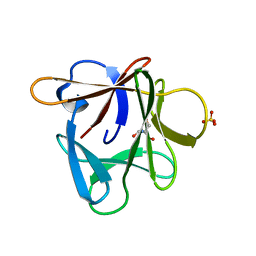
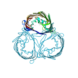
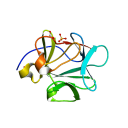
3Q7W
DownloadVisualize
BU of 3q7w by Molmil
Crystal structure of Symfoil-4P/PV1: de novo designed beta-trefoil architecture with symmetric primary structure, primitive version 1
Descriptor: SULFATE ION, de novo designed beta-trefoil architecture with symmetric primary structure
Authors:Blaber, M, Lee, J.
Deposit date:2011-01-05
Release date:2012-01-11
Last modified:2024-02-21
Method:X-RAY DIFFRACTION (1.5 Å)
Cite:Simplified protein design biased for prebiotic amino acids yields a foldable, halophilic protein.
Proc.Natl.Acad.Sci.USA, 110, 2013
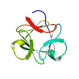
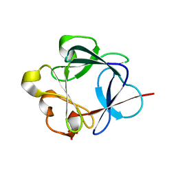
3Q7X
DownloadVisualize
BU of 3q7x by Molmil
Crystal structure of Symfoil-4P/PV1: de novo designed beta-trefoil architecture with symmetric primary structure, primitive version 1
Descriptor: 2-AMINO-2-HYDROXYMETHYL-PROPANE-1,3-DIOL, SULFATE ION, de novo designed beta-trefoil architecture with symmetric primary structure
Authors:Blaber, M, Lee, J.
Deposit date:2011-01-05
Release date:2012-01-11
Last modified:2024-02-21
Method:X-RAY DIFFRACTION (1.4 Å)
Cite:Simplified protein design biased for prebiotic amino acids yields a foldable, halophilic protein.
Proc.Natl.Acad.Sci.USA, 110, 2013
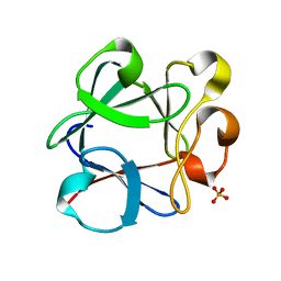
3P6I
DownloadVisualize
BU of 3p6i by Molmil
Crystal structure of Symfoil-4T Permutation #2: de novo designed beta-trefoil architecture with symmetric primary structure
Descriptor: 2-AMINO-2-HYDROXYMETHYL-PROPANE-1,3-DIOL, SULFATE ION, de novo designed beta-trefoil architecture with symmetric primary structure
Authors:Blaber, M, Lee, J.
Deposit date:2010-10-11
Release date:2011-10-12
Last modified:2024-02-21
Method:X-RAY DIFFRACTION (1.32 Å)
Cite:Permutations study of de novo designed symmetric beta-trefoil architecture
To be Published
3P6J
DownloadVisualize
BU of 3p6j by Molmil
Crystal structure of Symfoil-4T Permutation #3: de novo designed beta-trefoil architecture with symmetric primary structure
Descriptor: de novo designed beta-trefoil architecture with symmetric primary structure
Authors:Blaber, M, Lee, J.
Deposit date:2010-10-11
Release date:2011-10-12
Last modified:2024-02-21
Method:X-RAY DIFFRACTION (1.35 Å)
Cite:Permutations study of de novo designed symmetric beta-trefoil architecture
To be Published
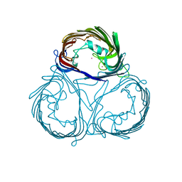
3VZW
DownloadVisualize
BU of 3vzw by Molmil
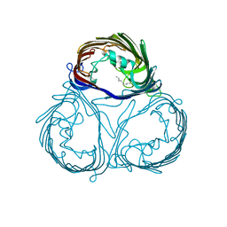
Crystal Structure of outer membrane protein PorB from Neisseria meningitidis in complex with galactose
Descriptor: CESIUM ION, beta-D-galactopyranose, outer membrane proteins
Authors:Tanabe, M, Iverson, T.M.
Deposit date:2012-10-17
Release date:2012-11-07
Last modified:2023-11-08
Method:X-RAY DIFFRACTION (3.2 Å)
Cite:Structural basis for solute transport, nucleotide regulation, and immunological recognition of Neisseria meningitidis PorB.
Proc.Natl.Acad.Sci.USA, 107, 2010
3VZU
DownloadVisualize
BU of 3vzu by Molmil
Crystal Structure of outer membrane protein PorB from Neisseria meningitidis in complex with AMP-PNP
Descriptor: PHOSPHOAMINOPHOSPHONIC ACID-ADENYLATE ESTER, outer membrane protein
Authors:Tanabe, M, Iverson, T.M.
Deposit date:2012-10-15
Release date:2013-03-13
Last modified:2023-11-08
Method:X-RAY DIFFRACTION (2.9 Å)
Cite:Structural basis for solute transport, nucleotide regulation, and immunological recognition of Neisseria meningitidis PorB.
Proc.Natl.Acad.Sci.USA, 107, 2010
3VZT
DownloadVisualize
BU of 3vzt by Molmil
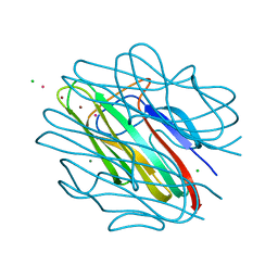
Crystal Structure of outer membrane protein PorB from Neisseria meningitidis
Descriptor: CESIUM ION, outer membrane protein
Authors:Tanabe, M, Iverson, T.M.
Deposit date:2012-10-15
Release date:2012-11-07
Last modified:2024-03-20
Method:X-RAY DIFFRACTION (2.3 Å)
Cite:Structural basis for solute transport, nucleotide regulation, and immunological recognition of Neisseria meningitidis PorB.
Proc.Natl.Acad.Sci.USA, 107, 2010
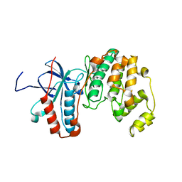
2LGC
DownloadVisualize
BU of 2lgc by Molmil
Joint NMR and X-ray refinement reveals the structure of a novel dibenzo[a,d]cycloheptenone inhibitor/p38 MAP kinase complex in solution
Descriptor: Mitogen-activated protein kinase 14
Authors:Habeck, M.
Deposit date:2011-07-25
Release date:2012-07-25
Last modified:2024-05-15
Method:SOLUTION NMR
Cite:Inferential NMR/X-ray-based structure determination of a dibenzo[a,d]cycloheptenone inhibitor-p38alpha MAP kinase complex in solution
Angew.Chem.Int.Ed.Engl., 51, 2012
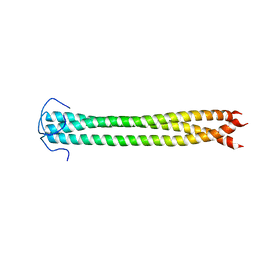
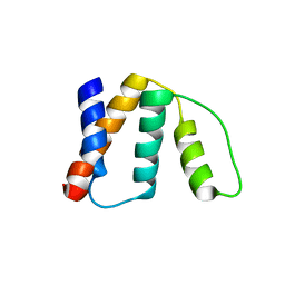
2N64
DownloadVisualize
BU of 2n64 by Molmil
NMR Structure of the C-terminal Coiled-Coil Domain of CIN85
Descriptor: SH3 domain-containing kinase-binding protein 1
Authors:Habeck, M, Becker, S, Griesinger, C, Wong, L.E.
Deposit date:2015-08-11
Release date:2016-07-13
Last modified:2024-05-01
Method:SOLUTION NMR
Cite:The adaptor protein CIN85 assembles intracellular signaling clusters for B cell activation.
Sci.Signal., 9, 2016
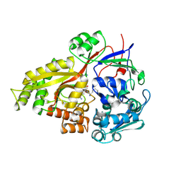
3A2S
DownloadVisualize
BU of 3a2s by Molmil
Crystal Structure of outer membrane protein PorB from Neisseria meningitidis in complex with sucrose
Descriptor: LAURYL DIMETHYLAMINE-N-OXIDE, Outer membrane protein II, beta-D-fructofuranose-(2-1)-alpha-D-glucopyranose
Authors:Tanabe, M, Iverson, T.M.
Deposit date:2009-06-04
Release date:2010-04-07
Last modified:2023-11-01
Method:X-RAY DIFFRACTION (2.2 Å)
Cite:Structural basis for solute transport, nucleotide regulation, and immunological recognition of Neisseria meningitidis PorB.
Proc.Natl.Acad.Sci.USA, 107, 2010
2ZZF
DownloadVisualize
BU of 2zzf by Molmil
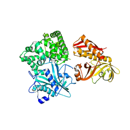
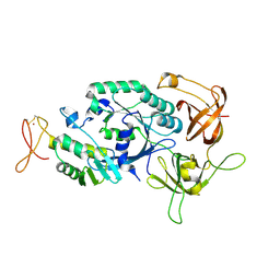
Crystal structure of alanyl-tRNA synthetase without oligomerization domain
Descriptor: Alanyl-tRNA synthetase, ZINC ION
Authors:Sokabe, M, Ose, T, Tokunaga, K, Nakamura, A, Nureki, O, Yao, M, Tanaka, I.
Deposit date:2009-02-10
Release date:2009-07-21
Last modified:2023-11-01
Method:X-RAY DIFFRACTION (2.7 Å)
Cite:The structure of alanyl-tRNA synthetase with editing domain.
Proc.Natl.Acad.Sci.USA, 106, 2009
2ZZG
DownloadVisualize
BU of 2zzg by Molmil
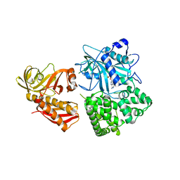
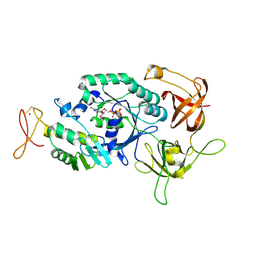
Crystal structure of alanyl-tRNA synthetase in complex with 5''-O-(N-(L-alanyl)-sulfamyoxyl) adenine without oligomerization domain
Descriptor: '5'-O-(N-(L-ALANYL)-SULFAMOYL)ADENOSINE, Alanyl-tRNA synthetase, ZINC ION
Authors:Sokabe, M, Ose, T, Tokunaga, K, Nakamura, A, Nureki, O, Yao, M, Tanaka, I.
Deposit date:2009-02-10
Release date:2009-07-21
Last modified:2023-11-01
Method:X-RAY DIFFRACTION (3.1 Å)
Cite:The structure of alanyl-tRNA synthetase with editing domain.
Proc.Natl.Acad.Sci.USA, 106, 2009
2ZZE
DownloadVisualize
BU of 2zze by Molmil
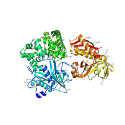
Crystal structure of alanyl-tRNA synthetase without oligomerization domain in lysine-methylated form
Descriptor: Alanyl-tRNA synthetase, ZINC ION
Authors:Sokabe, M, Ose, T, Tokunaga, K, Nakamura, A, Nureki, O, Yao, M, Tanaka, I.
Deposit date:2009-02-10
Release date:2009-07-21
Last modified:2023-11-15
Method:X-RAY DIFFRACTION (2.16 Å)
Cite:The structure of alanyl-tRNA synthetase with editing domain.
Proc.Natl.Acad.Sci.USA, 106, 2009
3BA4
DownloadVisualize
BU of 3ba4 by Molmil
Crystal structure of L26D mutant of Human acidic fibroblast growth factor
Descriptor: FORMIC ACID, Heparin-binding growth factor 1, SULFATE ION
Authors:Blaber, M, Lee, J.
Deposit date:2007-11-07
Release date:2008-04-15
Last modified:2023-08-30
Method:X-RAY DIFFRACTION (1.8 Å)
Cite:A logical OR redundancy within the Asx-Pro-Asx-Gly type I beta-turn motif.
J.Mol.Biol., 377, 2008
3BA5
DownloadVisualize
BU of 3ba5 by Molmil
Crystal structure of D28A mutant of Human acidic fibroblast growth factor
Descriptor: FORMIC ACID, Heparin-binding growth factor 1, SULFATE ION
Authors:Blaber, M, Lee, J.
Deposit date:2007-11-07
Release date:2008-04-15
Last modified:2023-08-30
Method:X-RAY DIFFRACTION (1.75 Å)
Cite:A logical OR redundancy within the Asx-Pro-Asx-Gly type I beta-turn motif.
J.Mol.Biol., 377, 2008
3BAD
DownloadVisualize
BU of 3bad by Molmil
Crystal structure of D70A/H93G mutant of Human acidic fibroblast growth factor
Descriptor: Heparin-binding growth factor 1, SULFATE ION
Authors:Blaber, M, Lee, J.
Deposit date:2007-11-07
Release date:2008-04-15
Last modified:2023-08-30
Method:X-RAY DIFFRACTION (2 Å)
Cite:A logical OR redundancy within the Asx-Pro-Asx-Gly type I beta-turn motif.
J.Mol.Biol., 377, 2008
3B9U
DownloadVisualize
BU of 3b9u by Molmil
Crystal structure of L26N/D28N/H93G mutant of Human acidic fibroblast growth factor
Descriptor: Heparin-binding growth factor 1, SULFATE ION
Authors:Blaber, M, Lee, J.
Deposit date:2007-11-06
Release date:2008-04-15
Last modified:2023-08-30
Method:X-RAY DIFFRACTION (1.55 Å)
Cite:A logical OR redundancy within the Asx-Pro-Asx-Gly type I beta-turn motif.
J.Mol.Biol., 377, 2008
3BA7
DownloadVisualize
BU of 3ba7 by Molmil
Crystal structure of L26N/D28A mutant of Human acidic fibroblast growth factor
Descriptor: FORMIC ACID, Heparin-binding growth factor 1, SULFATE ION
Authors:Blaber, M, Lee, J.
Deposit date:2007-11-07
Release date:2008-04-15
Last modified:2023-08-30
Method:X-RAY DIFFRACTION (1.6 Å)
Cite:A logical OR redundancy within the Asx-Pro-Asx-Gly type I beta-turn motif.
J.Mol.Biol., 377, 2008
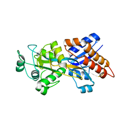
2Z23
DownloadVisualize
BU of 2z23 by Molmil
Crystal structure of Y.pestis oligo peptide binding protein OppA with tri-lysine ligand
Descriptor: Periplasmic oligopeptide-binding protein, peptide (LYS)(LYS)(LYS)
Authors:Tanabe, M, Bertland, T, Mirza, O, Byrne, B, Brown, K.A.
Deposit date:2007-05-17
Release date:2007-10-30
Last modified:2011-07-13
Method:X-RAY DIFFRACTION (2 Å)
Cite:Structures of OppA and PstS from Yersinia pestis indicate variability of interactions with transmembrane domains.
Acta Crystallogr.,Sect.D, 63, 2007
2Z22
DownloadVisualize
BU of 2z22 by Molmil
Crystal structure of phosphate preplasmic binding protein psts from yersinia pestis
Descriptor: PHOSPHATE ION, Periplasmic phosphate-binding protein
Authors:Tanabe, M, Byrne, B, Brown, K.A, Mirza, O, Bertland, T.
Deposit date:2007-05-17
Release date:2007-10-30
Last modified:2024-03-13
Method:X-RAY DIFFRACTION (2 Å)
Cite:Structures of OppA and PstS from Yersinia pestis indicate variability of interactions with transmembrane domains.
Acta Crystallogr.,Sect.D, 63, 2007
2FDQ
DownloadVisualize
BU of 2fdq by Molmil
crystal structure of ACBP from Armadillo Harderian Gland
Descriptor: Acyl-CoA-binding protein
Authors:Costabel, M.D, Guerin, D.M.A.
Deposit date:2005-12-14
Release date:2006-10-24
Last modified:2023-08-30
Method:X-RAY DIFFRACTION (3.5 Å)
Cite:Structure of armadillo ACBP: a new member of the acyl-CoA-binding protein family.
Acta Crystallogr.,Sect.F, 62, 2006
2D74
DownloadVisualize
BU of 2d74 by Molmil
Crystal structure of translation initiation factor aIF2betagamma heterodimer
Descriptor: Translation initiation factor 2 beta subunit, Translation initiation factor 2 gamma subunit, ZINC ION
Authors:Sokabe, M, Yao, M, Sakai, N, Toya, S, Tanaka, I.
Deposit date:2005-11-16
Release date:2006-07-25
Last modified:2024-10-30
Method:X-RAY DIFFRACTION (2.8 Å)
Cite:Structure of archaeal translational initiation factor 2 betagamma-GDP reveals significant conformational change of the beta-subunit and switch 1 region.
Proc.Natl.Acad.Sci.USA, 103, 2006
2DCU
DownloadVisualize
BU of 2dcu by Molmil
Crystal structure of translation initiation factor aIF2betagamma heterodimer with GDP
Descriptor: GUANOSINE-5'-DIPHOSPHATE, MAGNESIUM ION, Translation initiation factor 2 beta subunit, ...
Authors:Sokabe, M, Yao, M, Sakai, N, Toya, S, Tanaka, I.
Deposit date:2006-01-16
Release date:2006-07-25
Last modified:2024-10-23
Method:X-RAY DIFFRACTION (3.4 Å)
Cite:Structure of archaeal translational initiation factor 2 betagamma-GDP reveals significant conformational change of the beta-subunit and switch 1 region.
Proc.Natl.Acad.Sci.USA, 103, 2006
4D7Y
DownloadVisualize
BU of 4d7y by Molmil
Crystal structure of mouse C1QL1 globular domain
Descriptor: C1Q-RELATED FACTOR, CADMIUM ION, CHLORIDE ION, ...
Authors:Kakegawa, W, Mitakidis, N, Miura, E, Abe, M, Matsuda, K, Takeo, Y, Kohda, K, Motohashi, J, Takahashi, A, Nagao, S, Muramatsu, S, Watanabe, M, Sakimura, K, Aricescu, A.R, Yuzaki, M.
Deposit date:2014-12-01
Release date:2015-01-28
Last modified:2023-12-20
Method:X-RAY DIFFRACTION (1.44 Å)
Cite:Anterograde C1Ql1 Signaling is Required in Order to Determine and Maintain a Single-Winner Climbing Fiber in the Mouse Cerebellum
Neuron, 85, 2015

226707

數據於2024-10-30公開中

PDB statisticsPDBj update infoContact PDBjnumon