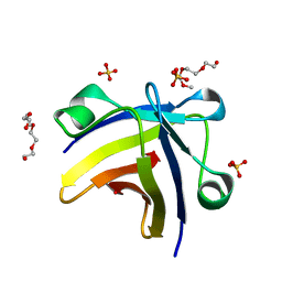
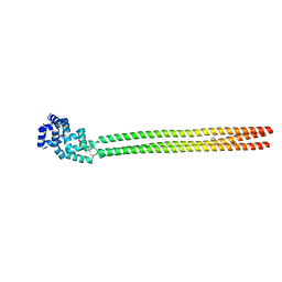
6U7E
 
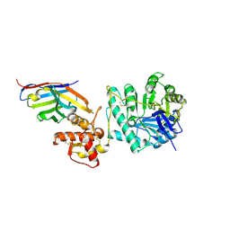
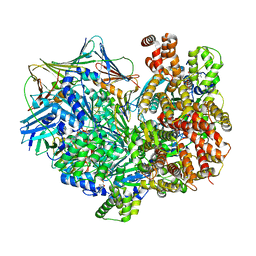
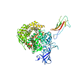 | | HCoV-229E RBD Class III in complex with human APN | | 分子名称: | 2-acetamido-2-deoxy-beta-D-glucopyranose, 2-acetamido-2-deoxy-beta-D-glucopyranose-(1-4)-2-acetamido-2-deoxy-beta-D-glucopyranose, Aminopeptidase N, ... | | 著者 | Tomlinson, A.C.A, Li, Z, Rini, J.M. | | 登録日 | 2019-09-02 | | 公開日 | 2019-11-13 | | 最終更新日 | 2023-10-11 | | 実験手法 | X-RAY DIFFRACTION (3 Å) | | 主引用文献 | The human coronavirus HCoV-229E S-protein structure and receptor binding.
Elife, 8, 2019
|
|
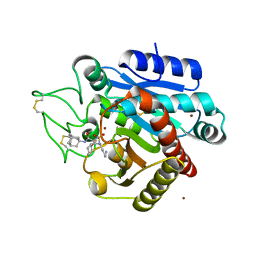
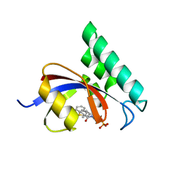
6YU1
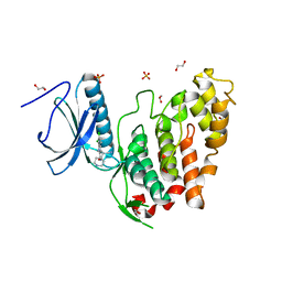 | | CLK3 bound with beta-carboline KH-CARB13 (Cpd 3) | | 分子名称: | (4~{S})-7,8-bis(chloranyl)-9-methyl-1-oxidanylidene-spiro[2,4-dihydropyrido[3,4-b]indole-3,4'-piperidine]-4-carbonitrile, 1,2-ETHANEDIOL, Dual specificity protein kinase CLK3, ... | | 著者 | Schroeder, M, Chaikuad, A, Knapp, S, Structural Genomics Consortium (SGC) | | 登録日 | 2020-04-25 | | 公開日 | 2020-07-15 | | 最終更新日 | 2024-01-24 | | 実験手法 | X-RAY DIFFRACTION (1.9 Å) | | 主引用文献 | DFG-1 Residue Controls Inhibitor Binding Mode and Affinity, Providing a Basis for Rational Design of Kinase Inhibitor Selectivity.
J.Med.Chem., 63, 2020
|
|
8AMJ
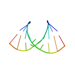
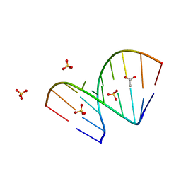 | | Crystal structure of unliganded AUGUGGCAU duplex | | 分子名称: | ACETATE ION, RNA (5'-R(*AP*UP*GP*UP*GP*GP*CP*AP*U)-3'), SULFATE ION | | 著者 | Kiliszek, A, Rypniewski, W. | | 登録日 | 2022-08-03 | | 公開日 | 2022-11-23 | | 最終更新日 | 2024-02-07 | | 実験手法 | X-RAY DIFFRACTION (2.02 Å) | | 主引用文献 | Structure and thermodynamics of a UGG motif interacting with Ba2+ and other metal ions: accommodating changes in the RNA structure and the presence of a G(syn)-G(syn) pair.
Rna, 29, 2022
|
|
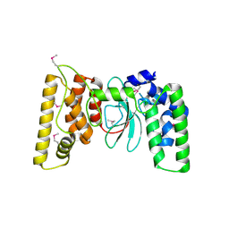
5JJW
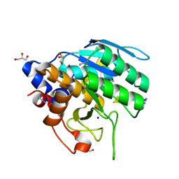
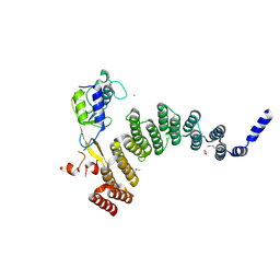 | | Crystal structure of the HAT domain of sart3 in complex with USP15 DUSP-UBL domain | | 分子名称: | 1,2-ETHANEDIOL, Squamous cell carcinoma antigen recognized by T-cells 3, UNKNOWN ATOM OR ION, ... | | 著者 | Dong, A, Zhang, Q, Walker, J.R, Bountra, C, Arrowsmith, C.H, Edwards, A.M, Tong, Y, Structural Genomics Consortium (SGC) | | 登録日 | 2016-04-25 | | 公開日 | 2016-05-04 | | 最終更新日 | 2016-07-06 | | 実験手法 | X-RAY DIFFRACTION (3.01 Å) | | 主引用文献 | Crystal structure of the HAT domain of sart3 in complex with USP15 DUSP-UBL domain
to be published
|
|
8AMI
 | |
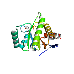
2KFB
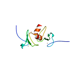
 | | The structure of the cataract causing P23T mutant of human gamma-D crystallin | | 分子名称: | Gamma-crystallin D | | 著者 | Jung, J, Byeon, I.L, Wang, Y, King, J, Gronenborn, A.M. | | 登録日 | 2009-02-12 | | 公開日 | 2009-07-28 | | 最終更新日 | 2024-05-22 | | 実験手法 | SOLUTION NMR | | 主引用文献 | The structure of the cataract-causing P23T mutant of human gammaD-crystallin exhibits distinctive local conformational and dynamic changes.
Biochemistry, 48, 2009
|
|
5LS9
 | |
8DGW
 | |
8PE1
 | | Crystal structure of Gel4 in complex with Nanobody 4 | | 分子名称: | 1,3-beta-glucanosyltransferase, 2-acetamido-2-deoxy-beta-D-glucopyranose, Nanobody 4, ... | | 著者 | Macias-Leon, J, Redrado-Hernandez, S, Castro-Lopez, J, Sanz, A.B, Arias, M, Farkas, V, Vincke, C, Muyldermans, S, Pardo, J, Arroyo, J, Galvez, E, Hurtado-Guerrero, R. | | 登録日 | 2023-06-13 | | 公開日 | 2024-06-19 | | 最終更新日 | 2024-08-21 | | 実験手法 | X-RAY DIFFRACTION (1.9 Å) | | 主引用文献 | Broad Protection against Invasive Fungal Disease from a Nanobody Targeting the Active Site of Fungal beta-1,3-Glucanosyltransferases.
Angew.Chem.Int.Ed.Engl., 63, 2024
|
|
8AMK
 | |
6ZHE
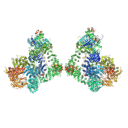 | | Cryo-EM structure of DNA-PK dimer | | 分子名称: | DNA (25-MER), DNA (26-MER), DNA (27-MER), ... | | 著者 | Chaplin, A.K, Hardwick, S.W, Chirgadze, D.Y, Blundell, T.L. | | 登録日 | 2020-06-23 | | 公開日 | 2020-10-21 | | 最終更新日 | 2024-05-01 | | 実験手法 | ELECTRON MICROSCOPY (7.24 Å) | | 主引用文献 | Dimers of DNA-PK create a stage for DNA double-strand break repair.
Nat.Struct.Mol.Biol., 28, 2021
|
|
6UBE
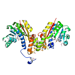
 | | Azide-triggered subtilisin SUBT_BACAM complexed with the peptide LFRAL | | 分子名称: | AZIDE ION, GLYCEROL, Peptide LFRAL, ... | | 著者 | Toth, E.A, Bryan, P.N, Orban, J, Gallagher, D.T, Custer, G. | | 登録日 | 2019-09-11 | | 公開日 | 2020-09-16 | | 最終更新日 | 2024-10-16 | | 実験手法 | X-RAY DIFFRACTION (1.6 Å) | | 主引用文献 | Engineering subtilisin proteases that specifically degrade active RAS.
Commun Biol, 4, 2021
|
|
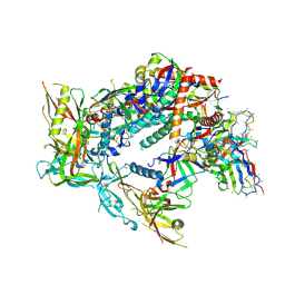
7TID
 | | Structure of the yeast clamp loader (Replication Factor C RFC) bound to the sliding clamp (Proliferating Cell Nuclear Antigen PCNA) and primer-template DNA | | 分子名称: | ADENOSINE-5'-DIPHOSPHATE, DNA (5'-D(*AP*GP*AP*CP*AP*CP*TP*AP*CP*GP*AP*GP*TP*AP*CP*AP*TP*A)-3'), DNA (5'-D(P*TP*TP*TP*TP*TP*TP*TP*AP*TP*GP*TP*AP*CP*TP*CP*GP*TP*AP*GP*TP*GP*TP*CP*T)-3'), ... | | 著者 | Gaubitz, C, Liu, X, Pajak, J, Stone, N, Hayes, J, Demo, G, Kelch, B.A. | | 登録日 | 2022-01-13 | | 公開日 | 2022-02-16 | | 最終更新日 | 2024-02-28 | | 実験手法 | ELECTRON MICROSCOPY (3.3 Å) | | 主引用文献 | Cryo-EM structures reveal high-resolution mechanism of a DNA polymerase sliding clamp loader.
Elife, 11, 2022
|
|
4QF3
 | | Crystal structure of human BAZ2B PHD zinc finger in the free form | | 分子名称: | Bromodomain adjacent to zinc finger domain protein 2B, ZINC ION | | 著者 | Tallant, C, Van Molle, I, Chirgadze, D.Y, Ciulli, A. | | 登録日 | 2014-05-19 | | 公開日 | 2014-07-02 | | 最終更新日 | 2024-04-03 | | 実験手法 | X-RAY DIFFRACTION (1.6 Å) | | 主引用文献 | Molecular basis of histone tail recognition by human TIP5 PHD finger and bromodomain of the chromatin remodeling complex NoRC.
Structure, 23, 2015
|
|
4HTF
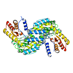
 | | Crystal structure of S-adenosylmethionine-dependent methyltransferase from Escherichia coli in complex with S-adenosylmethionine. | | 分子名称: | ACETATE ION, BETA-MERCAPTOETHANOL, S-ADENOSYLMETHIONINE, ... | | 著者 | Filippova, E.V, Minasov, G, Shuvalova, L, Kiryukhina, O, Jedrzejczak, R, Joachimiak, A, Anderson, W.F, Midwest Center for Structural Genomics (MCSG) | | 登録日 | 2012-11-01 | | 公開日 | 2012-11-21 | | 最終更新日 | 2017-11-15 | | 実験手法 | X-RAY DIFFRACTION (1.6 Å) | | 主引用文献 | Crystal structure of S-adenosylmethionine-dependent methyltransferase from Escherichia coli in complex with S-adenosylmethionine.
To be Published
|
|
6L1N
 | |
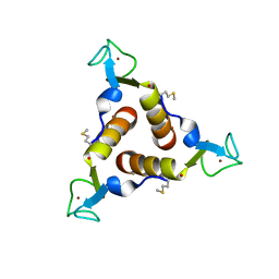
5ITJ
 | | The structure of histone-like protein | | 分子名称: | AbrB family transcriptional regulator, SULFATE ION, TETRAETHYLENE GLYCOL | | 著者 | Lin, B.L, Chen, C.Y, Huang, C.H, Ko, T.P, Chiang, C.H, Lin, K.F, Chang, Y.C, Lin, P.Y, Tsai, H.H.G, Wang, A.H.J. | | 登録日 | 2016-03-17 | | 公開日 | 2017-01-25 | | 最終更新日 | 2024-03-20 | | 実験手法 | X-RAY DIFFRACTION (1.63 Å) | | 主引用文献 | The Arginine Pairs and C-Termini of the Sso7c4 from Sulfolobus solfataricus Participate in Binding and Bending DNA.
PLoS ONE, 12, 2017
|
|
8AE1
 | |
5LYL
 | | Crystal structure of 1 in complex with tafCPB | | 分子名称: | (2~{S})-2-[[(2~{S})-1-(1-adamantylamino)-3-cyclohexyl-1-oxidanylidene-propan-2-yl]sulfamoylamino]-6-azanyl-hexanoic acid, Carboxypeptidase B, ZINC ION | | 著者 | Schreuder, H, Liesum, A, Loenze, P. | | 登録日 | 2016-09-28 | | 公開日 | 2016-10-26 | | 最終更新日 | 2016-12-21 | | 実験手法 | X-RAY DIFFRACTION (1.83 Å) | | 主引用文献 | Sulfamide as Zinc Binding Motif in Small Molecule Inhibitors of Activated Thrombin Activatable Fibrinolysis Inhibitor (TAFIa).
J. Med. Chem., 59, 2016
|
|
6TVH
 | |
1ZR3
 | | Crystal structure of the macro-domain of human core histone variant macroH2A1.1 (form B) | | 分子名称: | 2-(N-MORPHOLINO)-ETHANESULFONIC ACID, histone macroH2A1.1 | | 著者 | Kustatscher, G, Hothorn, M, Pugieux, C, Scheffzek, K, Ladurner, A.G. | | 登録日 | 2005-05-19 | | 公開日 | 2006-02-14 | | 最終更新日 | 2023-08-23 | | 実験手法 | X-RAY DIFFRACTION (1.66 Å) | | 主引用文献 | Splicing regulates NAD metabolite binding to histone macroH2A.
Nat.Struct.Mol.Biol., 12, 2005
|
|
6H9L
 | |
3IKG
 | | Structure-Based Design of Novel PIN1 Inhibitors (I) | | 分子名称: | (2R)-2-[(1-benzothiophen-2-ylcarbonyl)amino]-3-(3-methylphenyl)propyl phosphate, Peptidyl-prolyl cis-trans isomerase NIMA-interacting 1 | | 著者 | Parge, H, Ferre, R.A, Greasley, S, Matthews, D. | | 登録日 | 2009-08-05 | | 公開日 | 2009-09-22 | | 最終更新日 | 2023-09-06 | | 実験手法 | X-RAY DIFFRACTION (1.86 Å) | | 主引用文献 | Structure-based design of novel human Pin1 inhibitors (I).
Bioorg.Med.Chem.Lett., 19, 2009
|
|
2KO8
 | |
8T2E
 | | BG505 Boost2 SOSIP.664 in complex with NHP polyclonal antibody FP3 | | 分子名称: | 2-acetamido-2-deoxy-beta-D-glucopyranose, 2-acetamido-2-deoxy-beta-D-glucopyranose-(1-4)-2-acetamido-2-deoxy-beta-D-glucopyranose, FP3 Heavy Chain, ... | | 著者 | Pratap, P.P, Antansijevic, A, Ozorowski, G, Ward, A.B. | | 登録日 | 2023-06-05 | | 公開日 | 2024-06-12 | | 最終更新日 | 2024-07-24 | | 実験手法 | ELECTRON MICROSCOPY (3.5 Å) | | 主引用文献 | Priming antibody responses to the fusion peptide in rhesus macaques.
Npj Vaccines, 9, 2024
|
|


