7CLA
 
 | |
6J5I
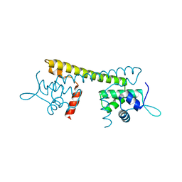
 | | Cryo-EM structure of the mammalian DP-state ATP synthase | | 分子名称: | ADENOSINE-5'-DIPHOSPHATE, ADENOSINE-5'-TRIPHOSPHATE, ATP synthase F1 subunit epsilon, ... | | 著者 | Gu, J, Zhang, L, Yi, J, Yang, M. | | 登録日 | 2019-01-11 | | 公開日 | 2019-06-26 | | 最終更新日 | 2024-03-27 | | 実験手法 | ELECTRON MICROSCOPY (3.34 Å) | | 主引用文献 | Cryo-EM structure of the mammalian ATP synthase tetramer bound with inhibitory protein IF1.
Science, 364, 2019
|
|
6J54
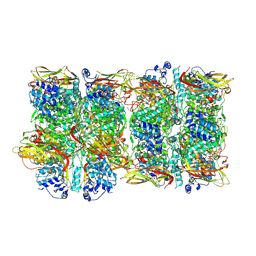
 | | Cryo-EM structure of the mammalian E-state ATP synthase FO section | | 分子名称: | ATP synthase membrane subunit 6.8PL, ATP synthase membrane subunit DAPIT, ATP synthase peripheral stalk-membrane subunit b, ... | | 著者 | Gu, J, Zhang, L, Yi, J, Yang, M. | | 登録日 | 2019-01-10 | | 公開日 | 2019-06-26 | | 最終更新日 | 2024-03-27 | | 実験手法 | ELECTRON MICROSCOPY (3.94 Å) | | 主引用文献 | Cryo-EM structure of the mammalian ATP synthase tetramer bound with inhibitory protein IF1.
Science, 364, 2019
|
|
6J5A
 | | Cryo-EM structure of the mammalian DP-state ATP synthase FO section | | 分子名称: | ATP synthase membrane subunit 6.8PL, ATP synthase membrane subunit DAPIT, ATP synthase peripheral stalk-membrane subunit b, ... | | 著者 | Gu, J, Zhang, L, Yi, J, Yang, M. | | 登録日 | 2019-01-10 | | 公開日 | 2019-06-26 | | 最終更新日 | 2024-03-27 | | 実験手法 | ELECTRON MICROSCOPY (4.35 Å) | | 主引用文献 | Cryo-EM structure of the mammalian ATP synthase tetramer bound with inhibitory protein IF1.
Science, 364, 2019
|
|
6IZO
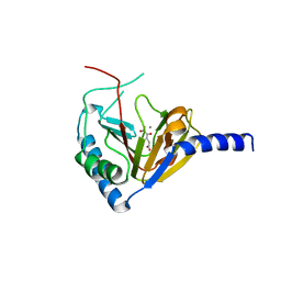
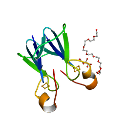
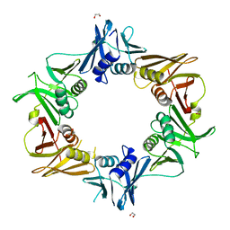 | | Crystal structure of DNA polymerase sliding clamp from Caulobacter crescentus | | 分子名称: | 1,2-ETHANEDIOL, Beta sliding clamp, DI(HYDROXYETHYL)ETHER | | 著者 | Jiang, X, Zhang, L, Teng, M, Li, X. | | 登録日 | 2018-12-20 | | 公開日 | 2019-11-27 | | 最終更新日 | 2023-11-22 | | 実験手法 | X-RAY DIFFRACTION (1.94 Å) | | 主引用文献 | Caulobacter crescentus beta sliding clamp employs a noncanonical regulatory model of DNA replication.
Febs J., 287, 2020
|
|
7CK6
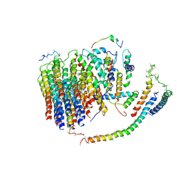
 | | Protein translocase of mitochondria | | 分子名称: | 1,2-DIACYL-SN-GLYCERO-3-PHOSPHOCHOLINE, Mitochondrial import receptor subunit TOM22 homolog, Mitochondrial import receptor subunit TOM40 homolog, ... | | 著者 | Yang, M, Wang, W, Zhang, L, Chen, X. | | 登録日 | 2020-07-15 | | 公開日 | 2020-11-04 | | 最終更新日 | 2024-03-27 | | 実験手法 | ELECTRON MICROSCOPY (3.4 Å) | | 主引用文献 | Atomic structure of human TOM core complex.
Cell Discov, 6, 2020
|
|
4KT8
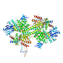
 | | The complex structure of Rv3378c-Y51FY90F with substrate, TPP | | 分子名称: | (2E)-3-methyl-5-[(1R,2S,8aS)-1,2,5,5-tetramethyl-1,2,3,5,6,7,8,8a-octahydronaphthalen-1-yl]pent-2-en-1-yl trihydrogen diphosphate, Diterpene synthase, PHOSPHATE ION | | 著者 | Chan, H.C, Feng, X, Ko, T.P, Huang, C.H, Hu, Y, Zheng, Y, Bogue, S, Nakano, C, Hoshino, T, Zhang, L, Lv, P, Liu, W, Crick, D.C, Liang, P.H, Wang, A.H, Oldfield, E, Guo, R.T. | | 登録日 | 2013-05-20 | | 公開日 | 2014-02-26 | | 最終更新日 | 2023-11-08 | | 実験手法 | X-RAY DIFFRACTION (2.4 Å) | | 主引用文献 | Structure and inhibition of tuberculosinol synthase and decaprenyl diphosphate synthase from Mycobacterium tuberculosis.
J.Am.Chem.Soc., 136, 2014
|
|
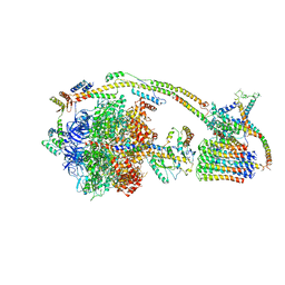
6J5K
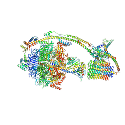
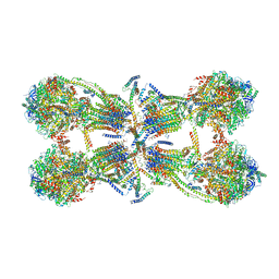 | | Cryo-EM structure of the mammalian ATP synthase tetramer bound with inhibitory protein IF1 | | 分子名称: | ADENOSINE-5'-DIPHOSPHATE, ADENOSINE-5'-TRIPHOSPHATE, ATP synthase F1 subunit alpha, ... | | 著者 | Gu, J, Zhang, L, Yi, J, Yang, M. | | 登録日 | 2019-01-11 | | 公開日 | 2019-06-26 | | 最終更新日 | 2024-03-27 | | 実験手法 | ELECTRON MICROSCOPY (6.2 Å) | | 主引用文献 | Cryo-EM structure of the mammalian ATP synthase tetramer bound with inhibitory protein IF1.
Science, 364, 2019
|
|
4KQX
 | | Mutant Slackia exigua KARI DDV in complex with NAD and an inhibitor | | 分子名称: | HISTIDINE, Ketol-acid reductoisomerase, MAGNESIUM ION, ... | | 著者 | Brinkmann-Chen, S, Flock, T, Cahn, J.K.B, Snow, C.D, Brustad, E.M, Mcintosh, J.A, Meinhold, P, Zhang, L, Arnold, F.H. | | 登録日 | 2013-05-15 | | 公開日 | 2013-06-26 | | 最終更新日 | 2023-09-20 | | 実験手法 | X-RAY DIFFRACTION (1.8 Å) | | 主引用文献 | General approach to reversing ketol-acid reductoisomerase cofactor dependence from NADPH to NADH.
Proc.Natl.Acad.Sci.USA, 110, 2013
|
|
6J5J
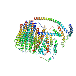
 | | Cryo-EM structure of the mammalian E-state ATP synthase | | 分子名称: | ADENOSINE-5'-DIPHOSPHATE, ADENOSINE-5'-TRIPHOSPHATE, ATP synthase F1 subunit epsilon, ... | | 著者 | Gu, J, Zhang, L, Yi, J, Yang, M. | | 登録日 | 2019-01-11 | | 公開日 | 2019-06-26 | | 最終更新日 | 2024-03-27 | | 実験手法 | ELECTRON MICROSCOPY (3.45 Å) | | 主引用文献 | Cryo-EM structure of the mammalian ATP synthase tetramer bound with inhibitory protein IF1.
Science, 364, 2019
|
|
4KQW
 | | The structure of the Slackia exigua KARI in complex with NADP | | 分子名称: | Ketol-acid reductoisomerase, L(+)-TARTARIC ACID, MAGNESIUM ION, ... | | 著者 | Brinkmann-Chen, S, Flock, T, Cahn, J.K.B, Snow, C.D, Brustad, E.M, Mcintosh, J.A, Meinhold, P, Zhang, L, Arnold, F.H. | | 登録日 | 2013-05-15 | | 公開日 | 2013-06-26 | | 最終更新日 | 2023-09-20 | | 実験手法 | X-RAY DIFFRACTION (1.39 Å) | | 主引用文献 | General approach to reversing ketol-acid reductoisomerase cofactor dependence from NADPH to NADH.
Proc.Natl.Acad.Sci.USA, 110, 2013
|
|
7CJX
 | |
4NPM
 | |
4NPL
 | |
3PLA
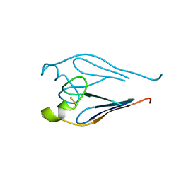
 | | Crystal structure of a catalytically active substrate-bound box C/D RNP from Sulfolobus solfataricus | | 分子名称: | 50S ribosomal protein L7Ae, C/D guide RNA, Fibrillarin-like rRNA/tRNA 2'-O-methyltransferase, ... | | 著者 | Lin, J, Lai, S, Jia, R, Xu, A, Zhang, L, Lu, J, Ye, K. | | 登録日 | 2010-11-15 | | 公開日 | 2011-01-26 | | 最終更新日 | 2023-11-01 | | 実験手法 | X-RAY DIFFRACTION (3.15 Å) | | 主引用文献 | Structural basis for site-specific ribose methylation by box C/D RNA protein complexes.
Nature, 469, 2011
|
|
3TBO
 | |
3TBN
 | |
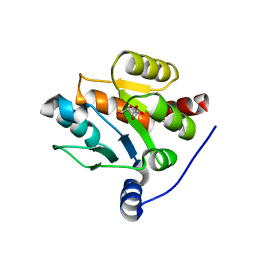
3TBM
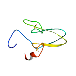
 | | Crystal structure of a type 4 CDGSH iron-sulfur protein. | | 分子名称: | FE2/S2 (INORGANIC) CLUSTER, L(+)-TARTARIC ACID, NONAETHYLENE GLYCOL, ... | | 著者 | Lin, J, Zhang, L, Ye, K. | | 登録日 | 2011-08-07 | | 公開日 | 2011-10-05 | | 最終更新日 | 2024-03-20 | | 実験手法 | X-RAY DIFFRACTION (1.797 Å) | | 主引用文献 | Structure and Molecular Evolution of CDGSH Iron-Sulfur Domains.
Plos One, 6, 2011
|
|
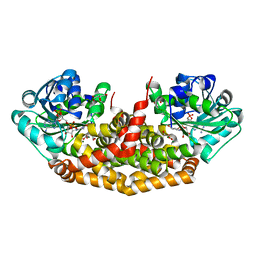
7FHA
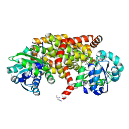
 | | Crystal structure of the ATP sulfurylase domain of human PAPSS2 in complex with APS | | 分子名称: | ADENOSINE-5'-PHOSPHOSULFATE, Bifunctional 3'-phosphoadenosine 5'-phosphosulfate synthase 2, POTASSIUM ION, ... | | 著者 | Zhang, P, Zhang, L, Zhang, L. | | 登録日 | 2021-07-29 | | 公開日 | 2021-12-01 | | 最終更新日 | 2023-11-29 | | 実験手法 | X-RAY DIFFRACTION (2 Å) | | 主引用文献 | Structural basis for the substrate recognition mechanism of ATP-sulfurylase domain of human PAPS synthase 2.
Biochem.Biophys.Res.Commun., 586, 2022
|
|
7FH3
 | |
8JI8
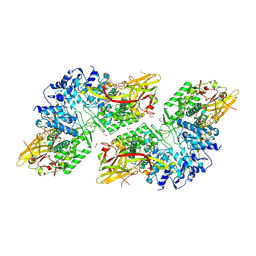 | | Crystal Structure of Prophenoloxidase PPO6 chimeric mutant (F215EASNRAIVD224 to G215DGPDSVVR223) from Aedes aegypti | | 分子名称: | 1,2-ETHANEDIOL, 2-AMINO-2-HYDROXYMETHYL-PROPANE-1,3-DIOL, COPPER (II) ION, ... | | 著者 | Zhu, X, Zhang, L, Yang, X, Bao, P, Ren, D, Han, Q. | | 登録日 | 2023-05-26 | | 公開日 | 2023-11-01 | | 実験手法 | X-RAY DIFFRACTION (2.65 Å) | | 主引用文献 | Mosquitoes have evolved two types of prophenoloxidases
To Be Published
|
|
8JIB
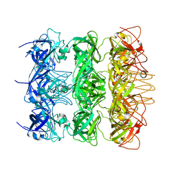
 | | Crystal Structure of Prophenoloxidase PPO6 from Aedes aegypti | | 分子名称: | 1,2-ETHANEDIOL, COPPER (II) ION, TK receptor | | 著者 | Zhu, X, Zhang, L, Yang, X, Bao, P, Ren, D, Han, Q. | | 登録日 | 2023-05-26 | | 公開日 | 2023-11-29 | | 実験手法 | X-RAY DIFFRACTION (3.15 Å) | | 主引用文献 | Mosquitoes have evolved two types of prophenoloxidases
To Be Published
|
|
7E6G
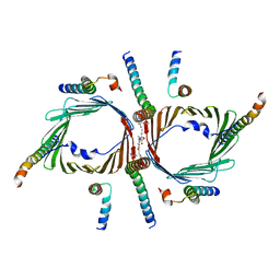
 | | Crystal structure of diguanylate cyclase SiaD in complex with its activator SiaC from Pseudomonas aeruginosa | | 分子名称: | DUF1987 domain-containing protein, MAGNESIUM ION, PHOSPHOMETHYLPHOSPHONIC ACID GUANYLATE ESTER, ... | | 著者 | Zhou, J.S, Zhang, L, Zhang, L. | | 登録日 | 2021-02-22 | | 公開日 | 2021-09-22 | | 最終更新日 | 2023-11-29 | | 実験手法 | X-RAY DIFFRACTION (2.65 Å) | | 主引用文献 | Structural basis for diguanylate cyclase activation by its binding partner in Pseudomonas aeruginosa .
Elife, 10, 2021
|
|
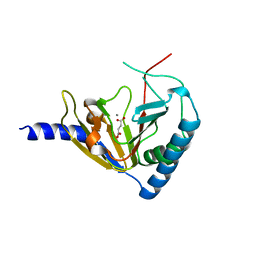
6KZ4
 | | YebT domain 5-7 | | 分子名称: | Intermembrane transport protein YebT | | 著者 | Wang, H.W, Liu, C, Zhang, L. | | 登録日 | 2019-09-23 | | 公開日 | 2020-01-15 | | 最終更新日 | 2024-03-27 | | 実験手法 | ELECTRON MICROSCOPY (3 Å) | | 主引用文献 | Cryo-EM Structure of a Bacterial Lipid Transporter YebT.
J.Mol.Biol., 432, 2020
|
|
6KZ3
 | | YebT domain1-4 | | 分子名称: | Intermembrane transport protein YebT | | 著者 | Wang, H.W, Liu, C, Zhang, L. | | 登録日 | 2019-09-23 | | 公開日 | 2020-01-15 | | 最終更新日 | 2024-03-27 | | 実験手法 | ELECTRON MICROSCOPY (3.1 Å) | | 主引用文献 | Cryo-EM Structure of a Bacterial Lipid Transporter YebT.
J.Mol.Biol., 432, 2020
|
|


