Loading
PDBj
MenuPDBj@FacebookPDBj@TwitterPDBj@YouTubewwPDB FoundationwwPDB
RCSB PDBPDBeBMRBAdv. SearchSearch help
Search by PDB author
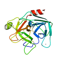
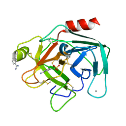
1BIT
DownloadVisualize
BU of 1bit by Molmil
THE CRYSTAL STRUCTURE OF ANIONIC SALMON TRYPSIN IN A SECOND CRYSTAL FORM
Descriptor: BENZAMIDINE, CALCIUM ION, SULFATE ION, ...
Authors:Berglund, G.I.
Deposit date:1994-08-26
Release date:1994-11-01
Last modified:2019-08-14
Method:X-RAY DIFFRACTION (1.83 Å)
Cite:Structure of anionic salmon trypsin in a second crystal form.
Acta Crystallogr.,Sect.D, 51, 1995
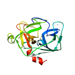
1ELT
DownloadVisualize
BU of 1elt by Molmil
STRUCTURE OF NATIVE PANCREATIC ELASTASE FROM NORTH ATLANTIC SALMON AT 1.61 ANGSTROMS RESOLUTION
Descriptor: CALCIUM ION, ELASTASE
Authors:Berglund, G.I, Smalaas, A.O.
Deposit date:1995-01-02
Release date:1996-01-01
Last modified:2018-04-18
Method:X-RAY DIFFRACTION (1.61 Å)
Cite:Structure of native pancreatic elastase from North Atlantic salmon at 1.61 A resolution.
Acta Crystallogr.,Sect.D, 51, 1995
2TBS
DownloadVisualize
BU of 2tbs by Molmil
COLD-ADAPTION OF ENZYMES: STRUCTURAL COMPARISON BETWEEN SALMON AND BOVINE TRYPSINS
Descriptor: BENZAMIDINE, CALCIUM ION, TRYPSIN
Authors:Smalas, A.O.
Deposit date:1994-01-14
Release date:1994-04-30
Last modified:2011-07-13
Method:X-RAY DIFFRACTION (1.8 Å)
Cite:Cold adaption of enzymes: structural comparison between salmon and bovine trypsins.
Proteins, 20, 1994
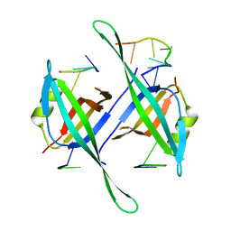
5ODP
DownloadVisualize
BU of 5odp by Molmil
Salinibacter ruber Single-Strand Binding protein D17K D71K mutant
Descriptor: DNA (5'-D(*TP*TP*TP*TP*TP*TP*TP*TP*T)-3'), Single-stranded DNA-binding protein
Authors:Pierechod, M, Rothweiler, U.
Deposit date:2017-07-06
Release date:2018-08-01
Last modified:2024-01-17
Method:X-RAY DIFFRACTION (2.535 Å)
Cite:Salinibacter ruber Single-Strand Binding protein D17K D71K mutant
To Be Published
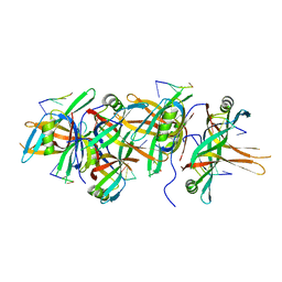
5ODN
DownloadVisualize
BU of 5odn by Molmil
Salinibacter ruber Single-Strand Binding protein
Descriptor: DNA (5'-D(*TP*TP*T)-3'), DNA (5'-D(P*TP*TP*TP*T)-3'), Single-stranded DNA-binding protein
Authors:Pierechod, M, Rothweiler, U.
Deposit date:2017-07-06
Release date:2018-08-01
Last modified:2024-01-17
Method:X-RAY DIFFRACTION (2.598 Å)
Cite:Salinibacter ruber Single-Strand Binding protein
To Be Published
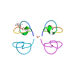
1LU0
DownloadVisualize
BU of 1lu0 by Molmil
Atomic Resolution Structure of Squash Trypsin Inhibitor: Unexpected Metal Coordination
Descriptor: (4R)-2-METHYLPENTANE-2,4-DIOL, GLYCEROL, SULFATE ION, ...
Authors:Thaimattam, R, Tykarska, E, Bierzynski, A, Sheldrick, G.M, Jaskolski, M.
Deposit date:2002-05-21
Release date:2002-08-28
Last modified:2021-10-27
Method:X-RAY DIFFRACTION (1.03 Å)
Cite:Atomic resolution structure of squash trypsin inhibitor: unexpected metal coordination.
Acta Crystallogr.,Sect.D, 58, 2002
<12


218853

PDB entries from 2024-04-24

PDB statisticsPDBj update infoContact PDBjnumon