Loading
PDBj
MenuPDBj@FacebookPDBj@TwitterPDBj@YouTubewwPDB FoundationwwPDB
RCSB PDBPDBeBMRBAdv. SearchSearch help
Search by PDB author
2NOK
DownloadVisualize
BU of 2nok by Molmil
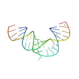
Crystal Structure of an RNA domain from Hepatitis C virus.
Descriptor: 5'-R(*CP*GP*GP*AP*GP*GP*AP*AP*CP*UP*AP*CP*UP*GP*UP*CP*UP*UP*CP*AP*CP*GP*CP*C)-3', 5'-R(*GP*CP*GP*UP*GP*UP*CP*GP*UP*GP*CP*AP*GP*CP*CP*UP*CP*CP*GP*G)-3', MAGNESIUM ION, ...
Authors:Dibrov, S.M, Johnston-Cos, H, Weng, Y.H.
Deposit date:2006-10-25
Release date:2007-02-13
Last modified:2023-12-27
Method:X-RAY DIFFRACTION (3 Å)
Cite:Functional architecture of HCV IRES domain II stabilized by divalent metal ions in the crystal and in solution.
Angew.Chem.Int.Ed.Engl., 46, 2007
3QBE
DownloadVisualize
BU of 3qbe by Molmil
Crystal structure of the 3-Dehydroquinate Synthase (aroB) from Mycobacterium tuberculosis
Descriptor: 3-dehydroquinate synthase, CHLORIDE ION, ZINC ION
Authors:Cheng, W.C, Wen, Y.H, Wang, W.C.
Deposit date:2011-01-13
Release date:2012-01-18
Last modified:2023-11-01
Method:X-RAY DIFFRACTION (2.07 Å)
Cite:Crystal structure of the 3-Dehydroquinate Synthase (aroB) from Mycobacterium tuberculosis
To be Published

222926

PDB entries from 2024-07-24

PDB statisticsPDBj update infoContact PDBjnumon