5T0N
 
 | |
5N6W
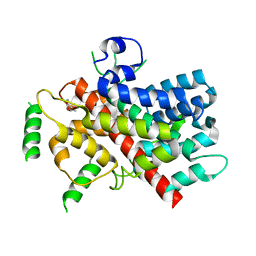
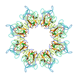 | | Retinoschisin R141H Mutant | | 分子名称: | Retinoschisin | | 著者 | Ramsay, E.P, Collins, R.F, Owens, T.W, Siebert, C.A, Jones, R.P.O, Roseman, A, Wang, T, Baldock, C. | | 登録日 | 2017-02-16 | | 公開日 | 2017-04-12 | | 最終更新日 | 2017-08-30 | | 実験手法 | ELECTRON MICROSCOPY (4.2 Å) | | 主引用文献 | Structural analysis of X-linked retinoschisis mutations reveals distinct classes which differentially effect retinoschisin function
Human Molecular Genetics, 25, 2016
|
|
5X57
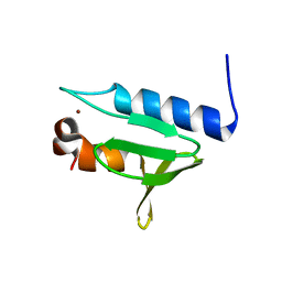 | | Structure of GAR domain of ACF7 | | 分子名称: | Microtubule-actin cross-linking factor 1, isoforms 1/2/3/5, NICKEL (II) ION | | 著者 | Yang, F, Wang, T, Zhang, Y, Wu, X.Y. | | 登録日 | 2017-02-15 | | 公開日 | 2017-07-05 | | 最終更新日 | 2024-03-27 | | 実験手法 | X-RAY DIFFRACTION (1.45 Å) | | 主引用文献 | ACF7 regulates inflammatory colitis and intestinal wound response by orchestrating tight junction dynamics.
Nat Commun, 8, 2017
|
|
7YH8
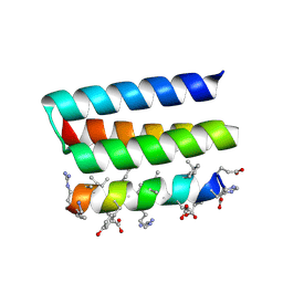 | | Crystal structure of a heterochiral protein complex | | 分子名称: | D-Pep-1, L-19437 | | 著者 | Liang, M, Li, S, Wang, T, Liu, L, Lu, P. | | 登録日 | 2022-07-13 | | 公開日 | 2023-07-19 | | 実験手法 | X-RAY DIFFRACTION (2.2 Å) | | 主引用文献 | Crystal structure of a heterochiral protein complex
To Be Published
|
|


