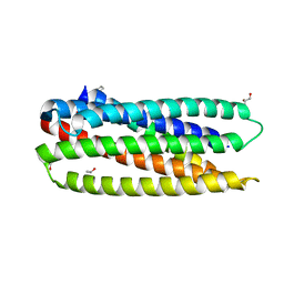
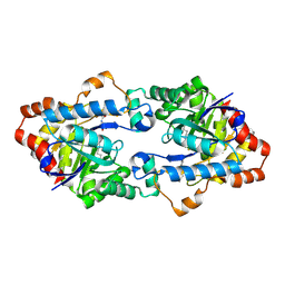
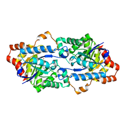
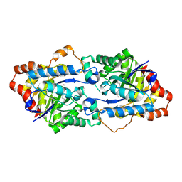
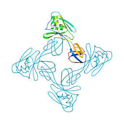
3NIT
 
 | | The structure of UBR box (native1) | | 分子名称: | E3 ubiquitin-protein ligase UBR1, ZINC ION | | 著者 | Choi, W.S, Jeong, B.-C, Lee, M.-R, Song, H.K. | | 登録日 | 2010-06-16 | | 公開日 | 2010-09-15 | | 最終更新日 | 2024-03-20 | | 実験手法 | X-RAY DIFFRACTION (2.6 Å) | | 主引用文献 | Structural basis for the recognition of N-end rule substrates by the UBR box of ubiquitin ligases
Nat.Struct.Mol.Biol., 17, 2010
|
|
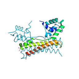
3O43
 | |
4H9V
 | | Structure of Geobacillus kaustophilus lactonase, mutant E101G/R230C with Zn2+ | | 分子名称: | FE (III) ION, HYDROXIDE ION, Phosphotriesterase, ... | | 著者 | Xue, B, Chow, J.Y, Yew, W.S, Robinson, R.C. | | 登録日 | 2012-09-25 | | 公開日 | 2012-11-07 | | 最終更新日 | 2013-05-22 | | 実験手法 | X-RAY DIFFRACTION (1.971 Å) | | 主引用文献 | Structural evidence of a productive active site architecture for an evolved quorum-quenching GKL lactonase.
Biochemistry, 52, 2013
|
|
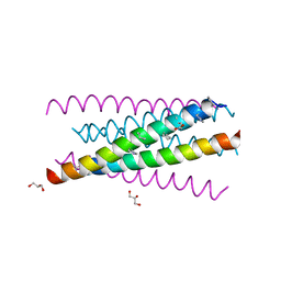
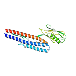
3O40
 | | Complex of a chimeric alpha/beta-peptide based on the gp41 CHR domain bound to gp41-5 | | 分子名称: | CHLORIDE ION, GLYCEROL, NONAETHYLENE GLYCOL, ... | | 著者 | Horne, W.S, Johnson, L.M, Gellman, S.H. | | 登録日 | 2010-07-26 | | 公開日 | 2011-07-27 | | 最終更新日 | 2023-11-15 | | 実験手法 | X-RAY DIFFRACTION (2.1 Å) | | 主引用文献 | Broad Distribution of Energetically Important Contacts across an Extended Protein Interface.
J.Am.Chem.Soc., 133, 2011
|
|
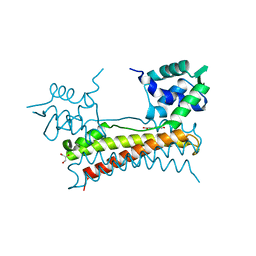
3O42
 | |
4H9T
 | | Structure of Geobacillus kaustophilus lactonase, mutant E101N with bound N-butyryl-DL-homoserine lactone | | 分子名称: | FE (III) ION, MANGANESE (II) ION, N-[(3S)-2-oxotetrahydrofuran-3-yl]butanamide, ... | | 著者 | Xue, B, Chow, J.Y, Yew, W.S, Robinson, R.C. | | 登録日 | 2012-09-24 | | 公開日 | 2012-11-07 | | 最終更新日 | 2013-05-22 | | 実験手法 | X-RAY DIFFRACTION (2.097 Å) | | 主引用文献 | Structural evidence of a productive active site architecture for an evolved quorum-quenching GKL lactonase.
Biochemistry, 52, 2013
|
|
3O3Z
 | |
3O3X
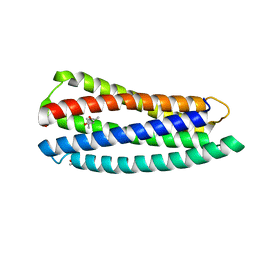 | | Crystal structure of gp41-5, a single-chain 5-helix-bundle based on HIV gp41 | | 分子名称: | (4S)-2-METHYL-2,4-PENTANEDIOL, ISOPROPYL ALCOHOL, gp41-5 | | 著者 | Horne, W.S, Johnson, L.M, Gellman, S.H. | | 登録日 | 2010-07-26 | | 公開日 | 2011-07-27 | | 最終更新日 | 2023-09-06 | | 実験手法 | X-RAY DIFFRACTION (1.45 Å) | | 主引用文献 | Broad Distribution of Energetically Important Contacts across an Extended Protein Interface.
J.Am.Chem.Soc., 133, 2011
|
|
4HA0
 | | Structure of Geobacillus kaustophilus lactonase, mutant R230D with Zn2+ | | 分子名称: | FE (III) ION, HYDROXIDE ION, Phosphotriesterase, ... | | 著者 | Xue, B, Chow, J.Y, Yew, W.S, Robinson, R.C. | | 登録日 | 2012-09-25 | | 公開日 | 2012-11-07 | | 最終更新日 | 2013-05-22 | | 実験手法 | X-RAY DIFFRACTION (1.902 Å) | | 主引用文献 | Structural evidence of a productive active site architecture for an evolved quorum-quenching GKL lactonase.
Biochemistry, 52, 2013
|
|
4H9U
 | | Structure of Geobacillus kaustophilus lactonase, wild-type with Zn2+ | | 分子名称: | FE (III) ION, HYDROXIDE ION, Phosphotriesterase, ... | | 著者 | Xue, B, Chow, J.Y, Yew, W.S, Robinson, R.C. | | 登録日 | 2012-09-25 | | 公開日 | 2012-11-07 | | 最終更新日 | 2013-05-22 | | 実験手法 | X-RAY DIFFRACTION (2.099 Å) | | 主引用文献 | Structural evidence of a productive active site architecture for an evolved quorum-quenching GKL lactonase.
Biochemistry, 52, 2013
|
|
4H9X
 | | Structure of Geobacillus kaustophilus lactonase, mutant E101G/R230C/D266N with Zn2+ and bound N-butyryl-DL-homoserine lactone | | 分子名称: | FE (III) ION, HYDROXIDE ION, N-[(3S)-2-oxotetrahydrofuran-3-yl]butanamide, ... | | 著者 | Xue, B, Chow, J.Y, Yew, W.S, Robinson, R.C. | | 登録日 | 2012-09-25 | | 公開日 | 2012-11-07 | | 最終更新日 | 2013-05-22 | | 実験手法 | X-RAY DIFFRACTION (2.201 Å) | | 主引用文献 | Structural evidence of a productive active site architecture for an evolved quorum-quenching GKL lactonase.
Biochemistry, 52, 2013
|
|
5X12
 | | Crystal structure of Bacillus subtilis PadR | | 分子名称: | Transcriptional regulator | | 著者 | Park, S.C, Kwak, Y.M, Song, W.S, Hong, M, Yoon, S.I. | | 登録日 | 2017-01-24 | | 公開日 | 2017-11-22 | | 最終更新日 | 2024-03-20 | | 実験手法 | X-RAY DIFFRACTION (1.7 Å) | | 主引用文献 | Structural basis of effector and operator recognition by the phenolic acid-responsive transcriptional regulator PadR
Nucleic Acids Res., 45, 2017
|
|
5X11
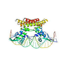 | | Crystal structure of Bacillus subtilis PadR in complex with operator DNA | | 分子名称: | DNA (28-MER), Transcriptional regulator | | 著者 | Park, S.C, Kwak, Y.M, Song, W.S, Hong, M, Yoon, S.I. | | 登録日 | 2017-01-24 | | 公開日 | 2017-11-22 | | 最終更新日 | 2023-11-22 | | 実験手法 | X-RAY DIFFRACTION (2.65 Å) | | 主引用文献 | Structural basis of effector and operator recognition by the phenolic acid-responsive transcriptional regulator PadR
Nucleic Acids Res., 45, 2017
|
|
5X14
 | | Crystal structure of Bacillus subtilis PadR in complex with ferulic acid | | 分子名称: | 3-(4-HYDROXY-3-METHOXYPHENYL)-2-PROPENOIC ACID, GLYCEROL, Transcriptional regulator | | 著者 | Park, S.C, Kwak, Y.M, Song, W.S, Hong, M, Yoon, S.I. | | 登録日 | 2017-01-24 | | 公開日 | 2017-11-22 | | 最終更新日 | 2023-11-22 | | 実験手法 | X-RAY DIFFRACTION (1.68 Å) | | 主引用文献 | Structural basis of effector and operator recognition by the phenolic acid-responsive transcriptional regulator PadR
Nucleic Acids Res., 45, 2017
|
|
5X13
 | | Crystal structure of Bacillus subtilis PadR in complex with p-coumaric acid | | 分子名称: | 4'-HYDROXYCINNAMIC ACID, GLYCEROL, Transcriptional regulator | | 著者 | Park, S.C, Kwak, Y.M, Song, W.S, Hong, M, Yoon, S.I. | | 登録日 | 2017-01-24 | | 公開日 | 2017-11-22 | | 最終更新日 | 2023-11-22 | | 実験手法 | X-RAY DIFFRACTION (1.7 Å) | | 主引用文献 | Structural basis of effector and operator recognition by the phenolic acid-responsive transcriptional regulator PadR
Nucleic Acids Res., 45, 2017
|
|
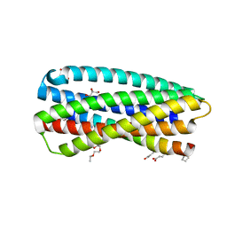
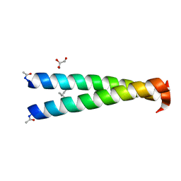
4NJ0
 | | GCN4-p1 single Val9 to Ile mutant | | 分子名称: | General control protein GCN4 | | 著者 | Oshaben, K.M, Horne, W.S. | | 登録日 | 2013-11-08 | | 公開日 | 2014-08-20 | | 最終更新日 | 2023-09-20 | | 実験手法 | X-RAY DIFFRACTION (1.9 Å) | | 主引用文献 | Tuning assembly size in Peptide-based supramolecular polymers by modulation of subunit association affinity.
Biomacromolecules, 15, 2014
|
|
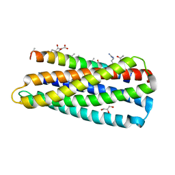
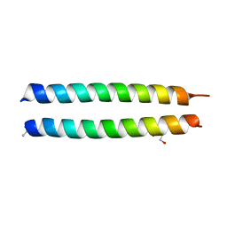
4NJ1
 | | GCN4-p1 double Val9, 23 to Ile mutant | | 分子名称: | General control protein GCN4 | | 著者 | Oshaben, K.M, Horne, W.S. | | 登録日 | 2013-11-08 | | 公開日 | 2014-08-20 | | 最終更新日 | 2023-09-20 | | 実験手法 | X-RAY DIFFRACTION (2 Å) | | 主引用文献 | Tuning assembly size in Peptide-based supramolecular polymers by modulation of subunit association affinity.
Biomacromolecules, 15, 2014
|
|
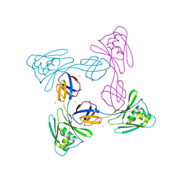
5XLJ
 | | Crystal structure of the flagellar cap protein flid D2-D3 domains from serratia marcescens in Space group P432 | | 分子名称: | CHLORIDE ION, Flagellar hook-associated protein 2, SODIUM ION | | 著者 | Cho, S.Y, Song, W.S, Hong, H.J, Yoon, S.I. | | 登録日 | 2017-05-10 | | 公開日 | 2017-06-14 | | 最終更新日 | 2023-11-22 | | 実験手法 | X-RAY DIFFRACTION (1.9 Å) | | 主引用文献 | Tetrameric structure of the flagellar cap protein FliD from Serratia marcescens.
Biochem. Biophys. Res. Commun., 489, 2017
|
|
4NIZ
 | |
4NJ2
 | | GCN4-p1 triple Val9, 23,30 to Ile mutant | | 分子名称: | GLYCEROL, General control protein GCN4 | | 著者 | Oshaben, K.M, Horne, W.S. | | 登録日 | 2013-11-08 | | 公開日 | 2014-08-20 | | 最終更新日 | 2023-09-20 | | 実験手法 | X-RAY DIFFRACTION (2.2 Å) | | 主引用文献 | Tuning assembly size in Peptide-based supramolecular polymers by modulation of subunit association affinity.
Biomacromolecules, 15, 2014
|
|
5XLK
 | | Crystal structure of the flagellar cap protein FliD D2-D3 domains from Serratia marcescens in Space group I422 | | 分子名称: | Flagellar hook-associated protein 2, ZINC ION | | 著者 | Cho, S.Y, Song, W.S, Hong, H.J, Yoon, S.I. | | 登録日 | 2017-05-10 | | 公開日 | 2017-06-14 | | 最終更新日 | 2023-11-22 | | 実験手法 | X-RAY DIFFRACTION (3.05 Å) | | 主引用文献 | Tetrameric structure of the flagellar cap protein FliD from Serratia marcescens.
Biochem. Biophys. Res. Commun., 489, 2017
|
|
4NX9
 | |
5KNR
 | | E. coli HPRT in complexed with 9-[(N-phosphonoethyl-N-phosphonoethoxyethyl)-2-aminoethyl]-guanine | | 分子名称: | (2-{[2-(2-amino-6-oxo-3,6-dihydro-9H-purin-9-yl)ethyl][2-(2-phosphonoethoxy)ethyl]amino}ethyl)phosphonic acid, Hypoxanthine-guanine phosphoribosyltransferase, MAGNESIUM ION | | 著者 | Eng, W.S, Keough, D.T, Hockova, D, Janeba, Z. | | 登録日 | 2016-06-28 | | 公開日 | 2017-07-19 | | 最終更新日 | 2023-09-27 | | 実験手法 | X-RAY DIFFRACTION (2.864 Å) | | 主引用文献 | Crystal Structures of Acyclic Nucleoside Phosphonates in Complex with Escherichia coli Hypoxanthine Phosphoribosyltransferase
Chemistryselect, 1, 2016
|
|
5KNU
 | | Crystal structure of E. coli hypoxanthine phosphoribosyltransferase in complexed with 9-[N,N-(Bis-3-phosphonopropyl)aminomethyl]-9-deazahypoxanthine | | 分子名称: | 3-[(4-oxidanylidene-3,5-dihydropyrrolo[3,2-d]pyrimidin-7-yl)methyl-(3-phosphonopropyl)amino]propylphosphonic acid, 4-(2-HYDROXYETHYL)-1-PIPERAZINE ETHANESULFONIC ACID, Hypoxanthine-guanine phosphoribosyltransferase, ... | | 著者 | Eng, W.S, Keough, D.T, Baszczynski, O, Hockova, D, Janeba, Z. | | 登録日 | 2016-06-28 | | 公開日 | 2017-07-19 | | 最終更新日 | 2023-09-27 | | 実験手法 | X-RAY DIFFRACTION (2.808 Å) | | 主引用文献 | Crystal Structures of Acyclic Nucleoside Phosphonates in Complex with Escherichia coli Hypoxanthine Phosphoribosyltransferase
Chemistryselect, 1, 2016
|
|
5KNX
 | | Crystal structure of E. coli hypoxanthine phosphoribosyltransferase in complexed with {[(2-[(Hypoxanthin-9H-yl)methyl]propane-1,3-diyl)bis(oxy)]bis- (methylene)}diphosphonic Acid | | 分子名称: | Hypoxanthine-guanine phosphoribosyltransferase, MAGNESIUM ION, [2-[(6-oxidanylidene-1~{H}-purin-9-yl)methyl]-3-(phosphonomethoxy)propoxy]methylphosphonic acid | | 著者 | Eng, W.S, Keough, D.T, Hockova, D, Janeba, Z, Guddat, L.W. | | 登録日 | 2016-06-28 | | 公開日 | 2017-07-19 | | 最終更新日 | 2024-03-06 | | 実験手法 | X-RAY DIFFRACTION (2.4 Å) | | 主引用文献 | Crystal Structures of Acyclic Nucleoside Phosphonates in Complex with Escherichia coli Hypoxanthine Phosphoribosyltransferase
Chemistryselect, 1, 2016
|
|


