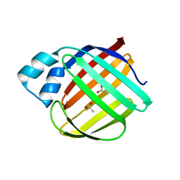
Loading
PDBj
メニューPDBj@FacebookPDBj@TwitterPDBj@YouTubewwPDB FoundationwwPDB
RCSB PDBPDBeBMRBAdv. SearchSearch help
Search by PDB author
4ZJ0
DownloadVisualize
BU of 4zj0 by Molmil
The crystal structure of monomer Q108K:K40L:Y60W CRBPII bound to all-trans-retinal
分子名称: ACETATE ION, RETINAL, Retinol-binding protein 2
著者Nossoni, Z, Assar, Z, Wang, W, Vasileiou, C, Borhan, B, Geiger, J.H.
登録日2015-04-28
公開日2016-05-11
最終更新日2023-09-27
実験手法X-RAY DIFFRACTION (1.5 Å)
主引用文献Domain-Swapped Dimers of Intracellular Lipid-Binding Proteins: Evidence for Ordered Folding Intermediates.
Structure, 24, 2016
4QYP
DownloadVisualize
BU of 4qyp by Molmil
The Crystal Structures of holo-wt human Cellular Retinol Binding protein II (hCRBPII) bound to Retinal
分子名称: ACETATE ION, RETINAL, Retinol-binding protein 2
著者Nossoni, Z, Assar, Z, Yapici, I, Nosrati, M, Wang, W, Berbasova, T, Vasileiou, C, Borhan, B, Geiger, H.
登録日2014-07-25
公開日2014-12-10
最終更新日2023-09-20
実験手法X-RAY DIFFRACTION (1.62 Å)
主引用文献Structures of holo wild-type human cellular retinol-binding protein II (hCRBPII) bound to retinol and retinal.
Acta Crystallogr.,Sect.D, 70, 2014
4QYN
DownloadVisualize
BU of 4qyn by Molmil
The Crystal Structures of holo-wt human Cellular Retinol Binding protein II (hCRBPII) bound to Retinol
分子名称: ACETATE ION, RETINOL, Retinol-binding protein 2
著者Nossoni, Z, Assar, Z, Yapici, I, Nosrati, M, Wang, W, Berbasova, T, Vasileiou, C, Borhan, B, Geiger, H.
登録日2014-07-24
公開日2014-12-31
最終更新日2024-02-28
実験手法X-RAY DIFFRACTION (1.19 Å)
主引用文献Structures of holo wild-type human cellular retinol-binding protein II (hCRBPII) bound to retinol and retinal.
Acta Crystallogr.,Sect.D, 70, 2014
3I17
DownloadVisualize
BU of 3i17 by Molmil
Crystal structure of the apo R132K:L121E mutant of cellular retinoic acid-binding protein II at 1.68 angstrom resolution
分子名称: Cellular retinoic acid-binding protein 2
著者Jia, X, Watson, C.T, Geiger, J.H.
登録日2009-06-25
公開日2009-07-28
最終更新日2023-09-06
実験手法X-RAY DIFFRACTION (1.68 Å)
主引用文献Elucidating the exact role of engineered CRABPII residues for the formation of a retinal protonated Schiff base.
Proteins, 77, 2009
2G78
DownloadVisualize
BU of 2g78 by Molmil
Crystal Structure of the R132K:Y134F Mutant of Cellular Retinoic Acid Binding Protein Type II in Complex with All-Trans-Retinoic Acid at 1.70 Angstroms Resolution
分子名称: Cellular retinoic acid-binding protein 2, RETINOIC ACID, SODIUM ION
著者Vaezeslami, S, Geiger, J.H.
登録日2006-02-27
公開日2007-04-17
最終更新日2024-02-14
実験手法X-RAY DIFFRACTION (1.7 Å)
主引用文献Protein design: reengineering cellular retinoic acid binding protein II into a rhodopsin protein mimic.
J.Am.Chem.Soc., 129, 2007
2G7B
DownloadVisualize
BU of 2g7b by Molmil
Crystal Structure of the R132K:R111L:L121E mutant of Cellular Retinoic Acid Binding Protein Type II In Complex With All-Trans-Retinal At 1.18 Angstroms Resolution
分子名称: ALL-TRANS AXEROPHTHENE, Cellular retinoic acid-binding protein 2, SODIUM ION
著者Vaezeslami, S, Geiger, J.H.
登録日2006-02-27
公開日2007-04-17
最終更新日2021-10-20
実験手法X-RAY DIFFRACTION (1.18 Å)
主引用文献Protein design: reengineering cellular retinoic acid binding protein II into a rhodopsin protein mimic.
J.Am.Chem.Soc., 129, 2007
2G79
DownloadVisualize
BU of 2g79 by Molmil
Crystal Structure of the R132K:Y134F Mutant of Cellular Retinoic Acid Binding Protein Type II in Complex with All-Trans-Retinal at 1.69 Angstroms Resolution
分子名称: Cellular retinoic acid-binding protein 2, RETINAL, SODIUM ION, ...
著者Vaezeslami, S, Geiger, J.H.
登録日2006-02-27
公開日2007-04-17
最終更新日2024-02-14
実験手法X-RAY DIFFRACTION (1.69 Å)
主引用文献Protein design: reengineering cellular retinoic acid binding protein II into a rhodopsin protein mimic.
J.Am.Chem.Soc., 129, 2007
5U6G
DownloadVisualize
BU of 5u6g by Molmil
Crystal Structure of the holo Domain-Swapped Dimer mutant Q108K:K40D Human Cellular Retinol Binding Protein II bound with all trans retinal
分子名称: RETINAL, Retinol-binding protein 2
著者Assar, Z, Geiger, J.H.
登録日2016-12-08
公開日2017-10-18
最終更新日2023-10-04
実験手法X-RAY DIFFRACTION (2.6 Å)
主引用文献Crystal Structure of the holo Domain-Swapped Dimer mutant Q108K:K40D Human Cellular Retinol Binding Protein II bound with all trans retinal
To Be Published
6ON5
DownloadVisualize
BU of 6on5 by Molmil
Crystal Structure of the Zn-bound Domain-Swapped Dimer Q108K:T51D:A28C:L36C:F57H Mutant of Human Cellular Retinol Binding Protein II
分子名称: Retinol-binding protein 2, ZINC ION
著者Ghanbarpour, A, Geiger, J.
登録日2019-04-20
公開日2019-10-16
最終更新日2023-10-11
実験手法X-RAY DIFFRACTION (1.638 Å)
主引用文献Engineering the hCRBPII Domain-Swapped Dimer into a New Class of Protein Switches.
J.Am.Chem.Soc., 141, 2019
6ON8
DownloadVisualize
BU of 6on8 by Molmil
Crystal Structure of the Reduced Form of Apo Domain-Swapped Dimer Q108K:T51D:A28C:L36C:F57H Mutant of Human Cellular Retinol Binding Protein II
分子名称: Retinol-binding protein 2
著者Ghanbarpour, A, Geiger, J.
登録日2019-04-20
公開日2019-10-16
最終更新日2023-10-11
実験手法X-RAY DIFFRACTION (2.395 Å)
主引用文献Engineering the hCRBPII Domain-Swapped Dimer into a New Class of Protein Switches.
J.Am.Chem.Soc., 141, 2019
6ON7
DownloadVisualize
BU of 6on7 by Molmil
Crystal Structure of Apo Domain-Swapped Dimer Q108K:T51D:A28C:L36C Mutant of Human Cellular Retinol Binding Protein II
分子名称: Retinol-binding protein 2
著者Ghanbarpour, A, Geiger, J.
登録日2019-04-20
公開日2019-10-16
最終更新日2023-10-11
実験手法X-RAY DIFFRACTION (1.981 Å)
主引用文献Engineering the hCRBPII Domain-Swapped Dimer into a New Class of Protein Switches.
J.Am.Chem.Soc., 141, 2019
6C7Z
DownloadVisualize
BU of 6c7z by Molmil
Crystal structure of the Q108K:K40L:T51V:R58F mutant of human Cellular Retinol Binding Protein II in complex with synthetic Ligand Julolidine
分子名称: (2E,4E)-3-methyl-5-(2,3,6,7-tetrahydro-1H,5H-pyrido[3,2,1-ij]quinolin-9-yl)penta-2,4-dienal, ACETATE ION, Retinol-binding protein 2
著者Nosrati, M, Geiger, J.H.
登録日2018-01-23
公開日2018-04-25
最終更新日2023-10-04
実験手法X-RAY DIFFRACTION (1.42 Å)
主引用文献A Genetically Encoded Ratiometric pH Probe: Wavelength Regulation-Inspired Design of pH Indicators.
Chembiochem, 19, 2018
4ZH6
DownloadVisualize
BU of 4zh6 by Molmil
Crystal Structure of the Domain-Swapped Dimer Y60L mutant of Human Cellular Retinol Binding Protein II
分子名称: ACETATE ION, Retinol-binding protein 2
著者Assar, Z, Nossoni, Z, Geiger, J.H.
登録日2015-04-24
公開日2016-06-08
最終更新日2023-09-27
実験手法X-RAY DIFFRACTION (1.5497 Å)
主引用文献Domain-Swapped Dimers of Intracellular Lipid-Binding Proteins: Evidence for Ordered Folding Intermediates.
Structure, 24, 2016
4ZH9
DownloadVisualize
BU of 4zh9 by Molmil
Crystal Structure of the Domain-Swapped Dimer Wild-Type of Human Cellular Retinol Binding Protein II
分子名称: Retinol-binding protein 2
著者Assar, Z, Nossoni, Z, Geiger, J.H.
登録日2015-04-24
公開日2016-06-08
最終更新日2023-09-27
実験手法X-RAY DIFFRACTION (2.66 Å)
主引用文献Domain-Swapped Dimers of Intracellular Lipid-Binding Proteins: Evidence for Ordered Folding Intermediates.
Structure, 24, 2016
4ZR2
DownloadVisualize
BU of 4zr2 by Molmil
Crystal Structure of the Domain-Swapped Dimer K40L:Q108K:Y60W mutant of Human Cellular Retinol Binding Protein II
分子名称: ACETATE ION, RETINAL, Retinol-binding protein 2
著者Assar, Z, Nossoni, Z, Geiger, J.H.
登録日2015-05-11
公開日2016-06-08
最終更新日2023-09-27
実験手法X-RAY DIFFRACTION (1.8024 Å)
主引用文献Domain-Swapped Dimers of Intracellular Lipid-Binding Proteins: Evidence for Ordered Folding Intermediates.
Structure, 24, 2016
5DG4
DownloadVisualize
BU of 5dg4 by Molmil
Crystal structure of monomer human cellular retinol binding protein II-Y60L
分子名称: ACETATE ION, Retinol-binding protein 2
著者Assar, Z, Nossoni, Z, Wang, W, Gieger, J.H, Borhan, B.
登録日2015-08-27
公開日2016-09-21
最終更新日2024-03-06
実験手法X-RAY DIFFRACTION (1.5 Å)
主引用文献Domain-Swapped Dimers of Intracellular Lipid-Binding Proteins: Evidence for Ordered Folding Intermediates.
Structure, 24, 2016
6VIT
DownloadVisualize
BU of 6vit by Molmil
The Crystal Structure of Apo Domain-Swapped dimer Q108K:T51D:I32C Variant of HCRBPII with an Engineered Disulfide Bond
分子名称: Retinol-binding protein 2
著者Ghanbarpour, A, Geiger, J.
登録日2020-01-13
公開日2020-08-19
最終更新日2023-10-11
実験手法X-RAY DIFFRACTION (3.2 Å)
主引用文献Human Cellular Retinol Binding Protein II Forms a Domain-Swapped Trimer Representing a Novel Fold and a New Template for Protein Engineering.
Chembiochem, 21, 2020
6VIS
DownloadVisualize
BU of 6vis by Molmil
The Crystal Structure of Domain-Swapped Trimer Q108K:K40D:T53A:R58L:Q38F:Q4F:V62E Variant of HCRBPII
分子名称: GLYCEROL, Retinol-binding protein 2
著者Ghanbarpour, A, Geiger, J.
登録日2020-01-13
公開日2020-09-09
最終更新日2023-10-11
実験手法X-RAY DIFFRACTION (2.79 Å)
主引用文献Human Cellular Retinol Binding Protein II Forms a Domain-Swapped Trimer Representing a Novel Fold and a New Template for Protein Engineering.
Chembiochem, 21, 2020
6WNF
DownloadVisualize
BU of 6wnf by Molmil
The Crystal Structure of Apo Domain-Swapped Dimer Q108K:K40D:T53A:R58L:Q38F:Q4F:F57H Variant of HCRBPII
分子名称: ACETATE ION, GLYCEROL, Retinol-binding protein 2
著者Ghanbarpour, A, Geiger, J.
登録日2020-04-22
公開日2020-08-19
最終更新日2023-10-18
実験手法X-RAY DIFFRACTION (1.67 Å)
主引用文献Human Cellular Retinol Binding Protein II Forms a Domain-Swapped Trimer Representing a Novel Fold and a New Template for Protein Engineering.
Chembiochem, 21, 2020
6WNJ
DownloadVisualize
BU of 6wnj by Molmil
The Crystal Structure of Apo Domain-Swapped Trimer Q108K:T51D:A28C:I32C of HCRBPII
分子名称: GLYCEROL, Retinol-binding protein 2, SULFATE ION, ...
著者Ghanbarpour, A, Geiger, J.
登録日2020-04-22
公開日2020-08-19
最終更新日2023-10-18
実験手法X-RAY DIFFRACTION (2.1 Å)
主引用文献Human Cellular Retinol Binding Protein II Forms a Domain-Swapped Trimer Representing a Novel Fold and a New Template for Protein Engineering.
Chembiochem, 21, 2020
6WP1
DownloadVisualize
BU of 6wp1 by Molmil
The Crystal Structure of Apo Domain-Swapped Trimer Q108K:K40L:T51K Variant of HCRBPII
分子名称: ACETATE ION, GLYCEROL, Retinol-binding protein 2
著者Ghanbarpour, A, Geiger, J.
登録日2020-04-26
公開日2020-08-19
最終更新日2023-10-18
実験手法X-RAY DIFFRACTION (2.99 Å)
主引用文献Human Cellular Retinol Binding Protein II Forms a Domain-Swapped Trimer Representing a Novel Fold and a New Template for Protein Engineering.
Chembiochem, 21, 2020
6WP0
DownloadVisualize
BU of 6wp0 by Molmil
The Crystal Structure of Domain-Swapped Trimer Q108K:T51D variant of HCRBPII
分子名称: ACETATE ION, GLYCEROL, Retinol-binding protein 2
著者Ghanbarpour, A, Geiger, J.
登録日2020-04-26
公開日2020-08-19
最終更新日2023-10-18
実験手法X-RAY DIFFRACTION (2.78 Å)
主引用文献Human Cellular Retinol Binding Protein II Forms a Domain-Swapped Trimer Representing a Novel Fold and a New Template for Protein Engineering.
Chembiochem, 21, 2020
6WP2
DownloadVisualize
BU of 6wp2 by Molmil
The Crystal Structure of Apo Zinc-Bound Domain Swapped-Trimer Q108K:K40D:T53A:R58L:Q38F:Q4F:F57H Variant of HCRBPII
分子名称: ACETATE ION, GLYCEROL, Retinol-binding protein 2, ...
著者Ghanbarpour, A, Geiger, J.
登録日2020-04-26
公開日2020-08-19
最終更新日2023-10-18
実験手法X-RAY DIFFRACTION (2.48 Å)
主引用文献Human Cellular Retinol Binding Protein II Forms a Domain-Swapped Trimer Representing a Novel Fold and a New Template for Protein Engineering.
Chembiochem, 21, 2020
8DB2
DownloadVisualize
BU of 8db2 by Molmil
Q108K:K40L:T51C:T53A:R58L:Q38F mutant of hCRBPII bound to synthetic fluorophore CM1V
分子名称: (2E)-3-[7-(diethylamino)-2-oxo-2H-1-benzopyran-3-yl]prop-2-enal, bound form, Retinol-binding protein 2
著者Bingham, C.R, Geiger, J.H, Borhan, B, Staples, R.
登録日2022-06-14
公開日2023-02-01
最終更新日2023-10-25
実験手法X-RAY DIFFRACTION (1.5 Å)
主引用文献Light controlled reversible Michael addition of cysteine: a new tool for dynamic site-specific labeling of proteins.
Analyst, 148, 2023
8DN1
DownloadVisualize
BU of 8dn1 by Molmil
Q108K:K40L:T51C:T53A:R58L:Q38F:Q4F mutant of hCRBPII bound to synthetic fluorophore CM1V at pH 7.2
分子名称: (2E)-3-[7-(diethylamino)-2-oxo-2H-1-benzopyran-3-yl]prop-2-enal, bound form, GLYCEROL, ...
著者Bingham, C.R, Borhan, B, Geiger, J.H.
登録日2022-07-10
公開日2023-02-01
最終更新日2023-10-25
実験手法X-RAY DIFFRACTION (1.32 Å)
主引用文献Light controlled reversible Michael addition of cysteine: a new tool for dynamic site-specific labeling of proteins.
Analyst, 148, 2023

218853

件を2024-04-24に公開中

PDB statisticsPDBj update infoContact PDBjnumon