Loading
PDBj
MenuPDBj@FacebookPDBj@TwitterPDBj@YouTubewwPDB FoundationwwPDB
RCSB PDBPDBeBMRBAdv. SearchSearch help
Search by PDB author
5FEN
DownloadVisualize
BU of 5fen by Molmil
Crystal structure of the Q108K:K40L:T53C mutant of human Cellular Retinol Binding Protein II in complex with All-trans-Retinal after 24 hours of incubation at 1.55 Angstrom Resolution
Descriptor: ACETATE ION, RETINAL, Retinol-binding protein 2
Authors:Nossoni, Z, Nosrati, M, Geiger, J.H.
Deposit date:2015-12-17
Release date:2016-12-21
Last modified:2023-09-27
Method:X-RAY DIFFRACTION (1.55 Å)
Cite:Crystal structure of the Q108K:K40L:T53C mutant of human Cellular Retinol Binding Protein II in complex with All-trans-Retinal after 24 hours of incubation at 1.55 Angstrom Resolution
To Be Published
5FFH
DownloadVisualize
BU of 5ffh by Molmil
Crystal structure of the Q108K:K40L:T51V:R58W:Y19W mutant of human Cellular Retinol Binding Protein II in complex with All-trans-Retinal at 1.68 Angstrom Resolution
Descriptor: ACETATE ION, RETINAL, Retinol-binding protein 2
Authors:Nossoni, Z, Nosrati, M, Geiger, J.H.
Deposit date:2015-12-18
Release date:2016-12-21
Last modified:2023-09-27
Method:X-RAY DIFFRACTION (1.68 Å)
Cite:Crystal structure of the Q108K:K40L:T51V:R58W:Y19W mutant of human Cellular Retinol Binding ProteinII in complex with All-trans-Retinal at 1.68 Angstrom Resolution
To Be Published
5F7G
DownloadVisualize
BU of 5f7g by Molmil
Crystal structure of the Q108K:K40L:T51V:R58Y:Y19W:Q38L mutant of human Cellular Retinol Binding Protein II in complex with All-trans-Retinal at 1.48 Angstrom Resolution
Descriptor: ACETATE ION, RETINAL, Retinol-binding protein 2
Authors:Nosrati, M, Nossoni, Z, Geiger, J.H.
Deposit date:2015-12-08
Release date:2016-12-14
Last modified:2023-09-27
Method:X-RAY DIFFRACTION (1.48 Å)
Cite:Crystal structure of the Q108K:K40L:T51V:R58F mutant of human Cellular Retinol Binding Protein II in complex with All-trans-Retinal after 24 hours of incubation at 1.54 Angstrom Resolution
To Be Published
2FRS
DownloadVisualize
BU of 2frs by Molmil
Crystal structure of the f15w mutant of apo-cellular retinoic acid binding protein type ii at 1.51 angstroms resolution
Descriptor: Cellular retinoic acid-binding protein 2, SODIUM ION
Authors:Vaezeslami, S, Geiger, J.H.
Deposit date:2006-01-20
Release date:2006-09-19
Last modified:2024-02-14
Method:X-RAY DIFFRACTION (1.51 Å)
Cite:The structure of Apo-wild-type cellular retinoic acid binding protein II at 1.4 A and its relationship to ligand binding and nuclear translocation.
J.Mol.Biol., 363, 2006
2FS6
DownloadVisualize
BU of 2fs6 by Molmil
Crystal Structure of Apo-Cellular Retinoic Acid Binding Protein Type II At 1.35 Angstroms Resolution
Descriptor: ACETATE ION, CHLORIDE ION, Cellular retinoic acid-binding protein 2, ...
Authors:Vaezeslami, S, Geiger, J.H.
Deposit date:2006-01-21
Release date:2006-09-19
Last modified:2024-02-14
Method:X-RAY DIFFRACTION (1.35 Å)
Cite:The structure of Apo-wild-type cellular retinoic acid binding protein II at 1.4 A and its relationship to ligand binding and nuclear translocation.
J.Mol.Biol., 363, 2006
2FR3
DownloadVisualize
BU of 2fr3 by Molmil
Crystal Structure of Cellular Retinoic Acid Binding Protein Type II in Complex with All-Trans-Retinoic Acid at 1.48 Angstroms Resolution
Descriptor: ACETATE ION, Cellular retinoic acid binding protein 2, RETINOIC ACID
Authors:Vaezeslami, S, Geiger, J.H.
Deposit date:2006-01-18
Release date:2006-09-19
Last modified:2024-02-14
Method:X-RAY DIFFRACTION (1.48 Å)
Cite:The structure of Apo-wild-type cellular retinoic acid binding protein II at 1.4 A and its relationship to ligand binding and nuclear translocation.
J.Mol.Biol., 363, 2006
2FS7
DownloadVisualize
BU of 2fs7 by Molmil
Crystal Structure of Apo-Cellular Retinoic Acid Binding Protein Type II At 1.55 Angstroms Resolution
Descriptor: ACETATE ION, CHLORIDE ION, Cellular retinoic acid-binding protein 2
Authors:Vaezeslami, S, Geiger, J.H.
Deposit date:2006-01-21
Release date:2006-09-19
Last modified:2024-02-14
Method:X-RAY DIFFRACTION (1.55 Å)
Cite:The structure of Apo-wild-type cellular retinoic acid binding protein II at 1.4 A and its relationship to ligand binding and nuclear translocation.
J.Mol.Biol., 363, 2006
3CWK
DownloadVisualize
BU of 3cwk by Molmil
Crystal Structure of the R132K:Y134F:R111L:T54V:L121E Mutant of Cellular Retinoic Acid Binding Protein Type II in Complex with All-trans-Retinoic Acid at 1.57 Angstroms Resolution
Descriptor: Cellular retinoic acid-binding protein 2, RETINOIC ACID, SULFATE ION
Authors:Vaezeslami, S, Geiger, J.H.
Deposit date:2008-04-22
Release date:2008-09-09
Last modified:2024-02-21
Method:X-RAY DIFFRACTION (1.6 Å)
Cite:Structural analysis of site-directed mutants of cellular retinoic acid-binding protein II addresses the relationship between structural integrity and ligand binding.
Acta Crystallogr.,Sect.D, 64, 2008
3D96
DownloadVisualize
BU of 3d96 by Molmil
Crystal Structure of the R132K:Y134F Mutant of Apo-Cellular Retinoic Acid Binding Protein Type II at 1.71 Angstroms Resolution
Descriptor: ACETATE ION, Cellular retinoic acid-binding protein 2
Authors:Vaezeslami, S, Geiger, J.H.
Deposit date:2008-05-26
Release date:2008-07-22
Last modified:2024-02-21
Method:X-RAY DIFFRACTION (1.71 Å)
Cite:Structural analysis of site-directed mutants of cellular retinoic acid-binding protein II addresses the relationship between structural integrity and ligand binding.
Acta Crystallogr.,Sect.D, 64, 2008
3D95
DownloadVisualize
BU of 3d95 by Molmil
Crystal Structure of the R132K:Y134F:R111L:L121E:T54V Mutant of Apo-Cellular Retinoic Acid Binding Protein Type II at 1.20 Angstroms Resolution
Descriptor: Cellular retinoic acid-binding protein 2
Authors:Vaezeslami, S, Geiger, J.H.
Deposit date:2008-05-26
Release date:2008-07-22
Last modified:2024-02-21
Method:X-RAY DIFFRACTION (1.2 Å)
Cite:Structural analysis of site-directed mutants of cellular retinoic acid-binding protein II addresses the relationship between structural integrity and ligand binding.
Acta Crystallogr.,Sect.D, 64, 2008
3D97
DownloadVisualize
BU of 3d97 by Molmil
Crystal Structure of the R132K:R111L:L121E Mutant of Apo-Cellular Retinoic Acid Binding Protein Type II At 1.50 Angstroms Resolution
Descriptor: 2-[3-(2-HYDROXY-1,1-DIHYDROXYMETHYL-ETHYLAMINO)-PROPYLAMINO]-2-HYDROXYMETHYL-PROPANE-1,3-DIOL, Cellular retinoic acid-binding protein 2, SODIUM ION
Authors:Vaezeslami, S, Geiger, J.H.
Deposit date:2008-05-26
Release date:2008-07-22
Last modified:2023-08-30
Method:X-RAY DIFFRACTION (1.5 Å)
Cite:Determining Crystal Structures of Proteins and Protein Complexes by X-Ray Crystallography: X-Ray Crystallographic Studies of the Mutants of Cellular Retinoic Acid Binding Protein Type II Toward Designing a Mimic of Rhodopsin.
Thesis
3FEL
DownloadVisualize
BU of 3fel by Molmil
Crystal structure of the R132K:R111L:T54E mutant of cellular retinoic acid-binding protein II at 1.85 angstrom resolution
Descriptor: 2-[3-(2-HYDROXY-1,1-DIHYDROXYMETHYL-ETHYLAMINO)-PROPYLAMINO]-2-HYDROXYMETHYL-PROPANE-1,3-DIOL, Cellular retinoic acid-binding protein 2
Authors:Jia, X, Geiger, J.H.
Deposit date:2008-11-30
Release date:2009-11-10
Last modified:2023-09-06
Method:X-RAY DIFFRACTION (1.85 Å)
Cite:Crystal structures of apo cellular retinoic acid-binding protein II mutants: Structural integrity investigated through multiple site mutations
To be Published
3FEP
DownloadVisualize
BU of 3fep by Molmil
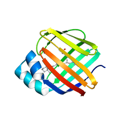
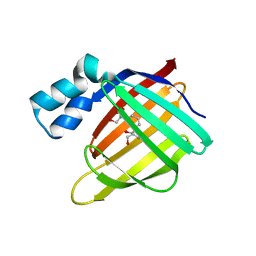
Crystal structure of the R132K:R111L:L121E:R59W-CRABPII mutant complexed with a synthetic ligand (merocyanin) at 2.60 angstrom resolution.
Descriptor: (2E,4E,6E)-3-methyl-6-(1,3,3-trimethyl-1,3-dihydro-2H-indol-2-ylidene)hexa-2,4-dienal, 2-(N-MORPHOLINO)-ETHANESULFONIC ACID, Cellular retinoic acid-binding protein 2
Authors:Jia, X, Geiger, J.H.
Deposit date:2008-11-30
Release date:2009-11-10
Last modified:2023-09-06
Method:X-RAY DIFFRACTION (2.6 Å)
Cite:"Turn-on" protein fluorescence: in situ formation of cyanine dyes.
J.Am.Chem.Soc., 137, 2015
3FA9
DownloadVisualize
BU of 3fa9 by Molmil
Crystal structure of the apo R132K:Y134F:R111L:L121D mutant of cellular retinoic acid-binding protein II at 1.94 angstrom resolution
Descriptor: ACETATE ION, Cellular retinoic acid-binding protein 2
Authors:Jia, X, Geiger, J.H.
Deposit date:2008-11-16
Release date:2009-10-27
Last modified:2023-12-27
Method:X-RAY DIFFRACTION (1.94 Å)
Cite:Crystal structures of apo cellular retinoic acid-binding protein II mutants: Structural integrity investigated through multiple site mutations
To be Published
3FA8
DownloadVisualize
BU of 3fa8 by Molmil
Crystal structure of the apo R132K:Y134F:R111L:L121E mutant of cellular retinoic acid-binding protein II at 1.78 angstrom resolution
Descriptor: Cellular retinoic acid-binding protein 2
Authors:Jia, X, Geiger, J.H.
Deposit date:2008-11-16
Release date:2009-10-27
Last modified:2023-09-06
Method:X-RAY DIFFRACTION (1.78 Å)
Cite:Crystal structures of apo cellular retinoic acid-binding protein II mutants: Structural integrity investigated through multiple site mutations
To be Published
3FEN
DownloadVisualize
BU of 3fen by Molmil
Crystal structure of the R132K:R111L:A32E mutant of cellular retinoic acid-binding protein II at 1.56 angstrom resolution
Descriptor: CHLORIDE ION, Cellular retinoic acid-binding protein 2
Authors:Jia, X, Geiger, J.H.
Deposit date:2008-11-30
Release date:2009-11-10
Last modified:2023-09-06
Method:X-RAY DIFFRACTION (1.56 Å)
Cite:Crystal structures of apo cellular retinoic acid-binding protein II mutants: Structural integrity investigated through multiple site mutations
To be Published
3FA7
DownloadVisualize
BU of 3fa7 by Molmil
Crystal structure of the apo R132K:R111L:L121E:R59E mutant of cellular retinoic acid-binding protein II at 1.90 angstrom resolution
Descriptor: 2-[3-(2-HYDROXY-1,1-DIHYDROXYMETHYL-ETHYLAMINO)-PROPYLAMINO]-2-HYDROXYMETHYL-PROPANE-1,3-DIOL, Cellular retinoic acid-binding protein 2
Authors:Jia, X, Geiger, J.H.
Deposit date:2008-11-15
Release date:2009-10-27
Last modified:2023-12-27
Method:X-RAY DIFFRACTION (1.9 Å)
Cite:Crystal structures of apo cellular retinoic acid-binding protein II mutants: Structural integrity investigated through multiple site mutations
To be Published
3FEK
DownloadVisualize
BU of 3fek by Molmil
Crystal structure of the R132K:Y134F:R111L:L121D:T54V mutant of cellular retinoic acid-binding protein II at 1.51 angstrom resolution
Descriptor: ACETATE ION, Cellular retinoic acid-binding protein 2, DI(HYDROXYETHYL)ETHER
Authors:Jia, X, Geiger, J.H.
Deposit date:2008-11-30
Release date:2009-11-10
Last modified:2023-09-06
Method:X-RAY DIFFRACTION (1.51 Å)
Cite:Crystal structures of apo cellular retinoic acid-binding protein II mutants: Structural integrity investigated through multiple site mutations
To be Published
4YGH
DownloadVisualize
BU of 4ygh by Molmil
Crystal Structure of R111K:Y134F:T54V:R132Q:P39Y:R59Y mutant of human Cellular Retinoic Acid Binding Protein II with Retinal at 2.1 Angstrom resolution - UV irradiated crystal - 2nd cycle
Descriptor: Cellular retinoic acid-binding protein 2, RETINAL
Authors:Nosrati, M, Geiger, J.H.
Deposit date:2015-02-26
Release date:2016-03-02
Last modified:2023-09-27
Method:X-RAY DIFFRACTION (2.1 Å)
Cite:A Photoisomerizing Rhodopsin Mimic Observed at Atomic Resolution.
J.Am.Chem.Soc., 138, 2016
4YH0
DownloadVisualize
BU of 4yh0 by Molmil
Crystal structure of the R111K:Y134F:T54V:R132Q:P39Y:R59Y mutant of human Cellular Retinoic Acid Binding Protein II in complex with Retinal at 2.14 angstrom resolution - UV irradiated crystal - 3rd cycle
Descriptor: Cellular retinoic acid-binding protein 2, RETINAL
Authors:Nosrati, M, Geiger, J.H.
Deposit date:2015-02-26
Release date:2016-03-02
Last modified:2023-09-27
Method:X-RAY DIFFRACTION (2.144 Å)
Cite:A Photoisomerizing Rhodopsin Mimic Observed at Atomic Resolution.
J.Am.Chem.Soc., 138, 2016
4YDA
DownloadVisualize
BU of 4yda by Molmil
Crystal structure of R111K:Y134F:T54V:R132Q:P39Q:R59Y mutant of human cellular retinoic acid binding proteinii with retinal at 1.95 angstrom - after 1hour visible light irradiation - 3rd cycle
Descriptor: Cellular retinoic acid-binding protein 2, RETINAL
Authors:Nosrati, M, Geiger, J.H.
Deposit date:2015-02-21
Release date:2016-02-24
Last modified:2023-09-27
Method:X-RAY DIFFRACTION (1.951 Å)
Cite:A Photoisomerizing Rhodopsin Mimic Observed at Atomic Resolution.
J.Am.Chem.Soc., 138, 2016
4YDB
DownloadVisualize
BU of 4ydb by Molmil
Crystal structure of the R111K:Y134F:T54V:R132Q:P39Q:R59Y mutant of human Cellular Retinoic Acid Binding Protein II in complex with Retinal at 2.03 angstrom -UV irradiated crystal- 3rd cycle
Descriptor: Cellular retinoic acid-binding protein 2, RETINAL
Authors:Nosrati, M, Geiger, J.H.
Deposit date:2015-02-21
Release date:2016-02-24
Last modified:2023-09-27
Method:X-RAY DIFFRACTION (2.03 Å)
Cite:A Photoisomerizing Rhodopsin Mimic Observed at Atomic Resolution.
J.Am.Chem.Soc., 138, 2016
4YCE
DownloadVisualize
BU of 4yce by Molmil
Crystal structure of the R111K:Y134F:T54V:R132Q:P39Q:R59Y mutant of human cellular retinoic acid binding proteinii with retinal at 1.95 angstrom- visible light irradiated crystal for 1 hour - 2nd cycle
Descriptor: Cellular retinoic acid-binding protein 2, RETINAL
Authors:Nosrati, M, Geiger, J.H.
Deposit date:2015-02-19
Release date:2016-02-24
Last modified:2023-09-27
Method:X-RAY DIFFRACTION (1.953 Å)
Cite:A Photoisomerizing Rhodopsin Mimic Observed at Atomic Resolution.
J.Am.Chem.Soc., 138, 2016
4YFQ
DownloadVisualize
BU of 4yfq by Molmil
CRYSTAL STRUCTURE OF THE R111K:Y134F:T54V:R132Q:P39Y:R59Y MUTANT OF HUMAN CELLULAR RETINOIC ACID BINDING PROTEINII WITH RETINAL AFTER 24 HOURS INCUBATION AT 1.62 ANGSTROM RESOLUTION - Thermodynamic product - 1st cycle
Descriptor: Cellular retinoic acid-binding protein 2, RETINAL
Authors:Nosrati, M, Geiger, J.H.
Deposit date:2015-02-25
Release date:2016-03-02
Last modified:2023-09-27
Method:X-RAY DIFFRACTION (1.62 Å)
Cite:A Photoisomerizing Rhodopsin Mimic Observed at Atomic Resolution.
J.Am.Chem.Soc., 138, 2016
4YFR
DownloadVisualize
BU of 4yfr by Molmil
Crystal structure of the R111K:Y134F:T54V:R132Q:P39Y:R59Y mutant of human Cellular Retinoic Acid Binding Protein II with Retinal at 1.95 Angstrom Resolution - UV irradiated crystal for 30 minutes - 1st Cycle
Descriptor: Cellular retinoic acid-binding protein 2, RETINAL
Authors:Nosrati, M, Geiger, J.H.
Deposit date:2015-02-25
Release date:2016-03-02
Last modified:2023-09-27
Method:X-RAY DIFFRACTION (1.952 Å)
Cite:A Photoisomerizing Rhodopsin Mimic Observed at Atomic Resolution.
J.Am.Chem.Soc., 138, 2016

218853

PDB entries from 2024-04-24

PDB statisticsPDBj update infoContact PDBjnumon