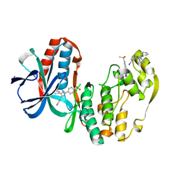
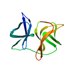
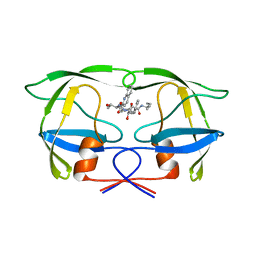
2WPO
 
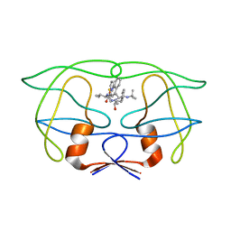
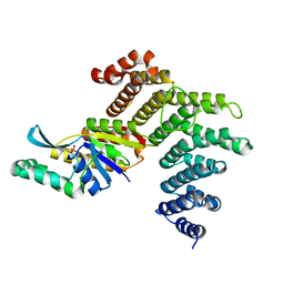
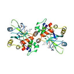 | | HCMV protease inhibitor complex | | 分子名称: | (2S)-2-(3,3-dimethylbutanoylamino)-N-[(2S)-1-[[(2S,3S)-3-hydroxy-4-[(4-iodophenyl)methylamino]-4-oxo-butan-2-yl]amino]- 1,4-dioxo-4-pyrrol-1-yl-butan-2-yl]-3,3-dimethyl-butanamide, HUMAN CYTOMEGALOVIRUS PROTEASE | | 著者 | Tong, L, Qian, C, Massariol, M.-J, Deziel, R, Yoakim, C, Lagace, L. | | 登録日 | 1998-08-04 | | 公開日 | 1999-08-04 | | 最終更新日 | 2023-08-09 | | 実験手法 | X-RAY DIFFRACTION (2.7 Å) | | 主引用文献 | Conserved mode of peptidomimetic inhibition and substrate recognition of human cytomegalovirus protease.
Nat.Struct.Biol., 5, 1998
|
|
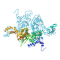
1BIM
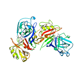 | | CRYSTALLOGRAPHIC STUDIES ON THE BINDING MODES OF P2-P3 BUTANEDIAMIDE RENIN INHIBITORS | | 分子名称: | (2S)-2-[(2-amino-1,3-thiazol-4-yl)methyl]-N~1~-{(1S,2S)-1-(cyclohexylmethyl)-2-hydroxy-2-[(3R)-1,5,5-trimethyl-2-oxopyrrolidin-3-yl]ethyl}-N~4~-[2-(dimethylamino)-2-oxoethyl]-N~4~-[(1S)-1-phenylethyl]butanediamide, Renin | | 著者 | Tong, L. | | 登録日 | 1995-09-27 | | 公開日 | 1996-01-29 | | 最終更新日 | 2022-03-09 | | 実験手法 | X-RAY DIFFRACTION (2.8 Å) | | 主引用文献 | Crystallographic studies on the binding modes of P2-P3 butanediamide renin inhibitors.
J.Biol.Chem., 270, 1995
|
|
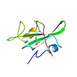
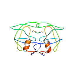
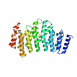
1BHF
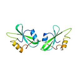
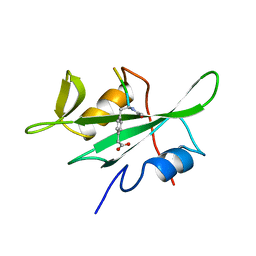 | | P56LCK SH2 DOMAIN INHIBITOR COMPLEX | | 分子名称: | INHIBITOR ACE-IPA-GLU-GLU-ILE, T-LYMPHOCYTE-SPECIFIC PROTEIN TYROSINE KINASE P56LCK | | 著者 | Tong, L, Warren, T.C, Lukas, S, Schembri-King, J, Betageri, R, Proudfoot, J.R, Jakes, S. | | 登録日 | 1998-06-08 | | 公開日 | 1998-10-21 | | 最終更新日 | 2012-12-12 | | 実験手法 | X-RAY DIFFRACTION (1.8 Å) | | 主引用文献 | Carboxymethyl-phenylalanine as a replacement for phosphotyrosine in SH2 domain binding.
J.Biol.Chem., 273, 1998
|
|
1BIL
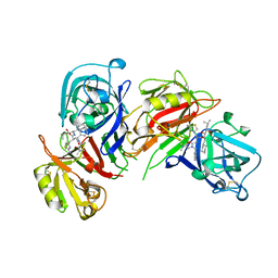 | | CRYSTALLOGRAPHIC STUDIES ON THE BINDING MODES OF P2-P3 BUTANEDIAMIDE RENIN INHIBITORS | | 分子名称: | (2S)-2-[(2-amino-1,3-thiazol-4-yl)methyl]-N~1~-[(1S,2R,3R)-1-(cyclohexylmethyl)-2,3-dihydroxy-5-methylhexyl]-N~4~-[2-(d imethylamino)-2-oxoethyl]-N~4~-[(1S)-1-phenylethyl]butanediamide, Renin | | 著者 | Tong, L. | | 登録日 | 1995-09-27 | | 公開日 | 1996-01-29 | | 最終更新日 | 2022-03-09 | | 実験手法 | X-RAY DIFFRACTION (2.4 Å) | | 主引用文献 | Crystallographic studies on the binding modes of P2-P3 butanediamide renin inhibitors.
J.Biol.Chem., 270, 1995
|
|
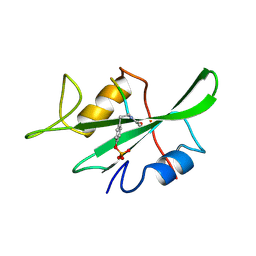
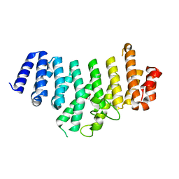
1BHH
 | | FREE P56LCK SH2 DOMAIN | | 分子名称: | P56 LCK TYROSINE KINASE SH2 DOMAIN, T-LYMPHOCYTE-SPECIFIC PROTEIN TYROSINE KINASE P56LCK | | 著者 | Tong, L, Warren, T.C, Lukas, S, Schembri-King, J, Betageri, R, Proudfoot, J.R, Jakes, S. | | 登録日 | 1998-06-08 | | 公開日 | 1998-10-21 | | 最終更新日 | 2024-02-07 | | 実験手法 | X-RAY DIFFRACTION (1.9 Å) | | 主引用文献 | Carboxymethyl-phenylalanine as a replacement for phosphotyrosine in SH2 domain binding.
J.Biol.Chem., 273, 1998
|
|
1IDA
 | |
1IDB
 | |
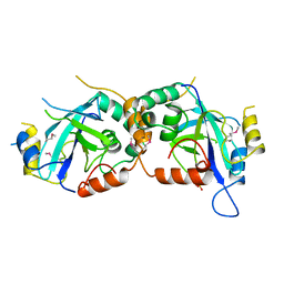
1HRN
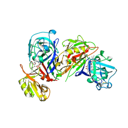 | | HIGH RESOLUTION CRYSTAL STRUCTURES OF RECOMBINANT HUMAN RENIN IN COMPLEX WITH POLYHYDROXYMONOAMIDE INHIBITORS | | 分子名称: | (2R,4S,5S)-N-[(2S,3R,4S)-1-cyclohexyl-3,4-dihydroxy-6-methylheptan-2-yl]-2-(cyclopropylmethyl)-4,5-dihydroxy-6-phenylhexanamide, 2-acetamido-2-deoxy-beta-D-glucopyranose, RENIN | | 著者 | Tong, L, Anderson, P.C. | | 登録日 | 1995-03-31 | | 公開日 | 1995-06-03 | | 最終更新日 | 2020-07-29 | | 実験手法 | X-RAY DIFFRACTION (1.8 Å) | | 主引用文献 | High resolution crystal structures of recombinant human renin in complex with polyhydroxymonoamide inhibitors.
J.Mol.Biol., 250, 1995
|
|
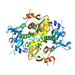
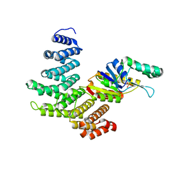
1IAN
 | | HUMAN P38 MAP KINASE INHIBITOR COMPLEX | | 分子名称: | 4-[5-(3-IODO-PHENYL)-2-(4-METHANESULFINYL-PHENYL)-1H-IMIDAZOL-4-YL]-PYRIDINE, P38 MAP KINASE | | 著者 | Tong, L. | | 登録日 | 1997-03-07 | | 公開日 | 1998-05-06 | | 最終更新日 | 2024-04-03 | | 実験手法 | X-RAY DIFFRACTION (2 Å) | | 主引用文献 | A highly specific inhibitor of human p38 MAP kinase binds in the ATP pocket.
Nat.Struct.Biol., 4, 1997
|
|
1LKK
 | |
1LKL
 | |
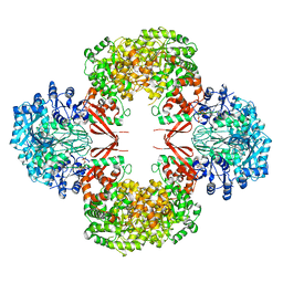
1WPO
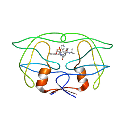
 | | HYDROLYTIC ENZYME HUMAN CYTOMEGALOVIRUS PROTEASE | | 分子名称: | HUMAN CYTOMEGALOVIRUS PROTEASE, SULFATE ION | | 著者 | Tong, L. | | 登録日 | 1996-07-23 | | 公開日 | 1997-10-15 | | 最終更新日 | 2021-11-03 | | 実験手法 | X-RAY DIFFRACTION (2 Å) | | 主引用文献 | A new serine-protease fold revealed by the crystal structure of human cytomegalovirus protease.
Nature, 383, 1996
|
|
2SNV
 | |
2MIP
 | | CRYSTAL STRUCTURE OF HUMAN IMMUNODEFICIENCY VIRUS (HIV) TYPE 2 PROTEASE IN COMPLEX WITH A REDUCED AMIDE INHIBITOR AND COMPARISON WITH HIV-1 PROTEASE STRUCTURES | | 分子名称: | HIV-2 PROTEASE, INHIBITOR BI-LA-398 | | 著者 | Tong, L, Pav, S, Pargellis, C, Do, F, Lamarre, D, Anderson, P.C. | | 登録日 | 1993-06-03 | | 公開日 | 1993-10-31 | | 最終更新日 | 2019-08-14 | | 実験手法 | X-RAY DIFFRACTION (2.2 Å) | | 主引用文献 | Crystal structure of human immunodeficiency virus (HIV) type 2 protease in complex with a reduced amide inhibitor and comparison with HIV-1 protease structures.
Proc.Natl.Acad.Sci.USA, 90, 1993
|
|
6O3P
 | |
6DKN
 | |
1JLD
 | |
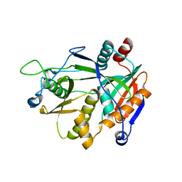
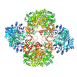
3PGQ
 | | Crystal Structure of the Carboxyltransferase Domain of S. cerevisiae Acetyl CoA Carboxylase in Complex with Pinoxaden | | 分子名称: | 8-(2-ethenyl-6-ethyl-4-methylphenyl)tetrahydro-7H-pyrazolo[1,2-d][1,4,5]oxadiazepine-7,9(8H)-dione, Acetyl-CoA carboxylase | | 著者 | Tong, L, Yu, L.P.C, Kim, Y.S. | | 登録日 | 2010-11-02 | | 公開日 | 2010-12-22 | | 最終更新日 | 2024-02-21 | | 実験手法 | X-RAY DIFFRACTION (2.8 Å) | | 主引用文献 | Mechanism for the inhibition of the carboxyltransferase domain of acetyl-coenzyme A carboxylase by pinoxaden.
Proc.Natl.Acad.Sci.USA, 107, 2010
|
|
3HBL
 | |
3HB9
 | |
3HO8
 | |
3O2T
 | |
3O2Q
 | |
3ODS
 | |
3O2S
 | |


