Loading
PDBj
MenuPDBj@FacebookPDBj@X(formerly Twitter)PDBj@BlueSkyPDBj@YouTubewwPDB FoundationwwPDB
RCSB PDBPDBeBMRBAdv. SearchSearch help
Search by PDB author
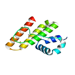
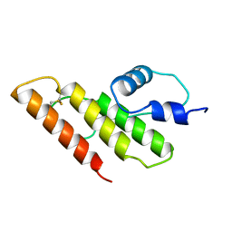
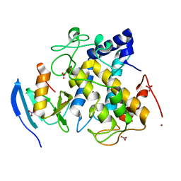
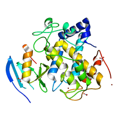
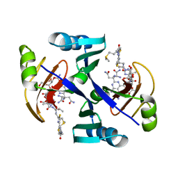
1JIF
DownloadVisualize
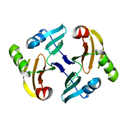
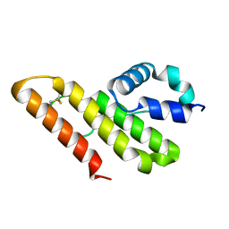
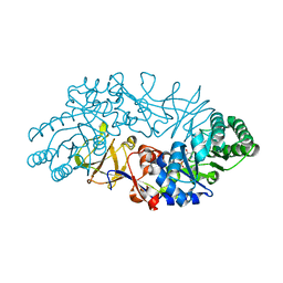
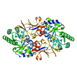
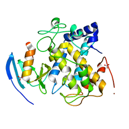
BU of 1jif by Molmil
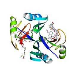
Crystal structure of bleomycin-binding protein from bleomycin-producing Streptomyces verticillus complexed with copper(II)-bleomycin
Descriptor: BLEOMYCIN A2, CHLORIDE ION, COPPER (II) ION, ...
Authors:Sugiyama, M, Kumagai, T, Hayashida, M, Maruyama, M, Matoba, Y.
Deposit date:2001-07-02
Release date:2002-02-06
Last modified:2024-03-13
Method:X-RAY DIFFRACTION (1.6 Å)
Cite:The 1.6-A crystal structure of the copper(II)-bound bleomycin complexed with the bleomycin-binding protein from bleomycin-producing Streptomyces verticillus.
J.Biol.Chem., 277, 2002
1JIE
DownloadVisualize
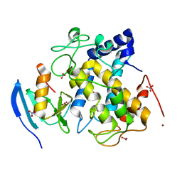
BU of 1jie by Molmil
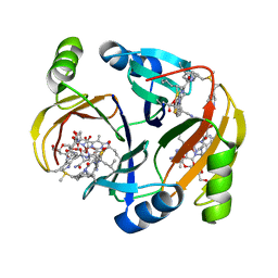
Crystal structure of bleomycin-binding protein from bleomycin-producing Streptomyces verticillus complexed with metal-free bleomycin
Descriptor: BLEOMYCIN A2, bleomycin-binding protein
Authors:Sugiyama, M, Kumagai, T, Hayashida, M, Maruyama, M, Matoba, Y.
Deposit date:2001-07-02
Release date:2002-02-06
Last modified:2023-10-25
Method:X-RAY DIFFRACTION (1.8 Å)
Cite:The 1.6-A crystal structure of the copper(II)-bound bleomycin complexed with the bleomycin-binding protein from bleomycin-producing Streptomyces verticillus.
J.Biol.Chem., 277, 2002
1NIQ
DownloadVisualize
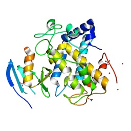
BU of 1niq by Molmil
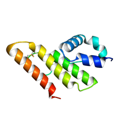
Solution Structure of the HOO-Bm bound BLMT, Transposon Tn5-encoding Bleomycin-binding Protein
Descriptor: BLEOMYCIN A2, Bleomycin Resistance Protein, COPPER (II) ION
Authors:Sugiyama, M.
Deposit date:2002-12-25
Release date:2003-12-25
Last modified:2022-02-23
Method:SOLUTION NMR
Cite:Solution Structure of the HOO-Bm bound BLMT, Transposon Tn5-encoding Bleomycin-binding Protein
To be published
1IT5
DownloadVisualize
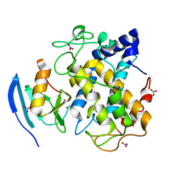
BU of 1it5 by Molmil
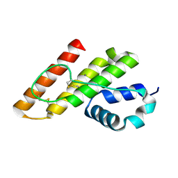
Solution structure of apo-type PLA2 from Streptomyces violaceruber A-2688.
Descriptor: Phospholipase A2
Authors:Sugiyama, M, Ohtani, K, Izuhara, M, Koike, T.
Deposit date:2002-01-09
Release date:2002-09-04
Last modified:2024-11-20
Method:SOLUTION NMR
Cite:A novel prokaryotic phospholipase A2. Characterization, gene cloning, and solution structure.
J.Biol.Chem., 277, 2002
1IT4
DownloadVisualize
BU of 1it4 by Molmil
Solution structure of the prokaryotic Phospholipase A2 from Streptomyces violaceoruber
Descriptor: CALCIUM ION, phospholipase A2
Authors:Ohtani, K, Sugiyama, M, Izuhara, M, Koike, T.
Deposit date:2002-01-08
Release date:2002-09-04
Last modified:2024-10-16
Method:SOLUTION NMR
Cite:A novel prokaryotic phospholipase A2. Characterization, gene cloning, and solution structure.
J.BIOL.CHEM., 277, 2002
1VFT
DownloadVisualize
BU of 1vft by Molmil
Crystal structure of L-cycloserine-bound form of alanine racemase from D-cycloserine-producing Streptomyces lavendulae
Descriptor: CHLORIDE ION, D-[3-HYDROXY-2-METHYL-5-PHOSPHONOOXYMETHYL-PYRIDIN-4-YLMETHYL]-N,O-CYCLOSERYLAMIDE, alanine racemase
Authors:Noda, M, Matoba, Y, Kumagai, T, Sugiyama, M.
Deposit date:2004-04-19
Release date:2004-09-14
Last modified:2023-11-15
Method:X-RAY DIFFRACTION (2.3 Å)
Cite:Structural evidence that alanine racemase from a D-cycloserine-producing microorganism exhibits resistance to its own product.
J.Biol.Chem., 279, 2004
1QTO
DownloadVisualize
BU of 1qto by Molmil
1.5 A CRYSTAL STRUCTURE OF A BLEOMYCIN RESISTANCE DETERMINANT FROM BLEOMYCIN-PRODUCING STREPTOMYCES VERTICILLUS
Descriptor: BLEOMYCIN-BINDING PROTEIN
Authors:Kawano, Y, Kumagai, T, Muta, K, Matoba, Y, Davies, J, Sugiyama, M.
Deposit date:1999-06-28
Release date:2000-06-28
Last modified:2024-02-14
Method:X-RAY DIFFRACTION (1.5 Å)
Cite:The 1.5 A crystal structure of a bleomycin resistance determinant from bleomycin-producing Streptomyces verticillus.
J.Mol.Biol., 295, 2000
1EWJ
DownloadVisualize
BU of 1ewj by Molmil
CRYSTAL STRUCTURE OF BLEOMYCIN-BINDING PROTEIN COMPLEXED WITH BLEOMYCIN
Descriptor: BLEOMYCIN A2, BLEOMYCIN RESISTANCE DETERMINANT
Authors:Maruyama, M, Kumagai, T, Matoba, Y, Hata, Y, Sugiyama, M.
Deposit date:2000-04-26
Release date:2001-04-26
Last modified:2024-02-07
Method:X-RAY DIFFRACTION (2.5 Å)
Cite:Crystal structures of the transposon Tn5-carried bleomycin resistance determinant uncomplexed and complexed with bleomycin.
J.Biol.Chem., 276, 2001
1ECS
DownloadVisualize
BU of 1ecs by Molmil
THE 1.7 A CRYSTAL STRUCTURE OF A BLEOMYCIN RESISTANCE DETERMINANT ENCODED ON THE TRANSPOSON TN5
Descriptor: BLEOMYCIN RESISTANCE PROTEIN, CALCIUM ION, TETRAETHYLENE GLYCOL
Authors:Maruyama, M, Matoba, Y, Kumagai, T, Sugiyama, M.
Deposit date:2000-01-25
Release date:2001-05-02
Last modified:2023-08-09
Method:X-RAY DIFFRACTION (1.7 Å)
Cite:Crystal structures of the transposon Tn5-carried bleomycin resistance determinant uncomplexed and complexed with bleomycin.
J.Biol.Chem., 276, 2001
1LWB
DownloadVisualize
BU of 1lwb by Molmil
Crystal structure of prokaryotic phospholipase A2 at atomic resolution
Descriptor: putative secreted protein
Authors:Matoba, Y, Sugiyama, M.
Deposit date:2002-05-31
Release date:2003-06-10
Last modified:2024-10-23
Method:X-RAY DIFFRACTION (1.05 Å)
Cite:Atomic resolution structure of prokaryotic phospholipase A2: Analysis of internal motion and implication for a catalytic mechanism.
PROTEINS: STRUCT.,FUNCT.,GENET., 51, 2003
1VFH
DownloadVisualize
BU of 1vfh by Molmil
Crystal structure of alanine racemase from D-cycloserine producing Streptomyces lavendulae
Descriptor: PYRIDOXAL-5'-PHOSPHATE, alanine racemase
Authors:Noda, M, Matoba, Y, Kumagai, T, Sugiyama, M.
Deposit date:2004-04-13
Release date:2004-09-14
Last modified:2023-11-15
Method:X-RAY DIFFRACTION (2 Å)
Cite:Structural evidence that alanine racemase from a D-cycloserine-producing microorganism exhibits resistance to its own product.
J.Biol.Chem., 279, 2004
1VFS
DownloadVisualize
BU of 1vfs by Molmil
Crystal structure of D-cycloserine-bound form of alanine racemase from D-cycloserine-producing Streptomyces lavendulae
Descriptor: CHLORIDE ION, D-[3-HYDROXY-2-METHYL-5-PHOSPHONOOXYMETHYL-PYRIDIN-4-YLMETHYL]-N,O-CYCLOSERYLAMIDE, alanine racemase
Authors:Noda, M, Matoba, Y, Kumagai, T, Sugiyama, M.
Deposit date:2004-04-19
Release date:2004-09-14
Last modified:2023-11-15
Method:X-RAY DIFFRACTION (1.9 Å)
Cite:Structural evidence that alanine racemase from a D-cycloserine-producing microorganism exhibits resistance to its own product.
J.Biol.Chem., 279, 2004
1KP4
DownloadVisualize
BU of 1kp4 by Molmil
CALCIUM-BOUND FORM OF PROKARYOTIC PHOSPHOLIPASE A2
Descriptor: CALCIUM ION, phospholipase A2
Authors:Matoba, Y, Katsube, Y, Sugiyama, M.
Deposit date:2001-12-28
Release date:2002-09-04
Last modified:2024-10-23
Method:X-RAY DIFFRACTION (1.6 Å)
Cite:The crystal structure of prokaryotic phospholipase A2.
J.Biol.Chem., 277, 2002
1WXC
DownloadVisualize
BU of 1wxc by Molmil
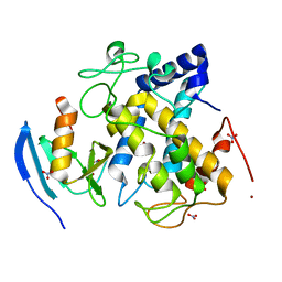
Crystal Structure of the copper-free Streptomyces castaneoglobisporus tyrosinase complexed with a caddie protein
Descriptor: MelC, NITRATE ION, tyrosinase
Authors:Matoba, Y, Kumagai, T, Yamamoto, A, Yoshitsu, H, Sugiyama, M.
Deposit date:2005-01-20
Release date:2006-01-31
Last modified:2024-03-13
Method:X-RAY DIFFRACTION (1.2 Å)
Cite:Crystallographic Evidence That the Dinuclear Copper Center of Tyrosinase Is Flexible during Catalysis
J.Biol.Chem., 281, 2006
1WX5
DownloadVisualize
BU of 1wx5 by Molmil
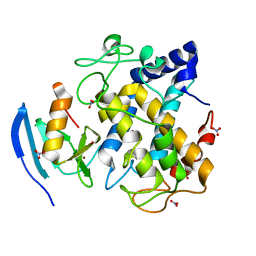
Crystal Structure of the copper-free Streptomyces castaneoglobisporus tyrosinase complexed with a caddie protein in the monoclinic crystal
Descriptor: CHLORIDE ION, MelC, SODIUM ION, ...
Authors:Matoba, Y, Kumagai, T, Yamamoto, A, Yoshitsu, H, Sugiyama, M.
Deposit date:2005-01-19
Release date:2006-01-31
Last modified:2023-10-25
Method:X-RAY DIFFRACTION (2.02 Å)
Cite:Crystallographic Evidence That the Dinuclear Copper Center of Tyrosinase Is Flexible during Catalysis
J.Biol.Chem., 281, 2006
1WX2
DownloadVisualize
BU of 1wx2 by Molmil
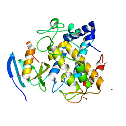
Crystal Structure of the oxy-form of the copper-bound Streptomyces castaneoglobisporus tyrosinase complexed with a caddie protein prepared by the addition of hydrogenperoxide
Descriptor: COPPER (II) ION, MelC, NITRATE ION, ...
Authors:Matoba, Y, Kumagai, T, Yamamoto, A, Yoshitsu, H, Sugiyama, M.
Deposit date:2005-01-19
Release date:2006-01-31
Last modified:2023-10-25
Method:X-RAY DIFFRACTION (1.8 Å)
Cite:Crystallographic Evidence That the Dinuclear Copper Center of Tyrosinase Is Flexible during Catalysis
J.Biol.Chem., 281, 2006
1WX4
DownloadVisualize
BU of 1wx4 by Molmil
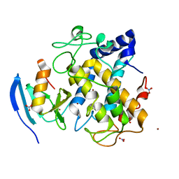
Crystal structure of the oxy-form of the copper-bound Streptomyces castaneoglobisporus tyrosinase complexed with a caddie protein prepared by the addition of dithiothreitol
Descriptor: COPPER (II) ION, MelC, NITRATE ION, ...
Authors:Matoba, Y, Kumagai, T, Yamamoto, A, Yoshitsu, H, Sugiyama, M.
Deposit date:2005-01-19
Release date:2006-01-31
Last modified:2023-10-25
Method:X-RAY DIFFRACTION (1.5 Å)
Cite:Crystallographic Evidence That the Dinuclear Copper Center of Tyrosinase Is Flexible during Catalysis
J.Biol.Chem., 281, 2006
1FAZ
DownloadVisualize
BU of 1faz by Molmil
THE CRYSTAL STRUCTURE OF PROKARYOTIC PHOSPHOLIPASE A2
Descriptor: PHOSPHOLIPASE A2
Authors:Matoba, Y, katsube, Y, Sugiyama, M.
Deposit date:2000-07-14
Release date:2001-07-18
Last modified:2024-11-20
Method:X-RAY DIFFRACTION (1.4 Å)
Cite:The crystal structure of prokaryotic phospholipase A2.
J.Biol.Chem., 277, 2002
1MH6
DownloadVisualize
BU of 1mh6 by Molmil
Solution Structure of the Transposon Tn5-encoding Bleomycin-binding Protein, BLMT
Descriptor: BLEOMYCIN RESISTANCE PROTEIN
Authors:Kumagai, T, Ohtani, K, Tsuboi, Y, Koike, T, Sugiyama, M.
Deposit date:2002-08-19
Release date:2003-02-19
Last modified:2024-05-29
Method:SOLUTION NMR
Cite:Solution structure of the transposon Tn5-encoding bleomycin-binding protein complexed with an activated bleomycin analogue.
To be published
5Y9N
DownloadVisualize
BU of 5y9n by Molmil
Crystal structure of Pyrococcus furiosus PbaA (monoclinic form), an archaeal homolog of proteasome-assembly chaperone
Descriptor: CHLORIDE ION, PbaA
Authors:Yagi-Utsumi, M, Sikdar, A, Kozai, T, Inoue, R, Sugiyama, M, Uchihashi, T, Satoh, T, Kato, K.
Deposit date:2017-08-26
Release date:2018-01-17
Last modified:2023-11-22
Method:X-RAY DIFFRACTION (2.55 Å)
Cite:Conversion of functionally undefined homopentameric protein PbaA into a proteasome activator by mutational modification of its C-terminal segment conformation
Protein Eng. Des. Sel., 31, 2018
2ZWF
DownloadVisualize
BU of 2zwf by Molmil
Crystal structure of the copper-bound tyrosinase in complex with a caddie protein from streptomyces castaneoglobisporus obtained by soaking the deoxy-form crystal in dioxygen-saturated solution for 80 minutes
Descriptor: COPPER (II) ION, MelC, NITRATE ION, ...
Authors:Matoba, Y, Sugiyama, M.
Deposit date:2008-12-03
Release date:2009-12-15
Last modified:2024-10-30
Method:X-RAY DIFFRACTION (1.4 Å)
Cite:Crystallographic evidence of drastic movement of a copper ion toward the substrate tyrosine for starting hydroxylation reaction of tyrosinase
To be Published
2ZWE
DownloadVisualize
BU of 2zwe by Molmil
Crystal structure of the copper-bound tyrosinase in complex with a caddie protein from streptomyces castaneoglobisporus obtained by soaking the deoxy-form crystal in dioxygen-saturated solution for 40 minutes
Descriptor: COPPER (II) ION, MelC, NITRATE ION, ...
Authors:Matoba, Y, Sugiyama, M.
Deposit date:2008-12-03
Release date:2009-12-15
Last modified:2024-11-06
Method:X-RAY DIFFRACTION (1.32 Å)
Cite:Crystallographic evidence of drastic movement of a copper ion toward the substrate tyrosine for starting hydroxylation reaction of tyrosinase
To be Published
2ZMZ
DownloadVisualize
BU of 2zmz by Molmil
The 1.37-A crystal structure of the hydroxylamine-induced deoxy-form of the copper-bound tyrosinase in complex with a caddie protein from Streptomyces castaneoglobisporus
Descriptor: CADDIE, COPPER (I) ION, NITRATE ION, ...
Authors:Matoba, Y, Sugiyama, M.
Deposit date:2008-04-21
Release date:2009-05-05
Last modified:2023-11-01
Method:X-RAY DIFFRACTION (1.37 Å)
Cite:X-Ray Snapshots of a Hydroxylation Mechanism of Tyrosinase
To be Published
2ZWD
DownloadVisualize
BU of 2zwd by Molmil
Crystal structure of the copper-bound tyrosinase in complex with a caddie protein from streptomyces castaneoglobisporus obtained by soaking the deoxy-form crystal in dioxygen-saturated solution for 5 minutes
Descriptor: COPPER (I) ION, MelC, NITRATE ION, ...
Authors:Matoba, Y, Sugiyama, M.
Deposit date:2008-12-03
Release date:2009-12-15
Last modified:2023-11-01
Method:X-RAY DIFFRACTION (1.35 Å)
Cite:Crystallographic evidence of drastic movement of a copper ion toward the substrate tyrosine for starting hydroxylation reaction of tyrosinase
To be Published
2ZMY
DownloadVisualize
BU of 2zmy by Molmil
Crystal structure of the met2-form of the copper-bound tyrosinase in complex with a caddie protein from Streptomyces castaneoglobisporus obtained by soaking in cupric sulfate solution for 80 hours
Descriptor: CADDIE, COPPER (II) ION, NITRATE ION, ...
Authors:Matoba, Y, Sugiyama, M.
Deposit date:2008-04-21
Release date:2009-05-05
Last modified:2023-11-01
Method:X-RAY DIFFRACTION (1.45 Å)
Cite:X-Ray Snapshots of a Hydroxylation Mechanism of Tyrosinase
To be Published


1234>

227561

PDB entries from 2024-11-20

PDB statisticsPDBj update infoContact PDBjnumon