Loading
PDBj
MenuPDBj@FacebookPDBj@TwitterPDBj@YouTubewwPDB FoundationwwPDB
RCSB PDBPDBeBMRBAdv. SearchSearch help
Search by PDB author
4R8Z
DownloadVisualize
BU of 4r8z by Molmil
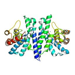
Crystal Structure of PA4781 HD-GYP domain from Pseudomonas aeruginosa at 2.2A resolution showing a bi-metallic Ni ion center
Descriptor: CHLORIDE ION, Cyclic di-GMP phosphodiesterase, NICKEL (II) ION
Authors:Giardina, G, Cutruzzolaa, F, Rinaldo, S, Stelitano, V.
Deposit date:2014-09-03
Release date:2015-03-04
Last modified:2024-02-28
Method:X-RAY DIFFRACTION (2.2 Å)
Cite:Structural basis of functional diversification of the HD-GYP domain revealed by the Pseudomonas aeruginosa PA4781 protein, which displays an unselective bimetallic binding site.
J.Bacteriol., 197, 2015
4IOB
DownloadVisualize
BU of 4iob by Molmil
Crystal structure of the GGDEF domain of PA1120 (YfiN or TpbB) from Pseudomonas aeruginosa at 2.7 Ang.
Descriptor: Diguanylate cyclase TpbB, GLYCEROL, TERTIARY-BUTYL ALCOHOL
Authors:Giardina, G, Cutruzzola, F.
Deposit date:2013-01-07
Release date:2013-12-04
Last modified:2023-09-20
Method:X-RAY DIFFRACTION (2.78 Å)
Cite:Investigating the Allosteric Regulation of YfiN from Pseudomonas aeruginosa: Clues from the Structure of the Catalytic Domain.
Plos One, 8, 2013

219869

PDB entries from 2024-05-15

PDB statisticsPDBj update infoContact PDBjnumon