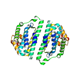
4RP7
 
 | |
4RP6
 | |
4QXX
 | |
2NPB
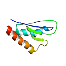 | | NMR solution structure of mouse SelW | | 分子名称: | Selenoprotein W | | 著者 | Aachmann, F.L, Fomenko, D.E, Soragni, A, Gladyshev, V.N, Dikiy, A. | | 登録日 | 2006-10-27 | | 公開日 | 2006-11-14 | | 最終更新日 | 2023-06-14 | | 実験手法 | SOLUTION NMR | | 主引用文献 | Solution structure of selenoprotein W and NMR analysis of its interaction with 14-3-3 proteins
J.Biol.Chem., 282, 2007
|
|
2LBU
 | | HADDOCK calculated model of Congo red bound to the HET-s amyloid | | 分子名称: | Small s protein, sodium 3,3'-(1E,1'E)-biphenyl-4,4'-diylbis(diazene-2,1-diyl)bis(4-aminonaphthalene-1-sulfonate) | | 著者 | Schutz, A.K, Soragni, A, Hornemann, S, Aguzzi, A, Ernst, M, Bockmann, A, Meier, B.H. | | 登録日 | 2011-04-07 | | 公開日 | 2011-06-01 | | 最終更新日 | 2011-07-13 | | 実験手法 | SOLID-STATE NMR | | 主引用文献 | The Amyloid-Congo Red Interface at Atomic Resolution.
Angew.Chem.Int.Ed.Engl., 2011
|
|
2KJ3
 | | High-resolution structure of the HET-s(218-289) prion in its amyloid form obtained by solid-state NMR | | 分子名称: | Small s protein | | 著者 | Van Melckebeke, H, Wasmer, C, Lange, A, AB, E, Loquet, A, Meier, B.H. | | 登録日 | 2009-05-20 | | 公開日 | 2010-06-02 | | 最終更新日 | 2011-07-13 | | 実験手法 | SOLID-STATE NMR | | 主引用文献 | Atomic-Resolution Three-Dimensional Structure of HET-s(218-289) Amyloid Fibrils by Solid-State NMR Spectroscopy
J.Am.Chem.Soc., 132, 2010
|
|
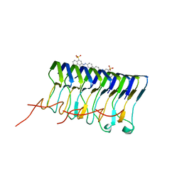
2WVN
 | | Structure of the HET-s N-terminal domain | | 分子名称: | SMALL S PROTEIN | | 著者 | Greenwald, J, Buhtz, C, Ritter, C, Kwiatkowski, W, Choe, S, Saupe, S.J, Riek, R. | | 登録日 | 2009-10-19 | | 公開日 | 2010-07-28 | | 最終更新日 | 2019-05-08 | | 実験手法 | X-RAY DIFFRACTION (2.62 Å) | | 主引用文献 | The Mechanism of Prion Inhibition by Het-S.
Mol.Cell, 38, 2010
|
|
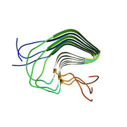
2WVQ
 | | Structure of the HET-s N-terminal domain. Mutant D23A, P33H | | 分子名称: | (2R,3S)-1,4-DIMERCAPTOBUTANE-2,3-DIOL, 2,3-DIHYDROXY-1,4-DITHIOBUTANE, SMALL S PROTEIN | | 著者 | Greenwald, J, Buhtz, C, Ritter, C, Kwiatkowski, W, Choe, S, Saupe, S.J, Riek, R. | | 登録日 | 2009-10-19 | | 公開日 | 2010-07-28 | | 最終更新日 | 2023-12-20 | | 実験手法 | X-RAY DIFFRACTION (2 Å) | | 主引用文献 | The mechanism of prion inhibition by HET-S.
Mol. Cell, 38, 2010
|
|
2WVO
 | | Structure of the HET-S N-terminal domain | | 分子名称: | CHLORIDE ION, SMALL S PROTEIN | | 著者 | Greenwald, J, Buhtz, C, Ritter, C, Kwiatkowski, W, Choe, S, Saupe, S.J, Riek, R. | | 登録日 | 2009-10-19 | | 公開日 | 2010-07-28 | | 最終更新日 | 2023-12-20 | | 実験手法 | X-RAY DIFFRACTION (2.3 Å) | | 主引用文献 | The Mechanism of Prion Inhibition by Het-S.
Mol.Cell, 38, 2010
|
|


