Loading
PDBj
MenuPDBj@FacebookPDBj@TwitterPDBj@YouTubewwPDB FoundationwwPDB
RCSB PDBPDBeBMRBAdv. SearchSearch help
Search by PDB author
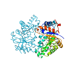
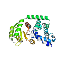
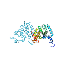
3SIB
DownloadVisualize
BU of 3sib by Molmil
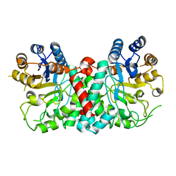
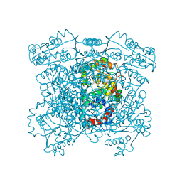
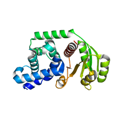
Crystal structure of URE3-binding protein, wild-type
Descriptor: CALCIUM ION, SODIUM ION, URE3-BP sequence specific DNA binding protein
Authors:Seattle Structural Genomics Center for Infectious Disease (SSGCID)
Deposit date:2011-06-17
Release date:2011-06-29
Last modified:2023-09-13
Method:X-RAY DIFFRACTION (1.9 Å)
Cite:Crystal structure of URE3-binding protein
To be Published
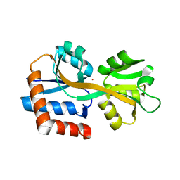
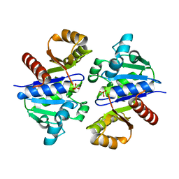
3SJS
DownloadVisualize
BU of 3sjs by Molmil
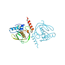
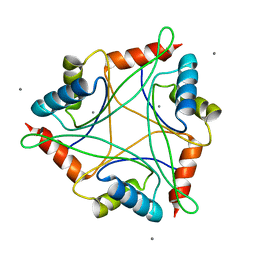
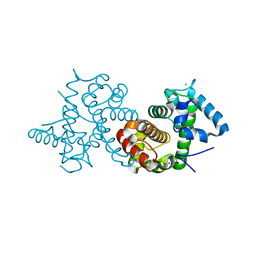
Crystal structure of URE3-binding protein from Entamoeba histolytica, (D127A,N129A) mutant, native form
Descriptor: CALCIUM ION, URE3-BP sequence specific DNA binding protein
Authors:Seattle Structural Genomics Center for Infectious Disease (SSGCID)
Deposit date:2011-06-21
Release date:2011-07-06
Last modified:2023-09-13
Method:X-RAY DIFFRACTION (1.9 Å)
Cite:Crystal structure of URE3-binding protein from Entamoeba histolytica
to be published
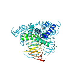
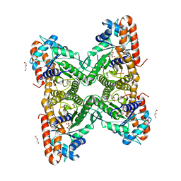
4G6C
DownloadVisualize
BU of 4g6c by Molmil
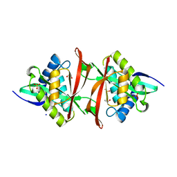
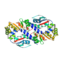
Crystal structure of beta-hexosaminidase 1 from Burkholderia cenocepacia J2315
Descriptor: Beta-hexosaminidase 1
Authors:Seattle Structural Genomics Center for Infectious Disease (SSGCID)
Deposit date:2012-07-18
Release date:2012-08-01
Last modified:2024-02-28
Method:X-RAY DIFFRACTION (1.38 Å)
Cite:Crystal structure of beta-hexosaminidase 1 from Burkholderia cenocepacia J2315
To be Published
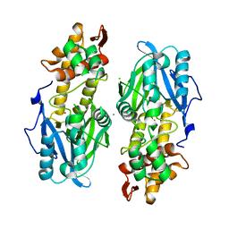
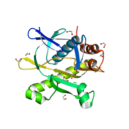
5U29
DownloadVisualize
BU of 5u29 by Molmil
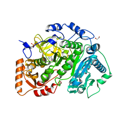
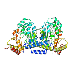
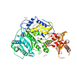
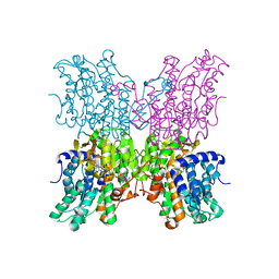
Crystal structure of Cryptococcus neoformans H99 Acetyl-CoA Synthetase in complex with Ac-AMS
Descriptor: 1,2-ETHANEDIOL, 5'-O-(acetylsulfamoyl)adenosine, Acetyl-coenzyme A synthetase, ...
Authors:Seattle Structural Genomics Center for Infectious Disease (SSGCID), Fox III, D, Edwards, T.E, Potts, K.T, Taylor, B.M.
Deposit date:2016-11-30
Release date:2017-12-06
Last modified:2023-10-04
Method:X-RAY DIFFRACTION (2.5 Å)
Cite:Crystal structure of Cryptococcus neoformans H99 Acetyl-CoA Synthetase in complex with Ac-AMS
To Be Published
3U04
DownloadVisualize
BU of 3u04 by Molmil
Crystal structure of peptide deformylase from ehrlichia chaffeensis in complex with actinonin
Descriptor: ACTINONIN, CHLORIDE ION, Peptide deformylase 1, ...
Authors:Seattle Structural Genomics Center for Infectious Disease (SSGCID)
Deposit date:2011-09-28
Release date:2011-10-12
Last modified:2023-09-13
Method:X-RAY DIFFRACTION (1.7 Å)
Cite:Crystal structure of peptide deformylase from ehrlichia chaffeensis in complex with actinonin
To be Published
3UJH
DownloadVisualize
BU of 3ujh by Molmil
Crystal structure of substrate-bound Glucose-6-phosphate isomerase from Toxoplasma gondii
Descriptor: 1,2-ETHANEDIOL, CHLORIDE ION, GLUCOSE-6-PHOSPHATE, ...
Authors:Seattle Structural Genomics Center for Infectious Disease (SSGCID)
Deposit date:2011-11-07
Release date:2011-11-16
Last modified:2023-09-13
Method:X-RAY DIFFRACTION (2.1 Å)
Cite:Crystal structure of substrate-bound Glucose-6-phosphate isomerase from Toxoplasma gondii
To be Published
4HWG
DownloadVisualize
BU of 4hwg by Molmil
Structure of UDP-N-acetylglucosamine 2-epimerase from Rickettsia bellii
Descriptor: PHOSPHATE ION, UDP-N-acetylglucosamine 2-epimerase
Authors:Seattle Structural Genomics Center for Infectious Disease (SSGCID)
Deposit date:2012-11-07
Release date:2012-11-28
Last modified:2023-09-20
Method:X-RAY DIFFRACTION (2 Å)
Cite:Structure of UDP-N-acetylglucosamine 2-epimerase from Rickettsia bellii
TO BE PUBLISHED
4IYQ
DownloadVisualize
BU of 4iyq by Molmil
Crystal structure of divalent ion tolerance protein CutA1 from Ehrlichia chaffeensis
Descriptor: CALCIUM ION, Divalent ion tolerance protein CutA1
Authors:Seattle Structural Genomics Center for Infectious Disease (SSGCID)
Deposit date:2013-01-29
Release date:2013-02-13
Last modified:2023-09-20
Method:X-RAY DIFFRACTION (2.55 Å)
Cite:Crystal structure of divalent ion tolerance protein CutA1 from Ehrlichia chaffeensis
To be Published
4J3G
DownloadVisualize
BU of 4j3g by Molmil
Crystal structure of Ribosomal-protein-alanine N-acetyltransferase from Brucella melitensis
Descriptor: 1,2-ETHANEDIOL, GCN5-related N-acetyltransferase, IMIDAZOLE, ...
Authors:Seattle Structural Genomics Center for Infectious Disease (SSGCID)
Deposit date:2013-02-05
Release date:2013-03-13
Last modified:2023-09-20
Method:X-RAY DIFFRACTION (1.75 Å)
Cite:Crystal structure of Ribosomal-protein-alanine N-acetyltransferase from Brucella melitensis
TO BE PUBLISHED
4IXO
DownloadVisualize
BU of 4ixo by Molmil
X-ray structure of NifS-like protein from Rickettsia africae ESF-5
Descriptor: 1,2-ETHANEDIOL, CALCIUM ION, NifS-like protein
Authors:Seattle Structural Genomics Center for Infectious Disease (SSGCID)
Deposit date:2013-01-27
Release date:2013-02-27
Last modified:2023-12-06
Method:X-RAY DIFFRACTION (2.2 Å)
Cite:X-ray structure of NifS-like protein from Rickettsia africae ESF-5
To be Published
4K3Z
DownloadVisualize
BU of 4k3z by Molmil
Crystal structure of D-erythrulose 4-phosphate dehydrogenase from Brucella melitensis, solved by iodide SAD
Descriptor: 1,2-ETHANEDIOL, D-erythrulose 4-phosphate dehydrogenase, GLYCEROL
Authors:Seattle Structural Genomics Center for Infectious Disease (SSGCID)
Deposit date:2013-04-11
Release date:2013-05-15
Last modified:2024-03-13
Method:X-RAY DIFFRACTION (1.95 Å)
Cite:Crystal structure of D-erythrulose 4-phosphate dehydrogenase from Brucella melitensis, solved by iodide SAD
To be Published
4F3P
DownloadVisualize
BU of 4f3p by Molmil
Crystal structure of a Glutamine-binding periplasmic protein from Burkholderia pseudomallei in complex with glutamine
Descriptor: Glutamine-binding periplasmic protein
Authors:Seattle Structural Genomics Center for Infectious Disease (SSGCID)
Deposit date:2012-05-09
Release date:2012-05-23
Last modified:2023-09-13
Method:X-RAY DIFFRACTION (2.4 Å)
Cite:Crystal structure of a Glutamine-binding periplasmic protein from Burkholderia pseudomallei in complex with glutamine
To be Published
3MC4
DownloadVisualize
BU of 3mc4 by Molmil
Crystal structure of WW/RSP5/WWP domain: bacterial transferase hexapeptide repeat: serine O-Acetyltransferase from Brucella Melitensis
Descriptor: 1,2-ETHANEDIOL, CHLORIDE ION, WW/Rsp5/WWP domain:Bacterial transferase hexapeptide repeat:Serine O-acetyltransferase
Authors:Seattle Structural Genomics Center for Infectious Disease (SSGCID), Seattle Structural Genomics Center for Infectious Disease (SSGCID)
Deposit date:2010-03-26
Release date:2010-04-21
Last modified:2023-11-22
Method:X-RAY DIFFRACTION (1.95 Å)
Cite:Crystal structure of WW/RSP5/WWP domain: bacterial transferase hexapeptide repeat: serine O-Acetyltransferase from Brucella Melitensis
To be Published
5U8O
DownloadVisualize
BU of 5u8o by Molmil
Crystal Structure of Beta-lactamase domain protein, from Burkholderia multivorans
Descriptor: CALCIUM ION, CHLORIDE ION, UNKNOWN ATOM OR ION, ...
Authors:Seattle Structural Genomics Center for Infectious Disease (SSGCID)
Deposit date:2016-12-14
Release date:2017-01-25
Last modified:2023-10-04
Method:X-RAY DIFFRACTION (2.4 Å)
Cite:Crystal Structure of Beta-lactamase domain protein, from Burkholderia multivorans
to be published
5VPV
DownloadVisualize
BU of 5vpv by Molmil
Crystal structure of Apo Cryptococcus neoformans H99 Acetyl-CoA Synthetase with an Acetylated Active Site Lysine
Descriptor: Acetyl-coenzyme A synthetase, GLYCEROL, PHOSPHATE ION
Authors:Seattle Structural Genomics Center for Infectious Disease (SSGCID), Fox III, D, Davies, D.R, Calhoun, B.
Deposit date:2017-05-05
Release date:2017-05-17
Method:X-RAY DIFFRACTION (2.6 Å)
Cite:Crystal structure of Apo Cryptococcus neoformans H99 Acetyl-CoA Synthetase with an Acetylated Active Site Lysine
to be published
3FVB
DownloadVisualize
BU of 3fvb by Molmil
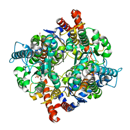
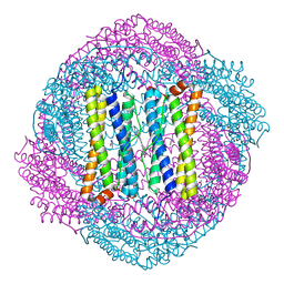
Crystal structure of ferritin (bacterioferritin) from Brucella melitensis
Descriptor: Bacterioferritin, CHLORIDE ION, FE (III) ION, ...
Authors:Seattle Structural Genomics Center for Infectious Disease (SSGCID)
Deposit date:2009-01-15
Release date:2009-02-03
Last modified:2023-09-06
Method:X-RAY DIFFRACTION (1.806 Å)
Cite:Crystal structure of ferritin from Brucella melitensis
To be Published
3GE4
DownloadVisualize
BU of 3ge4 by Molmil
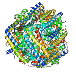
Crystal structure of ferritin:DNA-binding protein DPS from Brucella Melitensis
Descriptor: CALCIUM ION, DNA PROTECTION DURING STARVATION PROTEIN
Authors:Seattle Structural Genomics Center for Infectious Disease (SSGCID)
Deposit date:2009-02-25
Release date:2009-03-03
Last modified:2023-09-06
Method:X-RAY DIFFRACTION (1.7 Å)
Cite:Crystal structure of ferritin:DNA-binding protein DPS from Brucella Melitensis
To be Published
5UXX
DownloadVisualize
BU of 5uxx by Molmil
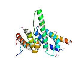
Co-crystal structure of the sigma factor RpoE in complex with the anti-sigma factor NepR from Bartonella quintana
Descriptor: Anti-sigma factor NepR, RNA polymerase sigma factor, SULFATE ION
Authors:Seattle Structural Genomics Center for Infectious Disease (SSGCID)
Deposit date:2017-02-23
Release date:2018-02-28
Method:X-RAY DIFFRACTION (2.45 Å)
Cite:Co-crystal structure of the sigma factor RpoE in complex with the anti-sigma factor NepR from Bartonella quintana
to be published
5UXW
DownloadVisualize
BU of 5uxw by Molmil
Crystal Structure of Anti-anti-sigma factor PhyR from Bartonella quintana
Descriptor: 1,2-ETHANEDIOL, Sensory transduction regulatory protein
Authors:Seattle Structural Genomics Center for Infectious Disease (SSGCID)
Deposit date:2017-02-23
Release date:2018-02-28
Last modified:2023-10-04
Method:X-RAY DIFFRACTION (2.3 Å)
Cite:Crystal Structure of Anti-anti-sigma factor PhyR from Bartonella quintana
to be published
3K9H
DownloadVisualize
BU of 3k9h by Molmil
Crystal structure of a plasmid partition protein from borrelia burgdorferi at 2.25A resolution
Descriptor: PF-32 protein, SULFATE ION
Authors:Seattle Structural Genomics Center for Infectious Disease (SSGCID)
Deposit date:2009-10-15
Release date:2009-11-03
Last modified:2023-09-06
Method:X-RAY DIFFRACTION (2.25 Å)
Cite:Crystal structure of a plasmid partition protein from borrelia burgdorferi at 2.25A resolution
To be Published
3KX6
DownloadVisualize
BU of 3kx6 by Molmil
Crystal structure of fructose-1,6-bisphosphate aldolase from Babesia bovis at 2.1A resolution
Descriptor: 1,2-ETHANEDIOL, CITRIC ACID, DI(HYDROXYETHYL)ETHER, ...
Authors:Seattle Structural Genomics Center for Infectious Disease (SSGCID)
Deposit date:2009-12-02
Release date:2009-12-22
Last modified:2024-03-13
Method:X-RAY DIFFRACTION (2.1 Å)
Cite:Crystal structure of fructose-1,6-bisphosphate aldolase from Babesia bovis at 2.1A resolution
To be Published
3KRE
DownloadVisualize
BU of 3kre by Molmil
Crystal structure of phosphoribosylaminoimidazole-succinocarboxamide synthase from Ehrlichia chaffeensis at 1.8A resolution
Descriptor: 1,2-ETHANEDIOL, CHLORIDE ION, Phosphoribosylaminoimidazole-succinocarboxamide synthase
Authors:Seattle Structural Genomics Center for Infectious Disease (SSGCID)
Deposit date:2009-11-18
Release date:2009-12-01
Last modified:2023-09-06
Method:X-RAY DIFFRACTION (1.8 Å)
Cite:Crystal structure of phosphoribosylaminoimidazole-succinocarboxamide synthase from Ehrlichia chaffeensis at 1.8A resolution
To be Published
3L0G
DownloadVisualize
BU of 3l0g by Molmil
Crystal structure of Nicotinate-nucleotide pyrophosphorylase from Ehrlichia chaffeensis at 2.05A resolution
Descriptor: 1,2-ETHANEDIOL, FORMIC ACID, Nicotinate-nucleotide pyrophosphorylase
Authors:Seattle Structural Genomics Center for Infectious Disease (SSGCID)
Deposit date:2009-12-09
Release date:2009-12-22
Last modified:2023-09-06
Method:X-RAY DIFFRACTION (2.05 Å)
Cite:Crystal structure of Nicotinate-nucleotide pyrophosphorylase from Ehrlichia chaffeensis at 2.05A resolution
To be Published
3GLQ
DownloadVisualize
BU of 3glq by Molmil
Crystal structure of S-adenosyl-L-homocysteine hydrolase from Burkholderia pseudomallei in complex with 9-beta-D-arabino-furansyl-adenine
Descriptor: 2-(6-AMINO-PURIN-9-YL)-5-HYDROXYMETHYL-TETRAHYDRO-FURAN-3,4-DIOL, Adenosylhomocysteinase, NICOTINAMIDE-ADENINE-DINUCLEOTIDE
Authors:Seattle Structural Genomics Center for Infectious Disease (SSGCID)
Deposit date:2009-03-12
Release date:2009-03-24
Last modified:2023-09-06
Method:X-RAY DIFFRACTION (2.3 Å)
Cite:Crystal structure of S-adenosyl-L-homocysteine hydrolase from Burkholderia pseudomallei in complex with 9-beta-D-arabino-furansyl-adenine
To be Published
5UXV
DownloadVisualize
BU of 5uxv by Molmil
Crystal Structure of Anti-anti-sigma factor PhyR I40V/S51C mutant from Bartonella quintana
Descriptor: 1,2-ETHANEDIOL, Sensory transduction regulatory protein
Authors:Seattle Structural Genomics Center for Infectious Disease (SSGCID)
Deposit date:2017-02-23
Release date:2018-02-28
Last modified:2023-10-04
Method:X-RAY DIFFRACTION (2.6 Å)
Cite:Crystal Structure of Anti-anti-sigma factor PhyR I40V/S51C mutant from Bartonella quintana
to be published

218853

PDB entries from 2024-04-24

PDB statisticsPDBj update infoContact PDBjnumon