3DAD
 
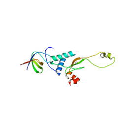
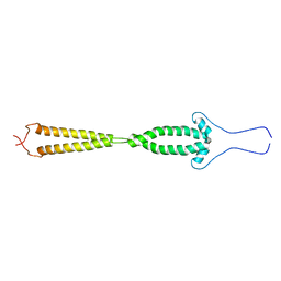
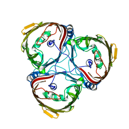
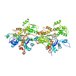
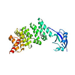 | | Crystal structure of the N-terminal regulatory domains of the formin FHOD1 | | 分子名称: | FH1/FH2 domain-containing protein 1 | | 著者 | Schulte, A, Stolp, B, Schonichen, A, Pylypenko, O, Rak, A, Fackler, O.T, Geyer, M. | | 登録日 | 2008-05-29 | | 公開日 | 2008-09-16 | | 最終更新日 | 2024-02-21 | | 実験手法 | X-RAY DIFFRACTION (2.3 Å) | | 主引用文献 | The Human Formin FHOD1 Contains a Bipartite Structure of FH3 and GTPase-Binding Domains Required for Activation.
Structure, 16, 2008
|
|
3REB
 | |
3REA
 | |
2PK2
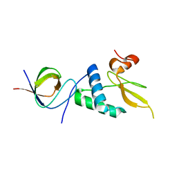
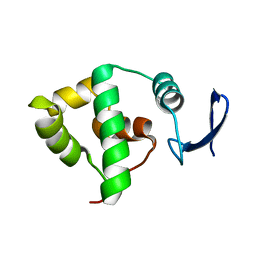
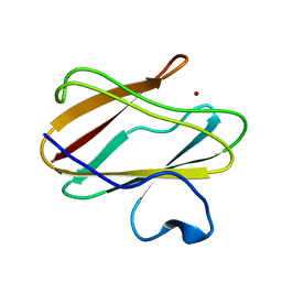
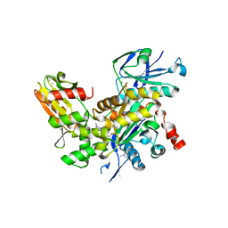
 | | Cyclin box structure of the P-TEFb subunit Cyclin T1 derived from a fusion complex with EIAV Tat | | 分子名称: | Cyclin-T1, Protein Tat | | 著者 | Anand, K, Schulte, A, Fujinaga, K, Scheffzek, K, Geyer, M. | | 登録日 | 2007-04-17 | | 公開日 | 2007-07-03 | | 最終更新日 | 2024-02-21 | | 実験手法 | X-RAY DIFFRACTION (2.67 Å) | | 主引用文献 | Cyclin Box Structure of the P-TEFb Subunit Cyclin T1 Derived from a Fusion Complex with EIAV Tat.
J.Mol.Biol., 370, 2007
|
|
3RBB
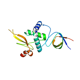
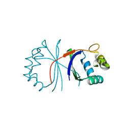
 | | HIV-1 NEF protein in complex with engineered HCK SH3 domain | | 分子名称: | 1,2-ETHANEDIOL, Protein Nef, Tyrosine-protein kinase HCK | | 著者 | Horenkamp, F.A, Schulte, A, Weyand, M, Geyer, M. | | 登録日 | 2011-03-29 | | 公開日 | 2011-04-27 | | 最終更新日 | 2023-09-13 | | 実験手法 | X-RAY DIFFRACTION (2.35 Å) | | 主引用文献 | Conformation of the Dileucine-Based Sorting Motif in HIV-1 Nef Revealed by Intermolecular Domain Assembly.
Traffic, 12, 2011
|
|
2HYB
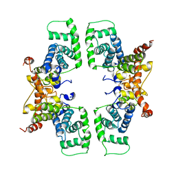
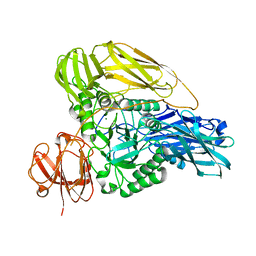
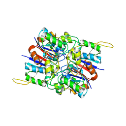 | | Crystal Structure of Hexameric DsrEFH | | 分子名称: | DsrH, Intracellular sulfur oxidation protein dsrF, Putative sulfurtransferase dsrE | | 著者 | Shin, D.H, Connie, H, Schulte, A, Dahl, C, Kim, R, Kim, S.H, Berkeley Structural Genomics Center (BSGC) | | 登録日 | 2006-08-04 | | 公開日 | 2007-07-03 | | 最終更新日 | 2024-02-21 | | 実験手法 | X-RAY DIFFRACTION (2.5 Å) | | 主引用文献 | Crystal structure of hexameric DsrEFH
To be Published
|
|
2HY5
 | | Crystal structure of DsrEFH | | 分子名称: | DsrH, Intracellular sulfur oxidation protein dsrF, Putative sulfurtransferase dsrE | | 著者 | Shin, D.H, Schulte, A, Dahl, C, Kim, R, Kim, S.H, Berkeley Structural Genomics Center (BSGC) | | 登録日 | 2006-08-04 | | 公開日 | 2006-09-19 | | 最終更新日 | 2024-02-14 | | 実験手法 | X-RAY DIFFRACTION (1.72 Å) | | 主引用文献 | Crystal structure of DsrEFH
To be Published
|
|
1YX3
 | | NMR structure of Allochromatium vinosum DsrC: Northeast Structural Genomics Consortium target OP4 | | 分子名称: | hypothetical protein DsrC | | 著者 | Cort, J.R, Dahl, C, Montelione, G.T, Kennedy, M.A, Northeast Structural Genomics Consortium (NESG) | | 登録日 | 2005-02-19 | | 公開日 | 2005-04-19 | | 最終更新日 | 2022-03-02 | | 実験手法 | SOLUTION NMR | | 主引用文献 | Allochromatium vinosum DsrC: solution-state NMR structure, redox properties, and interaction with DsrEFH, a protein essential for purple sulfur bacterial sulfur oxidation.
J.Mol.Biol., 382, 2008
|
|
2GD7
 | |
5MDP
 | |
5MDS
 | |
5MDR
 | |
5MDO
 | |
5MDQ
 | |
4P5S
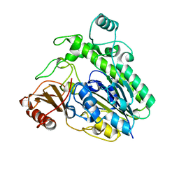
 | | Structure of reduced W45Y mutant of amicyanin | | 分子名称: | Amicyanin, COPPER (I) ION | | 著者 | Sukumar, N, Davidson, V.L. | | 登録日 | 2014-03-19 | | 公開日 | 2014-04-23 | | 最終更新日 | 2023-09-27 | | 実験手法 | X-RAY DIFFRACTION (1.02 Å) | | 主引用文献 | The sole tryptophan of amicyanin enhances its thermal stability but does not influence the electronic properties of the type 1 copper site.
Arch.Biochem.Biophys., 550-551, 2014
|
|
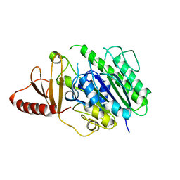
4P5R
 | | Structure of oxidized W45Y mutant of amicyanin | | 分子名称: | Amicyanin, COPPER (II) ION, SODIUM ION | | 著者 | Sukumar, N, Davidson, V.L. | | 登録日 | 2014-03-19 | | 公開日 | 2014-04-23 | | 最終更新日 | 2023-12-27 | | 実験手法 | X-RAY DIFFRACTION (1.09 Å) | | 主引用文献 | The sole tryptophan of amicyanin enhances its thermal stability but does not influence the electronic properties of the type 1 copper site.
Arch.Biochem.Biophys., 550-551, 2014
|
|
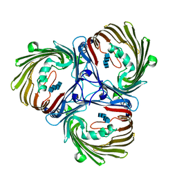
6HHM
 | | Crystal structure of the family S1_7 ulvan-specific sulfatase FA22070 from Formosa agariphila | | 分子名称: | Arylsulfatase, CALCIUM ION | | 著者 | Roret, T, Prechoux, A, Michel, G, Czjzek, M. | | 登録日 | 2018-08-28 | | 公開日 | 2019-06-26 | | 最終更新日 | 2024-01-17 | | 実験手法 | X-RAY DIFFRACTION (1.23 Å) | | 主引用文献 | A marine bacterial enzymatic cascade degrades the algal polysaccharide ulvan.
Nat.Chem.Biol., 15, 2019
|
|
6HR5
 | | Structure of the S1_25 family sulfatase module of the rhamnosidase FA22250 from Formosa agariphila | | 分子名称: | Alpha-L-rhamnosidase/sulfatase (GH78), CALCIUM ION | | 著者 | Roret, T, Prechoux, A, Czjzek, M, Michel, G. | | 登録日 | 2018-09-26 | | 公開日 | 2019-06-26 | | 最終更新日 | 2019-07-31 | | 実験手法 | X-RAY DIFFRACTION (2.912 Å) | | 主引用文献 | A marine bacterial enzymatic cascade degrades the algal polysaccharide ulvan.
Nat.Chem.Biol., 15, 2019
|
|
7WHG
 | |
7WHF
 | |
6HHN
 | |
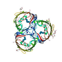
6HPD
 | | The structure of a beta-glucuronidase from glycoside hydrolase family 2 | | 分子名称: | BROMIDE ION, Beta-galactosidase (GH2), MAGNESIUM ION | | 著者 | Robb, C.S, Gerlach, N, Reisky, L, Bornshoeru, U, Hehemann, J.H. | | 登録日 | 2018-09-20 | | 公開日 | 2019-07-24 | | 最終更新日 | 2024-01-24 | | 実験手法 | X-RAY DIFFRACTION (2.43 Å) | | 主引用文献 | A marine bacterial enzymatic cascade degrades the algal polysaccharide ulvan.
Nat.Chem.Biol., 15, 2019
|
|
2W55
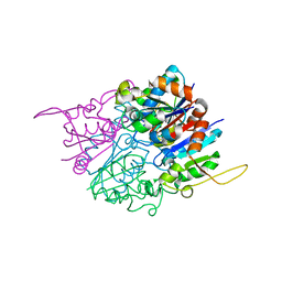
 | | Crystal Structure of Xanthine Dehydrogenase (E232Q variant) from Rhodobacter capsulatus in Complex with Hypoxanthine | | 分子名称: | BARIUM ION, FE2/S2 (INORGANIC) CLUSTER, FLAVIN-ADENINE DINUCLEOTIDE, ... | | 著者 | Doebbler, J.A, Truglio, J.J, Leimkuhler, S, Kisker, C. | | 登録日 | 2008-12-04 | | 公開日 | 2008-12-23 | | 最終更新日 | 2023-12-13 | | 実験手法 | X-RAY DIFFRACTION (3.4 Å) | | 主引用文献 | Mechanism of Substrate and Inhibitor Binding of Rhodobacter Capsulatus Xanthine Dehydrogenase.
J.Biol.Chem., 284, 2009
|
|
2W3R
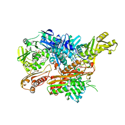 | | Crystal Structure of Xanthine Dehydrogenase (desulfo form) from Rhodobacter capsulatus in complex with hypoxanthine | | 分子名称: | CALCIUM ION, FE2/S2 (INORGANIC) CLUSTER, FLAVIN-ADENINE DINUCLEOTIDE, ... | | 著者 | Dietzel, U, Kuper, J, Leimkuhler, S, Kisker, C. | | 登録日 | 2008-11-14 | | 公開日 | 2008-12-23 | | 最終更新日 | 2023-12-13 | | 実験手法 | X-RAY DIFFRACTION (2.9 Å) | | 主引用文献 | Mechanism of Substrate and Inhibitor Binding of Rhodobacter Capsulatus Xanthine Dehydrogenase.
J.Biol.Chem., 284, 2009
|
|
2W3S
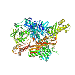 | | Crystal Structure of Xanthine Dehydrogenase (desulfo form) from Rhodobacter capsulatus in Complex with Xanthine | | 分子名称: | CALCIUM ION, FE2/S2 (INORGANIC) CLUSTER, FLAVIN-ADENINE DINUCLEOTIDE, ... | | 著者 | Dietzel, U, Kuper, J, Leimkuhler, S, Kisker, C. | | 登録日 | 2008-11-14 | | 公開日 | 2008-12-23 | | 最終更新日 | 2023-12-13 | | 実験手法 | X-RAY DIFFRACTION (2.6 Å) | | 主引用文献 | Mechanism of Substrate and Inhibitor Binding of Rhodobacter Capsulatus Xanthine Dehydrogenase.
J.Biol.Chem., 284, 2009
|
|


